![]()
Medical Terminology Daily (MTD) is a blog sponsored by Clinical Anatomy Associates, Inc. as a service to the medical community. We post anatomical, medical or surgical terms, their meaning and usage, as well as biographical notes on anatomists, surgeons, and researchers through the ages. Be warned that some of the images used depict human anatomical specimens.
You are welcome to submit questions and suggestions using our "Contact Us" form. The information on this blog follows the terms on our "Privacy and Security Statement" and cannot be construed as medical guidance or instructions for treatment.
We have 139 guests and no members online
")
Marcia Crocker Noyes
(1869 – 1946)
Further to my comment on old books and research that started with an interesting bookplate (Ex-Libris). I continued my research and found that the person in charge of the Osler library bookplate was a fascinating individual that today maybe a ghost in the MedChi library and building in Baltimore... This is certainly an article that can be called "A Moment in History"
Marcia Crocker Noyes was the librarian at The Maryland State Medical Society from 1896 to 1946 and was a founding member of the Medical Library Association.[1][2][3]
Sir William Osler, MD. a famous Johns Hopkins surgeon was a noted bibliophile and had a large personal collection of books on various topics. When he became the President of MedChi in 1896, he was dismayed at the condition of the library and knew that with the right person and some stewardship, it could become a significant collection. Sir William asked his friend, Dr. Bernard Steiner, a physician and President of the Enoch Pratt Free Library in Baltimore for suggestions of a librarian, and Dr. Steiner recommended Marcia Crocker Noyes. A native of New York, and a graduate of Hunter College, Marcia had moved to Baltimore for a lengthy visit with her sister, and took a “temporary” position at the Pratt Library, which turned into three years. Although she had no medical experience or background, she was enthusiastic, and most importantly, she was willing to move into the apartment provided for the librarian, who needed to be available 24 hours a day.
The image in this article is Ms. Noyes on her first year on the job. Marcia developed a book classification system for medical books, based on the Index Medicus, and called it the Classification for Medical Literature. The system uses the alphabet with capital letters for the major divisions of medicine and lower-case ones for the sub-sections. The system was used for many years, but it's now dated and the Faculty's original shelving scheme was never changed. The card catalogs still reflect her classification and many of the cards are written in Marcia's back-slanting handwriting.
Marcia knew enough to ask the Faculty's members about medical questions, terminology and literature. She gradually won over the predominantly male membership and they became her greatest allies; Sir William at the start, and then for nearly 40 years, Dr. John Ruhräh, a wealthy pediatrician with no immediate family of his own. She made a point of attending almost every Faculty function, and in 1904, under guidelines from the American Medical Association, Marcia was made the Faculty Secretary. For much of her first 10 years, she was the Faculty's only full-time employee, only being assisted by Mr. Caution, the Faculty's janitor. Later in life Marcia would say that she hired him because of his name!
Within ten years, the library had outgrown its space, and plans, spearheaded by Marcia and Sir William before his move to Oxford, were made to build a headquarters building, mainly to house the library's growing collection of medical books and journals.
Marcia was instrumental in the design and building of the new headquarters. She travelled to Philadelphia, New York and Boston to look at their medical society buildings, and eventually, the Philadelphia architectural firm, Ellicott & Emmart was selected to design and build the new Faculty building. Every detail of the building held her imprimatur, from the graceful staircase, to the light-filled reading room, and all of the myriad details of the millwork, marble tesserae, and most of all, the four-story cast iron stacks. She was on-site, climbing up unfinished staircases, checking out the progress of the building, which was built in less than one year at a cost of $90,000.
Among the features of the new building was a fourth-floor apartment for her. She referred to it as the "first penthouse in Baltimore" and it had a garden and rooftop terrace. The library collection eventually grew to more than 65,000 volumes from medical and specialty societies around the world. Journals were traded back and forth, and physicians eagerly anticipated the arrival of each new issue. At the same time, Marcia was involved in the Medical Library Association as one of eight founding members. The MLA promotes medical libraries and the exchange of information. One of the earliest mandates of the MLA was the Exchange, a distribution and trade service for those who had duplicates or little-used books in their collections. Initially, the Exchange was run out of the Philadelphia medical society, but in 1900 it was moved to Baltimore and Marcia oversaw it. Several hundred periodicals and journals were received and sent each month, a huge amount of work for a tiny staff. In 1904, the Faculty had run out of room to manage the Exchange, so it was moved to the Medical Society of the Kings County (Brooklyn). But without Marcia's excellent administrative skills, it floundered and in 1908, the MLA asked Marcia to take charge once again.
In 1909, when the new Faculty building opened, there was enough room to run the Exchange and with the help of MLA Treasurer, noted bibliophile and close friend, Dr. John Ruhräh, it once again became successful. Additionally, Marcia and Dr. Ruhräh combined forces to revive the MLA's bulletin, which had all but ceased publication in 1908, taking the Exchange with it. This duo maintained editorial control from 1911 until 1926. In 1934, around the time of Dr. Ruhräh's death, Marcia became the first “unmedicated” professional to head the MLA. During her tenure, the MLA incorporated, the first seal was adopted, and the annual meeting was held in Baltimore. Marcia wanted to write the history of the MLA once she retired from full-time work at the Faculty, but her health was beginning to fail. She had back problems and had suffered a serious burn on her shoulder as a young woman, possibly from her time running a summer camp, Camp Seyon, for young ladies in the Adirondack Mountains. In 1946, a celebration was planned to honor Marcia's 50 years at the Faculty. But she was adamant that the physicians wait until November, the actual date of her 50 years. However, they knew she was gravely ill, and might not make it until then, so a huge party was held in April. More than 250 physicians attended the celebration, but the ones she was closest to in the early years, were long gone. She was presented with a suitcase, a sum of money to use for travelling, and her favorite painting of Dr. John Philip Smith, a founder of the Medical College in Winchester, Virginia. It was painted by Edward Caledon Smith, a Virginia painter who had been a student of the painter Thomas Sully.[4] She adored this painting and vowed, jokingly, to take it with her wherever she went.
The painting was not to stay with her for very long, for she died in November 1946, and left it to the Faculty in her will. Her funeral was held in the Faculty's Osler Hall, named for her dear friend. More than 60 physicians served as her pallbearers, and she was buried at Baltimore's Green Mount Cemetery. In 1948, the MLA decided to establish an award in the name of Marcia Crocker Noyes. It was for outstanding achievement in medical library field and was to be awarded every two years, or when a truly worthy candidate was submitted. In 2014, the Faculty began giving a bouquet of flowers to the winner of the award in Marcia's name, and in honor of her work. Much evidence exists for this tradition, as we know that the physicians, especially Drs. Osler and Ruhräh, frequently gave her bouquets of flowers. Marcia also cultivated flower gardens at the Faculty and decorated the rooms with her work.
Today, the MedChi building is open for tours and if the rumors are to be believed Ms. Marcia Crocker Noyes is still at work in her beloved library as the "resident ghost" [1][5]

NOTE: This article has been modified from the original Wikipedia article on Marcia Crocker Noyes. The article itself is well-written with interesting images of the subject. I would encourage you to visit it. The second insert is from book 00736 in my personal library and shows in pencil, the incredibly small handwriting of Marsha C. Noyes.
Sources:
1. "Marcia, Marcia, Marcia" MedChi Archives blog.
2. "Marcia C. Noyes, Medical Librarian" (PDF). Bulletin of the Medical Library Association. 35 (1): 108–109. 1947. PMC 194645
3. Smith, Bernie Todd (1974). "Marcia Crocker Noyes, Medical Librarian: The Shaping of a Career" (PDF). Bulletin of the Medical Library Association. 62 (3): 314–324. PMC 198800Freely accessible. PMID 4619344.
4. Edward Caledon BRUCE (1825-1901)"
5. Behind the scenes tour MedChiBuilding
"Clinical Anatomy Associates, Inc., and the contributors of "Medical Terminology Daily" wish to thank all individuals who donate their bodies and tissues for the advancement of education and research”.
Click here for more information


- Details
Aaron Ruhalter, MD, FACS
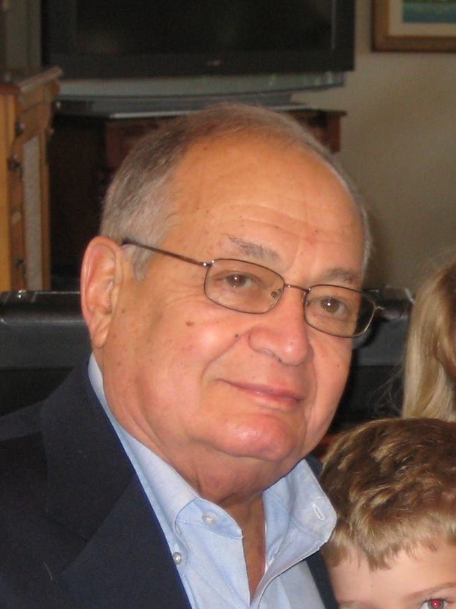
I am sad to let everybody know that Dr. Ruhalter passed on yesterday January 25th, 2016. A good friend, guide, and mentor, Dr. Ruhalter was always reminding me to keep on using drawings and sketches to teach human anatomy, a subject he loved, and at which he excelled. I will miss him dearly. May he rest in peace. Dr. Miranda
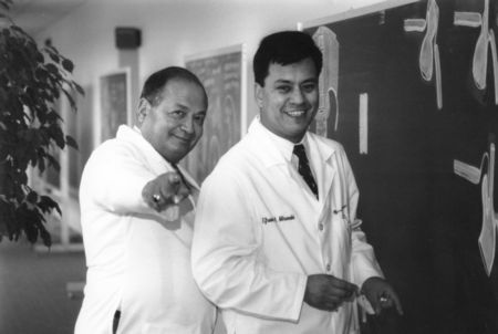
Dr. Aaron Ruhalter was for many years the Executive Director of Medical Education at the Johnson & Johnson Endo-Surgery Institute in Cincinnati. Dr. Ruhalter is a Professor of Anatomy and a former Professor of Surgery at the Robert Wood Johnson Memorial Hospital in New Jersey. He was also one of the founding members of the American Association of Clinical Anatomists (AACA).
His review on the surgical anatomy of the parotid gland, submandibular triangle, and floor of the mouth is outstanding. This review was published in 1997 in the book "Mastery of Surgery", third edition, by Drs. Lloyd M. Nyhus, Robert J. Baker, and Josef E. Fischer.
Dr. Miranda worked with Dr. Ruhalter for several years, both at the Endo-Surgery Institute and at the University of Cincinnati College of Medicine. The picture above shows both of them preparing an anatomy blackboard session at the Institute, back in 1994.
- Details
Refers to the internal lining of an artery or a vein. The tunica (layer) intima is composed of an inner layer known as endothelium (inner fabric or inner layer) and a subendothelial layer.
Also known as the [tunica intima vasorum], Latin for intimal (inner) layer of the vessels, the tunica inima is also known as "Bichat's tunic"
- Details

Click for a larger image
The term [bruxism] arises from the Greek [βρυγμός] (vrygm?s), meaning “a gnashing of the teeth”, or “bruxism”.
Bruxism is usually a subconscious problem, presenting most of the times at night while the individual is asleep, and does not continue into the waking hours. In some people bruxism does continue into the day and the person is constantly gnashing the teeth, but not conscious of the activity. Awake bruxism is sometimes called [bruxomania].
Bruxism is usually idiopathic (of unknown origin) or it can be secondary to other medical conditions.
Patients with bruxism usually present with damage to the teeth by the constant attrition of the teeth by laterally grinding of the teeth of the maxilla against the teeth of the jaw. This can lead to severe tooth damage with exposure of the dentin through and absent, fractured, or damaged enamel. A secondary problem is misalignment of the teeth. The accompanying image shows misalignment and tooth damage.
Note: The links to Google Translate include an icon that will allow you to hear the pronunciation of the word.
Image by DRosenbach (en:Image:Deviated midline.JPG) [Public domain], Wikimedia Commons
- Details

Abdominal regions
The transtubercular plane (also called the transtubercular line) is one of the surface reference lines used in surface anatomy and surgery to delineate the abdominal regions.
It is a horizontal or transverse plane that passes through the iliac tubercles, small elevations found in the iliac crest of the iliac bone. These tubercles can be palpated in a thin person, but their location can be approximated if you take the distance between the transpyloric plane and the superior aspect of the pubic symphysis and divide it in half.
The transtubercular plane marks the location of the body of the fifth lumbar vertebra and the location of the confluence of both common iliac veins to form the inferior vena cava.
Sources:
1. "Clinical Anatomy" Brantigan, OC 1963 McGraw Hill
2. "Tratado de Anatomia Humana" Testut et Latarjet 8th Ed. 1931 Salvat Editores, Spain
3. Davis, Gwilym G. "Applied Anatomy: The Construction of the Human Body Considered in Relation to Its Functions, Diseases, and Injuries"; Philadelphia: J.B. Lippincott Co., 1910
Image modified from the original Davis, 1910
- Details
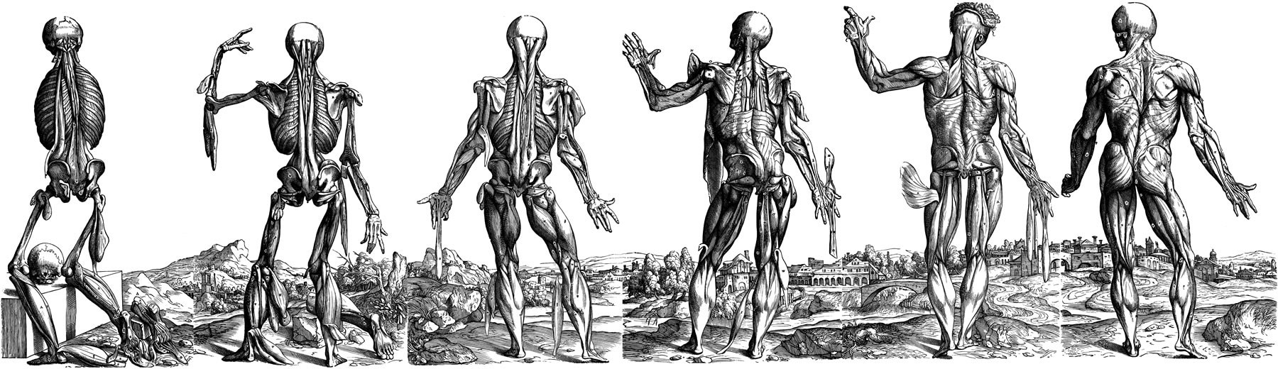
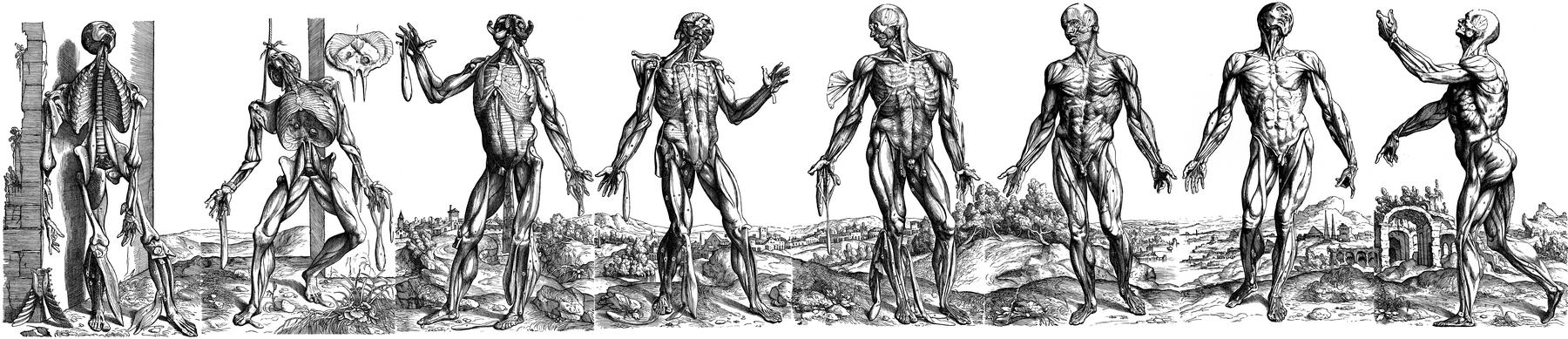
UPDATED: If you came to this article directly, you should probably start reading the first one here: "The landscape panorama of Vesalius' muscle men (1)", then come back.
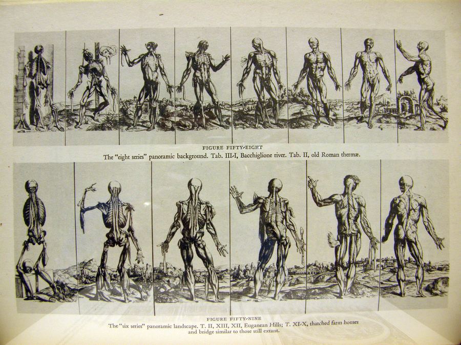
I had always looked at these panorama images as only one, the one that is formed by the anterior views (and one lateral view) of the muscle men plates which I shared with you in the first article. The image in the first article is what Harvey Cushing (1943) called the "eight-series". It was not until my friend Pascale Pollier sent me an interesting BBC article that I looked at the panorama formed by the posterior view images of the muscle men, called by Cushing the "six-series". After some work with original images that I own, I made the image shown here. If anything this panorama is even better!To see Cushing's original template click here.
In the enlarged version that appears when you click on the image on this article you can see how some houses' roofing and arches are shared between plates. Towards the right side of this panorama you can clearly see a meandering stream that passes through several plates. Another incredible fact of this masterpiece by Jan Stephan Van Calcar (the artist) and Andrea Vesalius (the anatomist).
The image shown here was done using original images from Vesalius’ Fabrica and composed using Adobe Fireworks CS5. Click on the image for a larger depiction. The larger image is 1800px wide.
Sources:
1. “A New View of the Vesalian Landscape” Cavanagh, GST Med Hist 1983, 27: 77-79
2. “The Panorama of Vesalius: A 'Lost' Design From Titian's Studio” Skandalakis, JE JAMA May 28, 1997, Vol 277, No. 20
3. “A Drawing for the Fabrica; and some Thoughts Upon the Vesalius Muscle-Men” Kemp. M. Med Hist. Jul 1970; 14(3): 277–288
4. “Andreas Vesalius: The Making, the Madman and the Myth” Joffe, SN. Persona Publishing 2009
5. "A Bio-Bibliography of Andreas Vesalius" Cushing, H. (1943) Schumann's
- Details
UPDATED: There is no doubt that the book “De Humani Corporis Fabrica Libri Septem” written by Andreas Vesalius and published by Johannes Oporinus in May, 1543 is one of the most important scientific anatomical and medical books ever written. Much has been said about the place this book has in medical history as part of the discarding of dogmas and the establishment of scientific observation and thinking.
Some of the most intriguing images published in the book are fourteen woodcuts in the second book. These amazing and detailed images show the muscles in a whole body as it is dissected. The text details the structures and the procedure of how the dissection is performed.
It is believed that these images were done by Jan Stephan Van Calcar, an artist from the Titian’s studio, although there are indications that these images may have been authored by somebody else, or even that they were the effort of more than one artist working under the close supervision of Andreas Vesalius.
Today’s anatomical images are very descriptive and the artistry is relegated to the technique used for the depiction of the image. In Vesalius’ muscle men plates each image has a background showing a landscape. It was not until 1903 that it was discovered that the landscapes of the different images were part of a complete landscape. This is evident if these images are placed side by side. There have been several books and articles published on these images. Interestingly, some of the images had to be reversed to be placed in the panorama. This is due to the process of cutting the woodblocks.
The image in this article is one of two identified and composed by Harvey Cushing in 1943 (see sources). He calls it the "eight-series". The "six-series" can be seen here. To see Cushing's original template click here. The panorama of the six-series was identified by Cushing to be an area of the Euganean hills near Venice and Padua, Italy. There landscape formed by the eight-series is an actual region near Padua, Italy showing the ruins of old Roman baths. Cavanagh (1938) adds more information on these images, for his .pdf article, click here.
The image shown at the top of this article was created using original images from Vesalius’ Fabrica and composed using Adobe Fireworks CS5. Click on the image for a larger depiction. The large image is 1800px wide.
NOTE: In June 2023, I was able to purchase a single leaf of the 1543 Fabrica, page 187, which depicts one of the "muscle men". In the panorama image in this article it is figure number 3, from left to right. For more information on this image click here. This image is now properly framed in my office. Dr. Miranda.
This article continues here: "The landscape panorama of Vesalius' Muscle Men (2)"
Sources:
1. “A New View of the Vesalian Landscape” Cavanagh, GST Med Hist 1983, 27: 77-79
2. “The Panorama of Vesalius: A 'Lost' Design From Titian's Studio” Skandalakis, JE JAMA May 28, 1997, Vol 277, No. 20
3. “A Drawing for the Fabrica; and some Thoughts Upon the Vesalius Muscle-Men” Kemp. M. Med Hist. Jul 1970; 14(3): 277–288
4. “Andreas Vesalius: The Making, the Madman and the Myth” Joffe, SN. Persona Publishing 2009
5. "A Bio-Bibliography of Andreas Vesalius" Cushing, H. (1943) Schumann's