![]()
Medical Terminology Daily (MTD) is a blog sponsored by Clinical Anatomy Associates, Inc. as a service to the medical community. We post anatomical, medical or surgical terms, their meaning and usage, as well as biographical notes on anatomists, surgeons, and researchers through the ages. Be warned that some of the images used depict human anatomical specimens.
You are welcome to submit questions and suggestions using our "Contact Us" form. The information on this blog follows the terms on our "Privacy and Security Statement" and cannot be construed as medical guidance or instructions for treatment.
We have 498 guests and no members online
")
Marcia Crocker Noyes
(1869 – 1946)
Further to my comment on old books and research that started with an interesting bookplate (Ex-Libris). I continued my research and found that the person in charge of the Osler library bookplate was a fascinating individual that today maybe a ghost in the MedChi library and building in Baltimore... This is certainly an article that can be called "A Moment in History"
Marcia Crocker Noyes was the librarian at The Maryland State Medical Society from 1896 to 1946 and was a founding member of the Medical Library Association.[1][2][3]
Sir William Osler, MD. a famous Johns Hopkins surgeon was a noted bibliophile and had a large personal collection of books on various topics. When he became the President of MedChi in 1896, he was dismayed at the condition of the library and knew that with the right person and some stewardship, it could become a significant collection. Sir William asked his friend, Dr. Bernard Steiner, a physician and President of the Enoch Pratt Free Library in Baltimore for suggestions of a librarian, and Dr. Steiner recommended Marcia Crocker Noyes. A native of New York, and a graduate of Hunter College, Marcia had moved to Baltimore for a lengthy visit with her sister, and took a “temporary” position at the Pratt Library, which turned into three years. Although she had no medical experience or background, she was enthusiastic, and most importantly, she was willing to move into the apartment provided for the librarian, who needed to be available 24 hours a day.
The image in this article is Ms. Noyes on her first year on the job. Marcia developed a book classification system for medical books, based on the Index Medicus, and called it the Classification for Medical Literature. The system uses the alphabet with capital letters for the major divisions of medicine and lower-case ones for the sub-sections. The system was used for many years, but it's now dated and the Faculty's original shelving scheme was never changed. The card catalogs still reflect her classification and many of the cards are written in Marcia's back-slanting handwriting.
Marcia knew enough to ask the Faculty's members about medical questions, terminology and literature. She gradually won over the predominantly male membership and they became her greatest allies; Sir William at the start, and then for nearly 40 years, Dr. John Ruhräh, a wealthy pediatrician with no immediate family of his own. She made a point of attending almost every Faculty function, and in 1904, under guidelines from the American Medical Association, Marcia was made the Faculty Secretary. For much of her first 10 years, she was the Faculty's only full-time employee, only being assisted by Mr. Caution, the Faculty's janitor. Later in life Marcia would say that she hired him because of his name!
Within ten years, the library had outgrown its space, and plans, spearheaded by Marcia and Sir William before his move to Oxford, were made to build a headquarters building, mainly to house the library's growing collection of medical books and journals.
Marcia was instrumental in the design and building of the new headquarters. She travelled to Philadelphia, New York and Boston to look at their medical society buildings, and eventually, the Philadelphia architectural firm, Ellicott & Emmart was selected to design and build the new Faculty building. Every detail of the building held her imprimatur, from the graceful staircase, to the light-filled reading room, and all of the myriad details of the millwork, marble tesserae, and most of all, the four-story cast iron stacks. She was on-site, climbing up unfinished staircases, checking out the progress of the building, which was built in less than one year at a cost of $90,000.
Among the features of the new building was a fourth-floor apartment for her. She referred to it as the "first penthouse in Baltimore" and it had a garden and rooftop terrace. The library collection eventually grew to more than 65,000 volumes from medical and specialty societies around the world. Journals were traded back and forth, and physicians eagerly anticipated the arrival of each new issue. At the same time, Marcia was involved in the Medical Library Association as one of eight founding members. The MLA promotes medical libraries and the exchange of information. One of the earliest mandates of the MLA was the Exchange, a distribution and trade service for those who had duplicates or little-used books in their collections. Initially, the Exchange was run out of the Philadelphia medical society, but in 1900 it was moved to Baltimore and Marcia oversaw it. Several hundred periodicals and journals were received and sent each month, a huge amount of work for a tiny staff. In 1904, the Faculty had run out of room to manage the Exchange, so it was moved to the Medical Society of the Kings County (Brooklyn). But without Marcia's excellent administrative skills, it floundered and in 1908, the MLA asked Marcia to take charge once again.
In 1909, when the new Faculty building opened, there was enough room to run the Exchange and with the help of MLA Treasurer, noted bibliophile and close friend, Dr. John Ruhräh, it once again became successful. Additionally, Marcia and Dr. Ruhräh combined forces to revive the MLA's bulletin, which had all but ceased publication in 1908, taking the Exchange with it. This duo maintained editorial control from 1911 until 1926. In 1934, around the time of Dr. Ruhräh's death, Marcia became the first “unmedicated” professional to head the MLA. During her tenure, the MLA incorporated, the first seal was adopted, and the annual meeting was held in Baltimore. Marcia wanted to write the history of the MLA once she retired from full-time work at the Faculty, but her health was beginning to fail. She had back problems and had suffered a serious burn on her shoulder as a young woman, possibly from her time running a summer camp, Camp Seyon, for young ladies in the Adirondack Mountains. In 1946, a celebration was planned to honor Marcia's 50 years at the Faculty. But she was adamant that the physicians wait until November, the actual date of her 50 years. However, they knew she was gravely ill, and might not make it until then, so a huge party was held in April. More than 250 physicians attended the celebration, but the ones she was closest to in the early years, were long gone. She was presented with a suitcase, a sum of money to use for travelling, and her favorite painting of Dr. John Philip Smith, a founder of the Medical College in Winchester, Virginia. It was painted by Edward Caledon Smith, a Virginia painter who had been a student of the painter Thomas Sully.[4] She adored this painting and vowed, jokingly, to take it with her wherever she went.
The painting was not to stay with her for very long, for she died in November 1946, and left it to the Faculty in her will. Her funeral was held in the Faculty's Osler Hall, named for her dear friend. More than 60 physicians served as her pallbearers, and she was buried at Baltimore's Green Mount Cemetery. In 1948, the MLA decided to establish an award in the name of Marcia Crocker Noyes. It was for outstanding achievement in medical library field and was to be awarded every two years, or when a truly worthy candidate was submitted. In 2014, the Faculty began giving a bouquet of flowers to the winner of the award in Marcia's name, and in honor of her work. Much evidence exists for this tradition, as we know that the physicians, especially Drs. Osler and Ruhräh, frequently gave her bouquets of flowers. Marcia also cultivated flower gardens at the Faculty and decorated the rooms with her work.
Today, the MedChi building is open for tours and if the rumors are to be believed Ms. Marcia Crocker Noyes is still at work in her beloved library as the "resident ghost" [1][5]

NOTE: This article has been modified from the original Wikipedia article on Marcia Crocker Noyes. The article itself is well-written with interesting images of the subject. I would encourage you to visit it. The second insert is from book 00736 in my personal library and shows in pencil, the incredibly small handwriting of Marsha C. Noyes.
Sources:
1. "Marcia, Marcia, Marcia" MedChi Archives blog.
2. "Marcia C. Noyes, Medical Librarian" (PDF). Bulletin of the Medical Library Association. 35 (1): 108–109. 1947. PMC 194645
3. Smith, Bernie Todd (1974). "Marcia Crocker Noyes, Medical Librarian: The Shaping of a Career" (PDF). Bulletin of the Medical Library Association. 62 (3): 314–324. PMC 198800Freely accessible. PMID 4619344.
4. Edward Caledon BRUCE (1825-1901)"
5. Behind the scenes tour MedChiBuilding
"Clinical Anatomy Associates, Inc., and the contributors of "Medical Terminology Daily" wish to thank all individuals who donate their bodies and tissues for the advancement of education and research”.
Click here for more information


- Details
The term [incarcerated] is composed by the prefix [in-] which is Latin and means “in”, and “into”; the Latin root [-carcer-] meaning “jail” and the adjectival suffix [-ated]. [Incarcerated] means “jailed” or “in jail”.
It is used in medicine to denote a hernia where a viscus or viscera are trapped inside its hernia sac, not allowing the visceral contents to move or slide in and out of the hernia sac. Incarcerated hernias have higher potential for morbidity as the protruding hernia is now prone to outside trauma and there is also the potential for vascular complications such as ischemia or hypoxia.
Note: The links to Google Translate include an icon that will allow you to hear the pronunciation of the word.
- Details
![By Government of the United Kingdom. (Transferred from en.wikipedia.) [Public domain], via Wikimedia Commons](https://commons.wikimedia.org/wiki/File:Letters_Patent_Australia.jpg "By Government of the United Kingdom. (Transferred from en.wikipedia.) [Public domain], via Wikimedia Commons")
Click for a larger image
The medical terms [patent] and [patency] use the root term [-pat-] which arises from the Latin word [patentum] and its nominative form [patens], meaning "open". It refers to a structure that is open allowing for free flow.
The suffixes [-ent] and [-cy] are abstract suffixes (similar to adjectival suffixes) that are used as "pertaining to", or simply ignored.
Note of interest: An initial look at this term used in medicine clashes with the legal meaning of [patent] which is defined as a "a government authority or license conferring a right or title for a set period, especially the sole right to exclude others from making, using, or selling an invention" (Google search). Fact is, they are similar and arise from the same origin.
In medieval times letters meant to be read by a single individual were sealed, but government letters, edicts or resolutions meant to be read by many or be the law of the land where written as [litteræ patentes] (medieval Latin) which can be loosely translated as "an open letter". These "Letters of Patent" where written in parchment with seals and looked impressive! The term later was reduced to "patent", thus the double meaning for this word both in the medical and legal arenas.
Note: The links to Google Translate include an icon that will allow you to hear the pronunciation of the word.
Clicking on the image will take you to an example of a patent (Australia)
- Details
- Written by: Prof. C. Uribe

Click for a larger image
Anatomy: The term [aortopulmonary window] is a radiological term that refers to a small space in the left mediastinal region. It is bounded anteriorly by the ascending aorta, posteriorly by the descending aorta, superiorly by the aortic arch, inferiorly by the left pulmonary artery, medially by the arterial ligament [ligamentum arteriosum] and left main bronchus, and laterally by the pleura and left lung. It contents the recurrent laryngeal nerve, lymph nodes, and adipose tissue.
Clinical: The aortopulmonary window is commonplace for lymphadenopathy in various inflammatory and neoplastic diseases.
Radiology: In a frontal projection, it corresponds to a focal concavity on the left border of the mediastinum, inferior to the aorta and superior to the left pulmonary artery. In the lateral projection (which is the proper image to identify this area), it is seen as a radiolucency inferior to the aortic arch and superior to the left pulmonary artery. Its appearance can be modified by tortuosity of the aorta.
Congenital heart disease: This term also refers to a congenital heart disease similar in appearance to patent ductus arteriosus, (or truncus arteriosus) with the difference that this involves septal defect. It is described as a communication between the ascending aorta and pulmonary trunk portion or right pulmonary artery. It is a rare anomaly that represents 0.2% -0.6% of congenital cardiac abnormalities.
Sources:
1. Heitzman ER. The infraaortic area. In: The mediastinum: With correlations radiologic anatomy and pathology. Berlin, Germany: Springer-Verlag, 1988; 151: -168.
2. Blank N, Castellino RA. Patterns of pleural reflections of the left upper mediastinum: Normal anatomy and distortions produced by adenopathy. Radiology 1972; 102: 585-589.
3. Marc Dewey, Donna Magid, Paul S. Wheeler and Bernd Hamm aortopulmonary Angle Window or on the Chest Radiograph? American Journal of Roentgenology. 2004; 182: 1085-1086.
4. SY Ho, Gerlis LM, Anderson C, Devine WA, Smith A. The morphology of aortopulmonary windows With regard to their classification and morphogenesis. Young Cardiol 1994; 4: 146-55.
5. Kutsche LM, Van Mierop LHS. Anatomy and pathogenesis of aorticopulmonary septal defect. Am J Cardiol 1987; 59: 443-7.
6. Stevenson, Roger E .; Hall, Judith G. (2006). Human malformations and related anomalies. Oxford University Press US. pp. 119-. ISBN 978-0-19-516568-5
7. Donoghue, Veronica B .; Bj?rnstad, Per G. Radiological Imaging of the Neonatal Chest. Springer. pp. 330-. ISBN 978-3-540-33748-5.
- Details
- Written by: Prof. C. Uribe
|
Anatomy:[Hilum] is a generic term which names area of an organ where all the structures that enter or leave the organ are found. In the lung it is depression in the mediastinal surface of the lung through which the vessels and bronchi enter and leave the lungs. This set of structures corresponds to the “root of the lung” or “pulmonary pedicle”. The term has a singular form, [hilum]; and a plural form, [hila]; the adjectival form is [hilar]. Radiographs and CAT Scan: The hilum appears as a composite opacity at the root of each lung produced by bronchi, arteries, veins, lymph nodes, nerves ,and other tissues. Sources: |
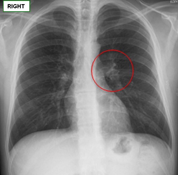 AP thoracic x-ray image showing the pulmonary hilum |
| Back to MTD Main Page | Subscribe to MTD |
- Details

Click for a larger image
We would like to welcome Professor Cristián Uribe as a contributor to Medical Terminology Daily.
Prof. Uribe is a Physical Therapist, has a Masters in Human Anatomy and is a Professor in the Human Anatomy Department of the Medical School at the University Finis Terrae in Santiago, Chile.
Prof. Uribe is also the Director of the Postgraduate Course on “Anatomical Bases of Normal Imaging”, as well as the Executive Secretary of the Postgraduate Office at the same University.
He published the book “Eponyms in Anatomical Nomenclature” (2011, Ed. U. Finis Terrae). For his LinkedIn page click here.
- Details

Click for a larger image
The [valve of the inferior vena cava] is probably better known by its eponym, the [Eustachian valve]. This is an incomplete valve found at the most distal end of the inferior vena cava, at the point where it opens into the right atrium of the heart. The valve is muscular at its base and composed mostly by membranous endocardium, and is normally the only valve present in the inferior vena cava.
It appears as a membranous semilunar fold extending posteriorly from the limbus fossa ovalis, anterior to the inferior caval orifice where it disappears. Its free border is usually membranous, concave, and directed anterosuperiorly. Its base is a transverse muscular ridge continuous with the anterior margin of the caval orifice.
It has no function in the adult, but in the fetus it helps to divert the flow of blood coming from the inferior vena cava towards the foramen ovale [fossa ovalis], as part of fetal circulation.
It has great anatomical variation in size, shape and thickness. On its medial aspect it joins with the Thebesian valve at the orifice of the coronary sinus.
First described by Bartolomeo Eustachius (c1500 - 1574) in 1653, the existence of the valve of the inferior vena cava was disputed by many until Winslow in 1717 confirmed its presence and suggested its function in fetal circulation.
Sources:
1. "The Valve of the Inferior Vena Cava" Hickie, JB Br Heart J. 1956 Jul; 18(3): 320–326
2. "Bartolommeo Eustachio; a great medical genius whose masterpiece remained hidden for 150 years" Wells, WA Arch Otolaring (1925) 48: 58