
Medical Terminology Daily (MTD) is a blog sponsored by Clinical Anatomy Associates, Inc. as a service to the medical community. We post anatomical, medical or surgical terms, their meaning and usage, as well as biographical notes on anatomists, surgeons, and researchers through the ages. Be warned that some of the images used depict human anatomical specimens.
You are welcome to submit questions and suggestions using our "Contact Us" form. The information on this blog follows the terms on our "Privacy and Security Statement" and cannot be construed as medical guidance or instructions for treatment.
We have 2236 guests online
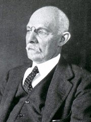
William S. Halsted, MD
(1852 – 1922)
American anatomist, teacher, and surgeon, William Stewart Halsted was born in New York City, USA to a wealthy family of English origin. His father was involved in charitable work and Governor and trustee to a city hospital. Not a brilliant student initially, Halsted took an undergraduate in Liberal Arts in Yale, CT., after which he entered the Medical College of Physicians at the Columbia College, where he excelled.
As a second-year medical student Halsted applied and obtained a position in surgery at a local hospital. In here he learned about Lister’s antiseptic technique and became an adamant proponent of it to reduce infection. In 1877 Halsted obtained his MD. After a short time as House Physician at the New York Hospital, Halsted traveled to Europe to further his education studying for two years at the Universities of Vienna, Leipzig, and W?rzburg.
Besides being at the forefront of surgical and antiseptic techniques (introducing the use of rubber gloves in surgery), Halsted was extremely concerned with the way medical students were taught in the US. He pioneered bedside clinical round discussions with the medical students after two years of basic sciences studies. Halsted developed the idea of a patient chart; he also developed the residency program for medical students in use today.
Halsted is probably the most influential researcher and surgeon at the turn of the century. He dedicated time to the study of intestinal anastomoses and the use of silk as a suture material. His experimental work in 1887 proved that the inclusion of the submucosa layer in an anastomosis was mandatory, as well that a single layered anastomosis was enough to attain closure. Perhaps Halsted’s most important contribution was the application and use of the scientific method to surgical questions. Halsted’s principles, also known as "Halsted's Rules of Surgery", set the standards used today in surgical suturing and surgical stapling.
He also pioneered the development and surgical techniques for radical mastectomy as a treatment for breast cancer.
As a side effect of this studied in anesthesia and the use of cocaine for anesthesia, Halsted became addicted to this substance, a problem that followed him through the years. Without impairing his capacity as a researcher and a surgeon, Halsted eventually recovered. He died in Baltimore in 1922 as a complication to surgery.
Sources:
1. Dubay, A. D., & Franz, G. M. (2003). Acute Wound Healing: The Biology of Acute Wound Failure. Surg Clin NA, 83, 463-481.
2. Halsted, W. S. (1887). Circular Suture of the Intestine - An Experimental Study. Am J Med Sci, 436-461.
3. “William Stewart Halsted: his life and contributions to surgery” Osborne, P. Lancet Oncol 2007; 8: 256–65
4. “William Stewart Halsted: Surgical pioneer” Burress, P Endoc Today (2010), 8: (2) 22
5. “William Stewart Halsted (1852–1922) Neurological stamp” Haas, LF J Neurol Neurosurg Psych 2000;69:641
Original image courtesy of "Images from the History of Medicine" at www.nih.gov

"Clinical Anatomy Associates, Inc., and the contributors of "Medical Terminology Daily" wish to thank all individuals who donate their bodies and tissues for the advancement of education and research”.
Click here for more information


- Details
Article by Dr. Sylviane Déderix, Pascale Pollier, and Theo Dirix
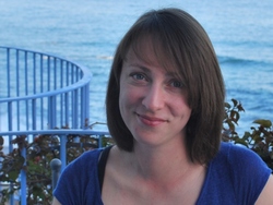
Sylviane Déderix. Ph.D.
From 4 until 8 September 2014, more than two hundred artists and scientists from more than 40 countries gathered on the Greek Island of Zakynthos to commemorate the quincentenary of the Flemish anatomist Andreas Vesalius who died on the island 450 years earlier. At this very moment when some start dreaming of a sequel of our Vesalius Continuum Conference, we continue to dream of the sequel of our search for his lost grave. The triennial of 2017 will be an ideal occasion to present a second phase in our search, on condition that the plan we are developing here succeeds.
The initial phase of the search for the Vesalius’s grave, started and presented in 2014, was based on recent re-examinations of historical sources that contest the traditional view that Vesalius was buried at Laganas. Research by the Flemish historians Omer Steeno, Maurits Biesbrouck, Theodoor Goddeeris and the local historical blogger Pavlos Plessas indeed suggest that the quest for his grave should rather focus on the town of Zakynthos, and more specifically on the courtyard of the church of Santa Maria delle Grazie.

Pascale Pollier
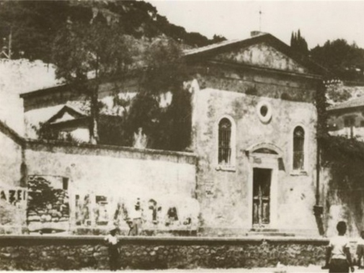
Unfortunately, the small church was destroyed along with most of the buildings in Zakynthos during the major earthquake that struck the Ionian Islands in 1953. Its ruins were then buried when the town was reconstructed, and its exact location was soon forgotten. Material evidence, local informants and cartographic data nevertheless point in the same direction: the church of Santa Maria delle Grazie would have been located in the northern sector of the modern town, around the current junction of Kolokotroni and Kolyva streets.
In order to assess the validity of this hypothesis, we called on the services of Geographic Information Systems (abbreviated GIS). GIS are computer-based tools used for the management, analysis, and display of geographically referenced information. Within the framework of the quest for Vesalius’ lost grave, they were used to overlay historical maps on modern cartographic data. The procedure, which is named geo-referencing, allows registering individual maps in a common geographic space so as to define their position in the real world. In the present case, the goal was to geo-reference a town map dated to 1892 and on which the church can be identified. See the accompanying photograph of the church.
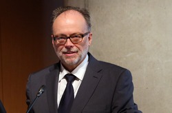
Theo Dirix
However, since the coastline and the town plan drastically changed after the earthquake, it was not possible to overlay the particular map directly onto modern satellite images: intermediary steps were necessary. The methodology consisted therefore in travelling back in time and geo-referencing three available maps from the most recent to the oldest. The result of the process confirmed that the ruins of the Santa Maria delle Grazie are to be found to the northwest of the intersection of the current Kolyva and Kolokotroni streets. The road that ran in front of the church in the late 19th and early 20th century followed a different orientation than Kolyva street, with the consequence that the church lies partly below the street and partly below private properties.
This small GIS project represents only a first phase in the quest for Vesalius’ grave. Phase 2 would be to conduct a geophysical prospection at the Kolyva/Kolokotroni intersection. By making use of non-destructive geophysical methods, we could get an idea of what is still lying under the modern surface, and at which depth. This would provide a fast and high resolution understanding of the area. In an urban environment, two techniques can be used: Ground Penetrating radar and Electrical Resistivity Tomography, which measure the propagation of electromagnetic waves and of the electrical current in the ground, respectively. If the geophysical results were conclusive, the possibility of small-scale excavations (Phase 3) could be considered.
Click for a larger image
The GIS was sponsored by Agfa HealthCare, the Greek subsidiary of the Belgian Agfa Gevaert Group, the Belgian University of Antwerp, and Theo Dirix. For the consequent phases, Pascale Pollier offers to sell five original wax models of her facial reconstruction of Andreas Vesalius. This inversed reconstruction of Vesalius’s skull, based on his portrait, will have to suffice until we find his skull, allowing her to reconstruct his real face. Vesalius Continuum, initially the conference where we launched the search of Vesalius’s grave, has evolved in a programme to which you can contribute.
Personal note: My sincere thanks to Dr. Déderix, Pascale Pollier, and Theo Dirix for contributing this article to "Medical Terminology Daily" and the quest to find and study Andreas Vesalius' grave. I am proud to have been one of the many international attendees to the 2014 meeting in the island of Zakynthos. Dr. Miranda.
- Details
UPDATED: This medical term is formed by the prefix [retr-] meaning “posterior”, the root term [-periton-] meaning “peritoneum”, and the adjectival suffix [-eal], meaning “pertaining to”. In the strictest sense, the term [retroperitoneal] means “posterior to the peritoneum”.
In practical terms in anatomy and surgery, this term refers to two situations:
In the first acception of the term, it refers to anatomical structures that are truly posterior to the peritoneum, between the peritoneum and the posterior abdominal wall, as are the kidneys, ureters, abdominal aorta, inferior vena cava, etc.
In the second acception of the term, it refers to digestive system structures that although posterior to the parietal peritoneum, they are also attached to the posterior abdominal wall by the peritoneum, fixating them to the posterior abdominal wall. These structures are immobilized in position by the peritoneum. When a surgeon needs to work on one of these “retroperitoneal” digestive system structures they need to render them mobile detaching them from the posterior abdominal wall by incising the peritoneum and “mobilizing “these structures.
There are three segments of the digestive system that are retroperitoneal (ergo, fixated to the posterior abdominal wall by the parietal peritoneum):
- the duodenum, except for its first inch
- the ascending colon
- the descending colon
- Details
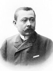
Dr. Jean Léo Testut
Jean Léo Testut (1849-1925) French physician, anatomist, historian, and anthropologist, Jean Léo Testut Deynat was born in Saint Avit Senier, France on March 22, 1849.
His early medical studies were interrupted by the Franco-Prussian war of 1870. He was awarded a medal for his courage and patriotism in this war, but declined to accept it. After the war Leo Testut finished his medical studies in 1878 at the Medical School in Bordeaux. His doctoral thesis received several awards, including the Silver Medal of the Paris Medical College.
During his medical studies at the Universities of Bordeaux and Paris, Testut was an assistant for both anatomy and physiology, eventually becoming the Chief of Anatomical Studies and Preparations in Bordeaux.
In 1887 he publishes his masterpiece: “Traité d'anatomie humaine” (Human Anatomy Treatise) in four volumes, which has a second publication in 1893.
Dr. Testut's assistant, Dr. André Latarjet (1877 – 1947) will later continue the work in this voluminous work taking it to five volumes, several editions, and translated into Spanish, German, and Italian. Smaller versions of the book as well as anatomical dissectors are published as companions to this superb book, becoming the standard of anatomical medical education in France and especially in Latin America for over 120 years.
In spite of this incredible publication, Leo Testut published well over 90 books and treatises, including an illustrated anatomical dissector. His work included anthropological research and comparative anatomy.
Dr. Testut worked as a military surgeon during World War I.
In his later life Dr. Testut received an incredible number of awards and decorations, including the Honor Legion Medal and Honorary Professor of the Lyon Medical School. He was also President of the World Association of Anatomists.
After he retired, he continued his historical studies publishing a further seven books!
Personal note: When I studied anatomy I was lucky to use the Testut and Latarjet “Compendio de Anatomía Humana”, the smaller version of the Treatise. The Treatise itself was available to us for study in the library of the Medical School at the University of Chile and I remember countless hours studying with this treasure of anatomy. Later I made it a point to own one of these incredible books, and I acquired the Spanish and the Italian version of the “Traité d'anatomie humaine”. A few years ago I added to my library a beautiful leather-bound French version of this book, which belonged to my dear friend Dr. Gonzalo Lopetegui Adams (1932 – 2004). Dr. Miranda.
Sources:
1. “Leo Testut (1849-1925)” Reverón, RR Int. J. Morphol., 29(4):1083-1086, 2011
2. “La anatomía de Testut y Latarjet” Reverón, RR R Soc Ven Hist Med (2013) 62:1; 62-72
3. “Jean Leo Testut (1849-1925): Anatomist and Anthropologist” Reverón, RR
4. “Huellas de un maestro de la Anatomía francesa: Jean Léo Testut, 1849-1925” Ledezma W.; P. Rev Inst Med Sucre LXXI: 128 (98-105), 2006
- Details
This article is part of the series "A Moment in History" where we honor those who have contributed to the growth of medical knowledge in the areas of anatomy, medicine, surgery, and medical research.
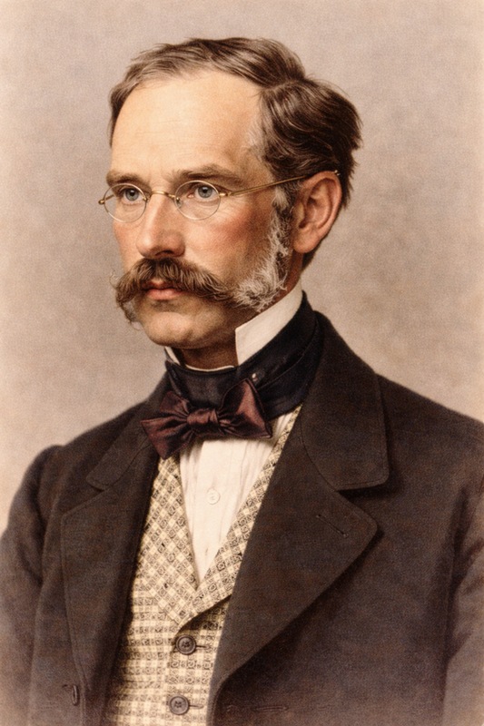
Dr. Václav Treitz
UPDATED: Dr. Václav Treitz (1819 - 1872). Also known as Wenzel Treitz, Dr. Václav Treitz was born in Hostomice, Bohemia. He attended the Charles-Ferdinand University in Prague studying humanities and medicine, receiving his medical degree in 1846. Treitz started postgraduate work at the Vienna General Hospital (Allgemeines Krankenhaus), where Joseph Skoda (1805-1881) was a proponent of “therapeutic nihilism” which stated that “drug treatment usually does more harm than good”, so a minimalistic or even pessimistic approach to diseases was used.
Large numbers of women at this hospital died of “puerperal fever” an postpartum uterine infection due to contamination by the unwashed hands of physicians and utter lack of cleanliness (septic technique had not been yet described). It was during Treitz’s time at the Vienna General Hospital that Ignaz Philipp Semmelweis (1818 – 1865) stated his initial observations on asepsis. Treitz later became a follower of Semmelweis’ and Lister’s teachings and techniques.
In 1852 Treitz was appointed Professor of Pathological Anatomy in the Jagellonian University in Prague.
In 1853 he published a paper ("Ueber einen neuen Muskel am Duodenum des Menschens" ) describing a new muscle he discovered at the duodenojejunal junction, later to be known as the eponymic “muscle of Treitz”; the fold of peritoneum over the muscle of Treitz is known today as the "ligament of Treitz". Treitz also described a paraduodenal retroperitoneal hernia that occurs at the paraduodenal recess, just lateral to the ligament of Treitz.
A staunch proponent of Czechoslovakian independence and language, Treitz was publicly attacked for his medical theories and nationalistic beliefs. Isolated and depressed, Treitz committed suicide in 1872.
The article on the "Ligament of Treitz" is the most popular article in "Medical Terminology Daily" with over 140 thousand hits!
Sources:
1. "Václav Treitz (1819-1872): Czechoslovakian Pathoanatomist and Patriot” Fox, RS; Fox, CG; Graham, WP. World J. Surg. 9, 361-366, 1985
2. "Treitz of the ligament of Treitz". Haubrich, W S. (2005) Gastroenterology, 128 (2), 279
3. "Preserving Treitz's muscle in hemorrhoidectomy". Gemsenj?ger, E Diseases of the Colon & Rectum (1982), 25 (7), p. 633.
4. “The Muscle Of Treitz And The Plica Duodeno-Jejunalis” Crymble, PT. The British Medical Journal, Vol. 2, No. 2598 (1910), 1156-1159
Original image, public domain, courtesy of Wikimedia Commons. Image in this article is AI enhanced.

- Details
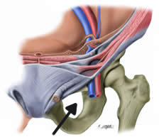
Obturator foramen
The word [foramen] is a Latin word meaning "opening, aperture, hole", from the Latin term [forare] meaning to bore a hole, to pierce. The plural form of the word is [foramina]. There are many foramina in the human body.
• Epiploic foramen (of Winslow): An opening bound by the lesser omentum, the inferior vena cava, duodenum and liver. It is a communication between the main peritoneal cavity and lesser sac, an area found posterior to the stomach.
• Nutritional foramina: Openings found in most bones allowing for passage of blood vessels.
• Obturator foramen: An opening in the pelvis bound by the following bones: ilium, ischium, and os pubis. Indicated in the accompanying image by an arrow.
• Foramen of Monro: A communication between the lateral ventricle and the third ventricle of the brain. There are actually two foramina of Monro (one on each side), and are important for cerebrospinal fluid circulation. Named after Alexander Monro Secundus (1733 - 1817).
- Details
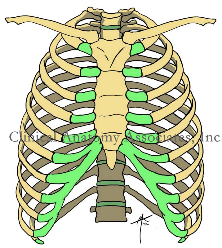
Click for a larger image
UPDATED: Root term meaning Thorax (Greek: [θώρακα] Chest plate or area covered by a chest plate). The thorax contains three areas, or regions, sometimes described as "spaces" (sic). These are two laterally situated pleural cavities, each containing a lung, and a median mediastinum, a large region containing the pericardium, heart, great vessels, thoracic duct, azygos venous system, esophagus, aorta, thymus, etc. The plural form for thorax is [thoraces].
• Thoracotomy: Opening of the thorax. Note that the term "thoratomy" en vogue nowadays is a misuse of the term
• Thoracocentesis: The suffix [-(o)centesis) means needle aspiration. Needle aspiration of the thorax
• Thoracoscopy: Visualization of the thorax
Personal note: There is a tendency, a trend to use the word [thorascopy] instead of the correct form [thoracoscopy]. The use of [thorascopy] is incorrect and should not be allowed in the clinical arena. In fact, it is not recognized by my medical spellchecker! Dr. Miranda