![]()
Medical Terminology Daily (MTD) is a blog sponsored by Clinical Anatomy Associates, Inc. as a service to the medical community. We post anatomical, medical or surgical terms, their meaning and usage, as well as biographical notes on anatomists, surgeons, and researchers through the ages. Be warned that some of the images used depict human anatomical specimens.
You are welcome to submit questions and suggestions using our "Contact Us" form. The information on this blog follows the terms on our "Privacy and Security Statement" and cannot be construed as medical guidance or instructions for treatment.
We have 1385 guests and no members online
")
Marcia Crocker Noyes
(1869 – 1946)
Further to my comment on old books and research that started with an interesting bookplate (Ex-Libris). I continued my research and found that the person in charge of the Osler library bookplate was a fascinating individual that today maybe a ghost in the MedChi library and building in Baltimore... This is certainly an article that can be called "A Moment in History"
Marcia Crocker Noyes was the librarian at The Maryland State Medical Society from 1896 to 1946 and was a founding member of the Medical Library Association.[1][2][3]
Sir William Osler, MD. a famous Johns Hopkins surgeon was a noted bibliophile and had a large personal collection of books on various topics. When he became the President of MedChi in 1896, he was dismayed at the condition of the library and knew that with the right person and some stewardship, it could become a significant collection. Sir William asked his friend, Dr. Bernard Steiner, a physician and President of the Enoch Pratt Free Library in Baltimore for suggestions of a librarian, and Dr. Steiner recommended Marcia Crocker Noyes. A native of New York, and a graduate of Hunter College, Marcia had moved to Baltimore for a lengthy visit with her sister, and took a “temporary” position at the Pratt Library, which turned into three years. Although she had no medical experience or background, she was enthusiastic, and most importantly, she was willing to move into the apartment provided for the librarian, who needed to be available 24 hours a day.
The image in this article is Ms. Noyes on her first year on the job. Marcia developed a book classification system for medical books, based on the Index Medicus, and called it the Classification for Medical Literature. The system uses the alphabet with capital letters for the major divisions of medicine and lower-case ones for the sub-sections. The system was used for many years, but it's now dated and the Faculty's original shelving scheme was never changed. The card catalogs still reflect her classification and many of the cards are written in Marcia's back-slanting handwriting.
Marcia knew enough to ask the Faculty's members about medical questions, terminology and literature. She gradually won over the predominantly male membership and they became her greatest allies; Sir William at the start, and then for nearly 40 years, Dr. John Ruhräh, a wealthy pediatrician with no immediate family of his own. She made a point of attending almost every Faculty function, and in 1904, under guidelines from the American Medical Association, Marcia was made the Faculty Secretary. For much of her first 10 years, she was the Faculty's only full-time employee, only being assisted by Mr. Caution, the Faculty's janitor. Later in life Marcia would say that she hired him because of his name!
Within ten years, the library had outgrown its space, and plans, spearheaded by Marcia and Sir William before his move to Oxford, were made to build a headquarters building, mainly to house the library's growing collection of medical books and journals.
Marcia was instrumental in the design and building of the new headquarters. She travelled to Philadelphia, New York and Boston to look at their medical society buildings, and eventually, the Philadelphia architectural firm, Ellicott & Emmart was selected to design and build the new Faculty building. Every detail of the building held her imprimatur, from the graceful staircase, to the light-filled reading room, and all of the myriad details of the millwork, marble tesserae, and most of all, the four-story cast iron stacks. She was on-site, climbing up unfinished staircases, checking out the progress of the building, which was built in less than one year at a cost of $90,000.
Among the features of the new building was a fourth-floor apartment for her. She referred to it as the "first penthouse in Baltimore" and it had a garden and rooftop terrace. The library collection eventually grew to more than 65,000 volumes from medical and specialty societies around the world. Journals were traded back and forth, and physicians eagerly anticipated the arrival of each new issue. At the same time, Marcia was involved in the Medical Library Association as one of eight founding members. The MLA promotes medical libraries and the exchange of information. One of the earliest mandates of the MLA was the Exchange, a distribution and trade service for those who had duplicates or little-used books in their collections. Initially, the Exchange was run out of the Philadelphia medical society, but in 1900 it was moved to Baltimore and Marcia oversaw it. Several hundred periodicals and journals were received and sent each month, a huge amount of work for a tiny staff. In 1904, the Faculty had run out of room to manage the Exchange, so it was moved to the Medical Society of the Kings County (Brooklyn). But without Marcia's excellent administrative skills, it floundered and in 1908, the MLA asked Marcia to take charge once again.
In 1909, when the new Faculty building opened, there was enough room to run the Exchange and with the help of MLA Treasurer, noted bibliophile and close friend, Dr. John Ruhräh, it once again became successful. Additionally, Marcia and Dr. Ruhräh combined forces to revive the MLA's bulletin, which had all but ceased publication in 1908, taking the Exchange with it. This duo maintained editorial control from 1911 until 1926. In 1934, around the time of Dr. Ruhräh's death, Marcia became the first “unmedicated” professional to head the MLA. During her tenure, the MLA incorporated, the first seal was adopted, and the annual meeting was held in Baltimore. Marcia wanted to write the history of the MLA once she retired from full-time work at the Faculty, but her health was beginning to fail. She had back problems and had suffered a serious burn on her shoulder as a young woman, possibly from her time running a summer camp, Camp Seyon, for young ladies in the Adirondack Mountains. In 1946, a celebration was planned to honor Marcia's 50 years at the Faculty. But she was adamant that the physicians wait until November, the actual date of her 50 years. However, they knew she was gravely ill, and might not make it until then, so a huge party was held in April. More than 250 physicians attended the celebration, but the ones she was closest to in the early years, were long gone. She was presented with a suitcase, a sum of money to use for travelling, and her favorite painting of Dr. John Philip Smith, a founder of the Medical College in Winchester, Virginia. It was painted by Edward Caledon Smith, a Virginia painter who had been a student of the painter Thomas Sully.[4] She adored this painting and vowed, jokingly, to take it with her wherever she went.
The painting was not to stay with her for very long, for she died in November 1946, and left it to the Faculty in her will. Her funeral was held in the Faculty's Osler Hall, named for her dear friend. More than 60 physicians served as her pallbearers, and she was buried at Baltimore's Green Mount Cemetery. In 1948, the MLA decided to establish an award in the name of Marcia Crocker Noyes. It was for outstanding achievement in medical library field and was to be awarded every two years, or when a truly worthy candidate was submitted. In 2014, the Faculty began giving a bouquet of flowers to the winner of the award in Marcia's name, and in honor of her work. Much evidence exists for this tradition, as we know that the physicians, especially Drs. Osler and Ruhräh, frequently gave her bouquets of flowers. Marcia also cultivated flower gardens at the Faculty and decorated the rooms with her work.
Today, the MedChi building is open for tours and if the rumors are to be believed Ms. Marcia Crocker Noyes is still at work in her beloved library as the "resident ghost" [1][5]

NOTE: This article has been modified from the original Wikipedia article on Marcia Crocker Noyes. The article itself is well-written with interesting images of the subject. I would encourage you to visit it. The second insert is from book 00736 in my personal library and shows in pencil, the incredibly small handwriting of Marsha C. Noyes.
Sources:
1. "Marcia, Marcia, Marcia" MedChi Archives blog.
2. "Marcia C. Noyes, Medical Librarian" (PDF). Bulletin of the Medical Library Association. 35 (1): 108–109. 1947. PMC 194645
3. Smith, Bernie Todd (1974). "Marcia Crocker Noyes, Medical Librarian: The Shaping of a Career" (PDF). Bulletin of the Medical Library Association. 62 (3): 314–324. PMC 198800Freely accessible. PMID 4619344.
4. Edward Caledon BRUCE (1825-1901)"
5. Behind the scenes tour MedChiBuilding
"Clinical Anatomy Associates, Inc., and the contributors of "Medical Terminology Daily" wish to thank all individuals who donate their bodies and tissues for the advancement of education and research”.
Click here for more information


- Details
Click on the image for a larger version
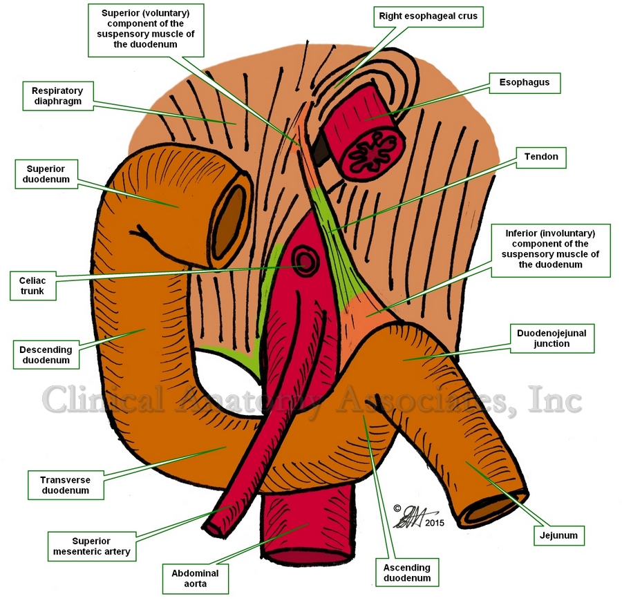
The duodenojejunal junction is the point where the fourth segment of the duodenum, the ascending duodenum, meets and is continuous with the most proximal portion of the jejunum.
The suspensory muscle of the duodenum attaches to the superior aspect of the duodenojejunal junction. This muscle is also known as the "muscle of Treitz" or "musculus suspensorius duodenii" and was first described in 1853 by Dr. Václav Treitz. The parietal peritoneum forms a fold over the suspensory muscle of the duodenum and this fold is known as the "ligament of Treitz".
The duodenojejunal junction (marked by the ligament of Treitz) is an important anatomical landmark used by anatomists and surgeons to denote the point where the small intestine passes from retroperitoneal duodenum to intraperitoneal jenunum. This means that while most of the duodenum is covered by parietal peritoneum, plastered to the posterior abdominal wall, and immobilized by it; the jejunum is mobile, meaning that the anatomist and surgeon can easily move it around because of the presence of a well-developed mesentery.
Sources:
1. "Clinically Oriented Anatomy" Moore, KL. 3r Ed. Williams & Wilkins 1992
2. "The origin of Medical Terms" Skinner, AH, 1970
3. "The suspensory muscle of the duodenum and its nerve supply" Jit, I.; Singh, S. J. Anat. (1977), 123, 2, pp. 397-405
4. "Anatomical and functional aspects of the human suspensory muscle of the duodenum." Costacurta, L. Acta Anat (Basel). 1972;82(1):34-46
Image property of: CAA.Inc. Artists: Dr. E. Miranda and D.M. Klein
- Details
This word arises from the Latin word [canna], meaning “reed” with the Latin suffix [-ula], used to denote something small. Since reeds are hollow, the word [cannula] can be translated as a “small, hollow reed”.
In medicine, a cannula is a small, hollow tube used to draw fluids or introduce drugs or fluids into the body. It can also be an instrument that is used as a guide for other instruments to be introduced in the body.
Cannulation is the act of using or placing a cannula. The Latin plural for cannula is [cannulae], although the English version [cannulas] is acceptable.
- Details

Click for a larger image
The [celiac trunk] is the first anterior unpaired branch of the abdominal aorta. Through its branches the celiac trunk provides arterial blood supply to the stomach, spleen, duodenum, and pancreas, as well as the liver. The celiac trunk is related on its left side to the suspensory muscle of the duodenum, which when covered by peritoneum forms the ligament of Treitz.
The celiac trunk is a very short artery which rapidly divides in its three branches:
- Left gastric artery: Provides blood supply to the stomach and is part of the lesser curvature vascular arcade.
- Splenic artery: Provides blood supply to the spleen, and to the stomach through a branch, the left gastroepiploic artery.
- Common hepatic artery: Provides blood supply to the liver, stomach and pancreas
Image property of: CAA.Inc. Photographer: David M. Klein
- Details
The medical term [septic] arises from the Greek word [σηπτικός] (siptikós) which means “rotting”, “decaying”, or "putrefact". It was later adopted in Latin as the word we use today: [septic].
Septic describes a condition of infection of the tissues or wound contamination by any means, including bacteria. When blood is contaminated or infected, we refer to it as [septicemia]
A medical terminology note: Although it would seem that the root terms for septal and septic are the same, they are not. In the first one it is [-sept-], while in the second one the whole word [septic] is also the root term.
Then, if the above is true, why do we say [sepsis]? It is because the base of the Greek word [σηπτικός] (siptikós) is [σήψις] (sipsis) meaning “to rot”
Note: The links to Google Translate include an icon that will allow you to hear the pronunciation of the word.
- Details
The root term [-sept-] arises from the Latin word [septum] which means “partition”, referring to a wall or division between areas or compartments. The addition of the adjectival suffix [-al] gives us the word [septal], meaning “pertaining to a septum”. The plural for [septum] is [septa]
In human anatomy the term is used in:
- Nasal septum: The osteocartilaginous division between both sides of the nose
- Interatrial septum: The wall or division between the atria of the heart
- Interventricular septum: The partition between the heart ventricles
- Septum pellucidum: A membranous separation between the lateral ventricles of the brain
- Intermuscular septa: There are several fibrous septa between the muscles in both the upper and lower extremities.
There are other septa in the human body not listed here.
Interestingly, the Greek counterpart of the Latin term [septum] is [διάφραγμα] (diáfragma) meaning “diaphragm”.
Note: The links to Google Translate include an icon that will allow you to hear the pronunciation of the word.
- Details

Click for a larger image
The root term [-hepat-] arises from the Greek word [ηπαρ] (ipar) which means “liver”. It is used in many medical terms:
- Hepatic: The adjectival suffix [-ic] means “pertaining to”. Pertaining to the liver, as in “common hepatic artery”
- Hepatitis: The suffix [-itis] means “inflammation” or “infection”. Inflammation of the liver
- Hepatectomy: The suffix [-ectomy] means “removal of”. Removal of the liver
- Hepatomegaly: The suffix [-(o)megaly] means “enlargement”. An enlarged liver
Note: The links to Google Translate include an icon that will allow you to hear the pronunciation of the word.
Image in Public Domain, by Henry Vandyke Carter, MD - Gray's Anatomy.