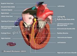
Conduction system of the heart
[UPDATED] The conduction system of the heart is part of a complex intrinsic heartbeat and rhythm control system that includes a cardiomyocyte-based component which acts as an automatic base, and an extrinsic and intrinsic autonomic nervous system component which acts as a modulator.
The classic description of the conduction system of the heart emphasizes only the cardiomyocyte-based component and refers to a group of specialized cardiac muscle structures that serve as pacemakers and distributors of the electrical stimuli that make the heart beat coordinately. It is important to stress the fact that this primary "conduction system of the heart" is not formed by nerves but rather by specialized cardiac muscle cells.
Components of the cardiomyocyte-based conduction system of the heart:
• SA node: The sinoatrial (SA) node is a small nodule of cardiac muscle tissue, somewhat horseshoe-shaped that is found at the junction of the superior vena cava and the right atrium. It receives blood supply from the SA node artery, a branch of the right coronary artery. Later research indicates that the pacemaker function of the SA node includes areas of the lateral wall of the right atrium which are involved in different heart rate speeds. It is also known eponymically as the "node of Keith and Flack" after Sir Arthur Keith (1866 - 1955) and Martin Flack, CBE (1882 - 1931).
The electrical impulses propagate between the right and left atria by way of the interatrial bundle, also known as "Bachmann's bundle", and between the SA node and the atrioventricular node by way of three internodal tracts, described by James Wenckebach, and Christen Thorel. More information on these internodal tracts here.
• AV node: The atrioventricular (AV) node is found at the junction of atria and ventricles in an area known as the "Triangle of Koch". Its function is to delay the electrical impulse passing from the atria to the ventricles by 1/10th of a second, enabling the sequential pumping action of the heart. The eponymic name for the AV node is "node of Aschoff-Tawara", and it receives its blood supply by way of the AV node artery, a branch that usually arises from the right coronary artery
• AV bundle: Also known as the "Bundle of His", this thick bundle of specialized myocardial cells is found in the interventricular septum. It divides into the right and left bundle branches
• Bundle branches: Sometimes known as the "crura" of the bundle of His, these two divisions of the AV bundle help distribute the electrical stimuli to the ventricular walls. The right bundle branch has an extension that crosses the lumen of the right ventricle, from the base of the anterior papillary muscle to the interventricular septum, forming a cord of tissue known as the "moderator band" or "septomarginal trabecula"
• Purkinje Fibers: These thin fibers are the terminal end of the conduction system of the heart and finish the distribution of the electrical stimuli to all parts of the ventricular walls
Although the structural components of the conduction system of the heart were known, it was Dr. Sunao Tawara (1873-1952) who discovered the AV node and described the connections between the components of what he called the "Reitzleitungssytem" (conduction system) of the heart.
The conduction system of the heart is part of a more complex rhythm control system of the heart. For more information click here.
Click on the image for a larger version. Image modified from the original: "3D Human Anatomy: Regional Edition DVD-ROM." Courtesy of Primal Pictures.


