Medical Terminology Daily (MTD) is a blog sponsored by Clinical Anatomy Associates, Inc. as a service to the medical community. We post anatomical, medical or surgical terms, their meaning and usage, as well as biographical notes on anatomists, surgeons, and researchers through the ages. Be warned that some of the images used depict human anatomical specimens.

You are welcome to submit questions and suggestions using our "Contact Us" form. The information on this blog follows the terms on our "Privacy and Security Statement" and cannot be construed as medical guidance or instructions for treatment.


We have 275 guests and no members online


A Moment in History

Andreas Vesalius Bruxellensis (1514- 1564)

A Flemish anatomist and surgeon, Andreas Vesalius was born on December 31, 1514 in Brussels, Belgium. He is considered to be the father of the science of Anatomy. Up until his studies and publications human anatomy studies consisted only on the confirmation of the old doctrines of Galen of Pergamon (129AD - 200AD). Anatomy professors would read to the students from Galen's work and a demonstrator would point in a body to the area being described, if a body was used at all. The reasoning was that there was no need to dissect since all that was needed to know was already written in Galen's books. Vesalius, Fallopius, and others started the change by describing what they actually saw in a dissection as opposed to what was supposed to be there. 

Vesalius had a notorious career, both as an anatomist and as a surgeon. His revolutionary book "De Humani Corporis Fabrica: Libri Septem" was published in May 26, 1543. One of the most famous anatomical images is his plate 22 of the book, called sometimes "The Hamlet". You can see this image if you hover over Vesalius' only known portrait which accompanies this article. Sir William Osler said of this book "... it is the greatest book ever printed, from which modern medicine dates" 

After the original 1543 printing, the Fabrica was reprinted in 1555. It was re-reprinted and translated in many languages, although many of these printings were low-quality copies with no respect for copyright or authorship.

The story of the wood blocks with the carved images used for the original printing extends into the 20th century. In 1934 these original wood blocks were used to print 617 copies of the book "Iconaes Anatomica". This book is rare and no more can be printed because, sadly, during a 1943 WWII bombing raid over Munich all the wood blocks were burnt.

One interesting aspect of the book was the landscape panorama in some of his most famous woodcuts which was only "discovered" until 1903.

Vesalius was controversial in life and he still is in death. We know that he died on his way back from a pilgrimage to Jerusalem, but how he died, and exactly where he died is lost in controversy. We do know he was alive when he set foot on the port of Zakynthos in the island of the same name in Greece. He is said to have suddenly collapsed and die at the gates of the city, presumably as a consequence of scurvy. Records show that he was interred in the cemetery of the Church of Santa Maria delle Grazie, but the city and the church were destroyed by an earthquake and Vesalius' grave lost to history. Modern researchers are looking into finding the lost grave and have identified the location of the cemetery. This story has not ended yet.

For a detailed biography of Andreas Vesalius CLICK HERE.

Personal note: To commemorate Andrea Vesalius' 500th birthday in 2014, there were many scientific meetings throughout the world, one of them was the "Vesalius Continuum" anatomical meeting on the island of Zakynthos, Greece on September 4-8, 2014. This is the island where Vesalius died in 1564. I had the opportunity to attend and there are several articles in this website on the presence of Andreas Vesalius on Zakynthos island. During 2015 I also attended a symposium on "Vesalius and the Invention of the Modern Body" at the St. Louis University. At this symposium I had the honor of meeting of Drs. Garrison and Hast, authors of the "New Fabrica". Dr. Miranda


 "Clinical Anatomy Associates, Inc., and the contributors of "Medical Terminology Daily" wish to thank all individuals who donate their bodies and tissues for the advancement of education and research”.

Click here for more information


Rare & Collectible Books at AbeBooks.com 

bookplateink.com

Azygos venous system

Azygos system
Azygos system

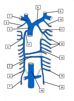
 The azygos venous system drains the posterior aspect of the thorax via the posterior intercostal veins It also connects the vascular territories of the superior vena cava and the  inferior vena cava, and is the superior continuation of the lumbar veins. The azygos system was first described by Bartolomeo Eustachius (c1500 - 1574).

The name azygos comes from the Greek [ζεύγος] and means “unyoked” or better “asymmetrical”. This system is different on each side of the body, also having important anatomical variations.

The azygos vein (Lat: vena azygos major) is the larger vein of the azygos system and is found on the right side of the body. It begins at the level of the first or second lumbar vertebra as a continuation of the right ascending lumbar vein; sometimes by a branch from the right renal vein or from the inferior vena cava. It enters the thoracic cavity through the aortic hiatus of the respiratory diaphragm, and ascends along the right side of the vertebral column to level of the fourth thoracic vertebra, where it arches forward over the root of the right lung, at this point the vein is called the azygos arch, which terminates in the posterior aspect of the superior vena cava (SVC) just superior to the point where the SVC enters the pericardium.

In the thorax, the azygos vein is found to the right of the thoracic duct on the right side of the descending aorta; it lies upon the intercostal arteries and is partly covered by the parietal pleura.

The azygos vein receives the right subcostal vein, nine or ten right posterior intercostal veins, the hemiazygos vein, the accessory hemiazygos vein, the right superior intercostal vein, and several minor esophageal, mediastinal, and pericardial veins.

The left side of this system is more complex and presents with more anatomical variations. Its main component is the hemiazygos vein (Lat: vena azygos minor), also known as the left lower azygos vein. It is a continuation of the left ascending lumbar vein, and it sometimes may arise from the left renal vein and passes into the thorax usually through the left aortic crus of the respiratory diaphragm. It ascends to the level of the 7th or 8th thoracic vertebra where it crosses the midline posterior to the esophagus, descending aorta and thoracic duct to empty into the right-sided azygos vein.  It receives the left subcostal vein and three to four lower posterior intercostal veins, and some esophageal and mediastinal veins.

The second component of the left azygos system is the accessory hemiazygos vein, also known as the left upper hemiazygos. This component varies in size depending on the third venous drainage component of the left posterior thoracic wall. This is the left superior intercostal vein (see attached diagram).

The accessory hemiazygos, similar to the hemiazygos vein will cross the midline posterior to the esophagus, descending aorta and thoracic duct to empty into the right-sided azygos vein. It may do so by a common vein or by a separate vein as shown in the attached diagram. If there is a common vein the hemiazygos is considered to be the inferior component and the hemiazygos is considered to be the superior component.

The left superior intercostal vein receives three or four posterior intercostal veins, and empties into the left brachiocephalic vein. In rare cases of absence of the hemiazygos vein, this left superior intercostal vein will extend as low as the fifth or sixth intercostal space.

Although not considered to be part of the azygos system, the drainage of the posterior thoracic wall is completed by the right and left supreme intercostal veins which empty the posterior aspect of the first intercostal space into the left and right brachiocephalic veins respectively.

The azygos system of veins constitute an important collateral venous circulation pathway which can be seen in action in cases of blockage of the superior or inferior vena cavæ.
 
Sources:
1. “Gray’s Anatomy” Henry Gray, 1918
2. "Tratado de Anatomia Humana" Testut et Latarjet 8th Ed. 1931 Salvat Editores, Spain
3. "Gray's Anatomy" 38th British Ed. Churchill Livingstone 1995

4. "Reconstructive Anatomy: A Method for the Study of Human Structure: Arnold, M WB Saunders1968
Image modified from the original from Arnold (4)