
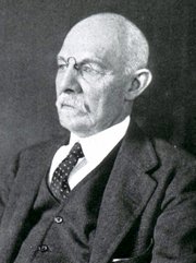




Dr. José Manuel Revuelta
Personal Note: Through my good friend Tito Estrada, I read an very interesting article in Spanish by Dr. Jose Manuel Revuelta. Dr. Revuelta is a Professor of Surgery and Professor Emeritus at the University of Cantabria. Former Head of Cardiovascular Surgery at Valdecilla Hospital in Santander, Spain. Dr. Revuelta contributed the article on "The Little Brain Inside the Heart" which we published in 2025.
The article's title (in Spanish) is "La ingeniería invisible que nos mantiene vivos" (The invisible engineering that keeps us alive), the incredible activity of the cardiac cells and the anatomical description of a helical heart muscle pioneered by Dr. Francisco Torrent-Guasp (1931 - 2005).
He has graciously granted us permission to translate and publish his article in “Medical Terminology Daily”. Dr. Miranda.
The Invisible Engineering That Keeps Us Alive
José Manuel Revuelta Soba
We are talking about a self-exciting, autonomous electrical system, a precision biochemical engine, a power plant capable of changing its "fuel" on the fly to power a very peculiar muscular architecture.
Millennium after millennium, humankind has gazed in awe at this constant pulse that marks the rhythm of life. When we try to repeatedly clench and unclench our fist tightly, in just a few minutes, the fatigue in our forearm forces us to stop. However, just inches away, a muscle the size of that fist contracts rhythmically 100,000 times a day without stopping. For the heart, muscle fatigue is not an option.
How does this organ manage to defy the laws of wear and tear that govern the rest of our biology? There is no man-made engine capable of withstanding such a level of friction and mechanical stress without external maintenance for eight or nine decades. Maintaining that uninterrupted heartbeat is no trivial feat; it's the result of a masterpiece of natural engineering. We're talking about an autonomous, self-exciting electrical system, a precision biochemical engine, a power plant capable of changing its "fuel" on the fly to power a unique muscular architecture.
The Engine That Generates Its Own Electricity
This marvel of endurance begins with an astonishing phenomenon: the heart doesn't wait for orders; it commands itself. Unlike the rest of our muscles, which depend on instructions from the brain, the heart contains a self-sufficient power plant.
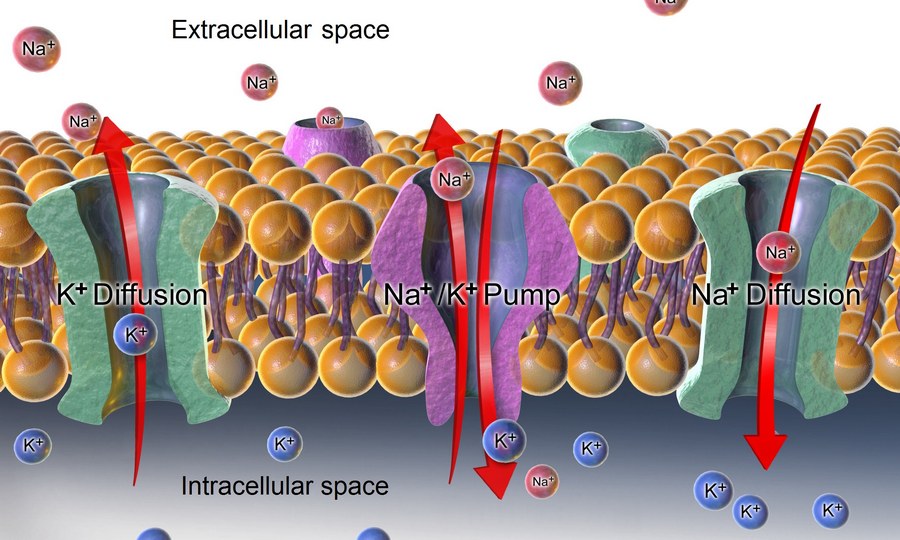
Sodium-Potassium Pump
The secret of this "miracle" lies in a coordinated exchange of minerals. Through microscopic gates located on the surface of the heart cells (ion channels), sodium and potassium ions rhythmically enter and exit. This flow, known as the sodium-potassium pump, creates an electrical potential difference. The result is tiny millivolt discharges that travel through the organ like a controlled shock wave. Each of these impulses—normally between 70 and 80 per minute—propagates through a network of specialized cardiac cells that function like the wiring in a building. This current is what the electrodes of an electrocardiogram (ECG) capture, mapping the activity of our internal "electrical network" on paper.
What is truly remarkable about the heart's electrical system is its redundancy. The main generator (sinoatrial node) sets the pace, but if it fails, the system doesn't shut down; immediately, another backup generator (atrioventricular node) kicks in, capable of activating in milliseconds to maintain the heartbeat. This energy is transmitted through an intracardiac conduction network until it reaches the Purkinje network, the final stretch of wires that makes the muscle contract and keeps life going.
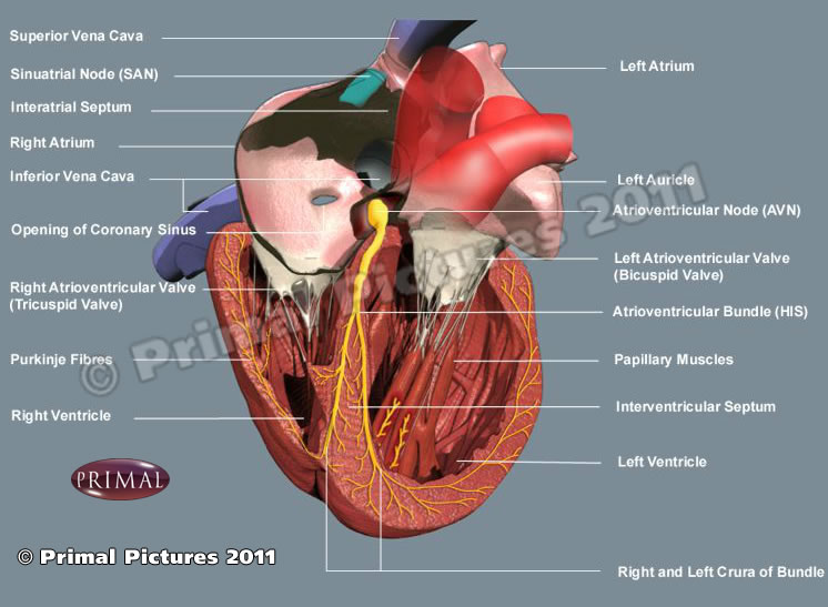
Conduction system of the heart
Amazing Biochemical Engine
If the electrical system generates the spark for ignition, calcium is the inductor that generates the movement. For the heart to contract with the force necessary to pump blood throughout the body, its cells must be flooded with this mineral at high speed. However, managing this flow is not simple: it requires precise biological engineering.
Within the heart cell, there is a specialized reservoir called the sarcoplasmic reticulum. Its function is to store, release, and recover calcium in fractions of a second. It is a closed-loop recycling system that ensures nothing is wasted and that the engine is always ready for the next cycle. When the electrical impulse arrives, ultrasensitive gates burst open, and calcium is released, activating the proteins that trigger contraction (systole). But for the heart to relax and refill with blood (diastole), that calcium must disappear immediately. This is when SERCA (sarcoplasmic/endoplasmic reticulum Ca2+-ATPase) proteins come into play. These proteins act as powerful suction pumps and, in a matter of milliseconds, draw calcium back into the sarcoplasmic reticulum, leaving the muscle relaxed and ready for the next movement.
This cycle of calcium delivery and retrieval occurs about 70 times per minute, but during intense exertion, the system can accelerate to more than 180 cycles per minute without losing synchronization. This ability to manage calcium so quickly and efficiently is what prevents the heart from cramping or becoming fatigued, as happens to our leg muscles after a strenuous run. While other engines might overheat or seize up, the heart uses this perfect recycling cycle to keep running.
A High-Performance Power Plant
If calcium is the messenger of contraction, mitochondria are the boiler that keeps the entire system running. In a typical muscle, mitochondria occupy barely 5% of the cell volume, while in the heart they constitute 35%. They are strategically located on the surface of the cardiac muscle fibers (cardiomyocytes) so that energy transport is practically instantaneous. Unlike other organs that store energy for later, “the heart lives for the day”; it produces and consumes its fuel, a molecule called ATP (adenosine triphosphate), in intervals of 8 to 10 seconds. To move this fuel from the mitochondria, the heart uses phosphocreatine, an ultrafast transport vehicle that guarantees a continuous and uninterrupted flow of energy.
While muscles can work briefly without oxygen—generating lactic acid, responsible for muscle soreness—the heart is an “oxygen addict.” Its metabolism is purely aerobic, allowing it to extract energy from every molecule. The most surprising aspect of this engineering is its flexibility; while the brain only accepts oxygen and glucose, the heart is an “efficient omnivore.” Its preferred fuel is fatty acids, but if the situation demands it, it can burn glucose or even lactate. This ability to switch fuels, depending on availability, ensures that the cell powerhouse (mitochondria) never runs out of supply, whether due to prolonged fasting or intense stress. In short, a perfect balance between energy production and expenditure without any rest.
The Helical Muscular Architecture
For decades, it was believed that the heart was a simple muscular sac that inflated and deflated autonomously. However, thanks to the pioneering vision of the Valencian (from Valencia, Spain) cardiologist Francisco Torrent-Guasp (1931 -2005), we now know that cardiac anatomy is much more sophisticated: the heart is a unique muscular band that coils upon itself in a spiral shape.
To understand how it works, let's forget the idea of a balloon being compressed. Instead, think of a wet towel we want to wring out: we don't press it from the sides, but rather twist it. The heart's engineering follows precisely this principle; its fibers are arranged in a spiral. During systole, the heart rotates on its own axis, performing a "corkscrew" motion. This rotation allows it to expel blood with a force and hemodynamic efficiency that a simple radial contraction could never achieve. But the most ingenious thing happens right at the end of the heartbeat: the elastic unwinding of this muscular band generates a vacuum effect that draws the blood back in. Instead of expending extra energy to fill up, the heart uses its own elastic architecture, it's energy efficiency in its purest form.
In this video, Dr, Torrent-Guasp demonstrates the helical architecture of the musculature of the human heart
Torrent-Guasp spent decades analyzing the hearts of various species in his small home laboratory in Denia, ignored by the scientific community. His luck changed when the prestigious surgeon Gerald Buckberg, from the University of California, Los Angeles (UCLA), recognized the brilliance of his discovery. Buckberg not only disseminated this theory internationally, but also designed a ventricular remodeling surgical technique, which he named "Pacopexy" in honor of his friend Paco Torrent. (Paco is the Spanish nickname for Francisco)
Currently, Torrent-Guasp's concept of the myocardial muscular band is studied at the world's leading universities. It is definitive proof that the heart is not just a muscle, but a marvel of human biology; a continuous muscular structure, without attachment points or independent elements that can fail, designed to adapt to changing hemodynamic pressures throughout a lifetime.
The true genius of the heart's invisible engineering lies not only in its electrical power or its elegant helical geometry, but in a capacity that any engineer would envy: constant self-repair while fully functioning. At the molecular level, proteins damaged by the uninterrupted effort are replaced by new copies without the rhythm being altered in the slightest. It is a unique resilience; we could say that "the heart never rests because it never stops renewing itself."
Ultimately, this organ is a testament to a brilliant biological technology that has perfected the art of enduring, a machine that refuses to surrender to fatigue. Each heartbeat is not just a movement; it is the triumph of the heart's biological engineering.
"What lies behind us and what lies before us are small things
compared with what lives within us"
Ralph Waldo Emerson (1803-1882)
American philosopher and poet.
Notes: The Sodium-potassium pump image public domain, courtesy of Blausen.com staff (2014). "Medical gallery of Blausen Medical 2014". WikiJournal of Medicine (2). DOI:10.15347/wjm/2014.010 ISSN. 2002-4436. Derivative by Mikael Häggström
Resources:
1. Bers, D. Cardiac excitation–contraction coupling. Nature 415, 198–205 (2002).
2. Mora, Vicente, Roldán, Ildefonso, Saurí, Assumpció, Fernández-Galera, Rubén, Monteagudo, Marta, Romero, Elena, Cabadés, Claudia, Cosín, Juan A., Trainini, Jorge C., & Lowenstein, Jorge A. Correspondencia de la deformación miocárdica con la teoría de Torrent-Guasp. Aporte de nuevos parámetros ecocardiográficos. Revista argentina de cardiología, 84(6), 1-2.
3. Website of Dr. Torrent-Guasp and family
4. Buckberg, G., Hoffman, J., Mahajan, A., Saleh S., Coghlan, C. Cardiac Mechanics Revisited: The Relationship of Cardiac Architecture to Ventricular Function. Circulation 118, 24
5. Buckberg G, Mahajan A, Saleh S. Structure and function relationships of the helical ventricular myocardial band. J Thorac Cardiovasc Surg, 2008; 136, 578-589.e11
6. The following 37 minute video was published in 2005 features Dr. Torrent-Guasp and is available on YouTube: