![]()
Medical Terminology Daily (MTD) is a blog sponsored by Clinical Anatomy Associates, Inc. as a service to the medical community. We post anatomical, medical or surgical terms, their meaning and usage, as well as biographical notes on anatomists, surgeons, and researchers through the ages. Be warned that some of the images used depict human anatomical specimens.
You are welcome to submit questions and suggestions using our "Contact Us" form. The information on this blog follows the terms on our "Privacy and Security Statement" and cannot be construed as medical guidance or instructions for treatment.
We have 951 guests and no members online
")
Marcia Crocker Noyes
(1869 – 1946)
Further to my comment on old books and research that started with an interesting bookplate (Ex-Libris). I continued my research and found that the person in charge of the Osler library bookplate was a fascinating individual that today maybe a ghost in the MedChi library and building in Baltimore... This is certainly an article that can be called "A Moment in History"
Marcia Crocker Noyes was the librarian at The Maryland State Medical Society from 1896 to 1946 and was a founding member of the Medical Library Association.[1][2][3]
Sir William Osler, MD. a famous Johns Hopkins surgeon was a noted bibliophile and had a large personal collection of books on various topics. When he became the President of MedChi in 1896, he was dismayed at the condition of the library and knew that with the right person and some stewardship, it could become a significant collection. Sir William asked his friend, Dr. Bernard Steiner, a physician and President of the Enoch Pratt Free Library in Baltimore for suggestions of a librarian, and Dr. Steiner recommended Marcia Crocker Noyes. A native of New York, and a graduate of Hunter College, Marcia had moved to Baltimore for a lengthy visit with her sister, and took a “temporary” position at the Pratt Library, which turned into three years. Although she had no medical experience or background, she was enthusiastic, and most importantly, she was willing to move into the apartment provided for the librarian, who needed to be available 24 hours a day.
The image in this article is Ms. Noyes on her first year on the job. Marcia developed a book classification system for medical books, based on the Index Medicus, and called it the Classification for Medical Literature. The system uses the alphabet with capital letters for the major divisions of medicine and lower-case ones for the sub-sections. The system was used for many years, but it's now dated and the Faculty's original shelving scheme was never changed. The card catalogs still reflect her classification and many of the cards are written in Marcia's back-slanting handwriting.
Marcia knew enough to ask the Faculty's members about medical questions, terminology and literature. She gradually won over the predominantly male membership and they became her greatest allies; Sir William at the start, and then for nearly 40 years, Dr. John Ruhräh, a wealthy pediatrician with no immediate family of his own. She made a point of attending almost every Faculty function, and in 1904, under guidelines from the American Medical Association, Marcia was made the Faculty Secretary. For much of her first 10 years, she was the Faculty's only full-time employee, only being assisted by Mr. Caution, the Faculty's janitor. Later in life Marcia would say that she hired him because of his name!
Within ten years, the library had outgrown its space, and plans, spearheaded by Marcia and Sir William before his move to Oxford, were made to build a headquarters building, mainly to house the library's growing collection of medical books and journals.
Marcia was instrumental in the design and building of the new headquarters. She travelled to Philadelphia, New York and Boston to look at their medical society buildings, and eventually, the Philadelphia architectural firm, Ellicott & Emmart was selected to design and build the new Faculty building. Every detail of the building held her imprimatur, from the graceful staircase, to the light-filled reading room, and all of the myriad details of the millwork, marble tesserae, and most of all, the four-story cast iron stacks. She was on-site, climbing up unfinished staircases, checking out the progress of the building, which was built in less than one year at a cost of $90,000.
Among the features of the new building was a fourth-floor apartment for her. She referred to it as the "first penthouse in Baltimore" and it had a garden and rooftop terrace. The library collection eventually grew to more than 65,000 volumes from medical and specialty societies around the world. Journals were traded back and forth, and physicians eagerly anticipated the arrival of each new issue. At the same time, Marcia was involved in the Medical Library Association as one of eight founding members. The MLA promotes medical libraries and the exchange of information. One of the earliest mandates of the MLA was the Exchange, a distribution and trade service for those who had duplicates or little-used books in their collections. Initially, the Exchange was run out of the Philadelphia medical society, but in 1900 it was moved to Baltimore and Marcia oversaw it. Several hundred periodicals and journals were received and sent each month, a huge amount of work for a tiny staff. In 1904, the Faculty had run out of room to manage the Exchange, so it was moved to the Medical Society of the Kings County (Brooklyn). But without Marcia's excellent administrative skills, it floundered and in 1908, the MLA asked Marcia to take charge once again.
In 1909, when the new Faculty building opened, there was enough room to run the Exchange and with the help of MLA Treasurer, noted bibliophile and close friend, Dr. John Ruhräh, it once again became successful. Additionally, Marcia and Dr. Ruhräh combined forces to revive the MLA's bulletin, which had all but ceased publication in 1908, taking the Exchange with it. This duo maintained editorial control from 1911 until 1926. In 1934, around the time of Dr. Ruhräh's death, Marcia became the first “unmedicated” professional to head the MLA. During her tenure, the MLA incorporated, the first seal was adopted, and the annual meeting was held in Baltimore. Marcia wanted to write the history of the MLA once she retired from full-time work at the Faculty, but her health was beginning to fail. She had back problems and had suffered a serious burn on her shoulder as a young woman, possibly from her time running a summer camp, Camp Seyon, for young ladies in the Adirondack Mountains. In 1946, a celebration was planned to honor Marcia's 50 years at the Faculty. But she was adamant that the physicians wait until November, the actual date of her 50 years. However, they knew she was gravely ill, and might not make it until then, so a huge party was held in April. More than 250 physicians attended the celebration, but the ones she was closest to in the early years, were long gone. She was presented with a suitcase, a sum of money to use for travelling, and her favorite painting of Dr. John Philip Smith, a founder of the Medical College in Winchester, Virginia. It was painted by Edward Caledon Smith, a Virginia painter who had been a student of the painter Thomas Sully.[4] She adored this painting and vowed, jokingly, to take it with her wherever she went.
The painting was not to stay with her for very long, for she died in November 1946, and left it to the Faculty in her will. Her funeral was held in the Faculty's Osler Hall, named for her dear friend. More than 60 physicians served as her pallbearers, and she was buried at Baltimore's Green Mount Cemetery. In 1948, the MLA decided to establish an award in the name of Marcia Crocker Noyes. It was for outstanding achievement in medical library field and was to be awarded every two years, or when a truly worthy candidate was submitted. In 2014, the Faculty began giving a bouquet of flowers to the winner of the award in Marcia's name, and in honor of her work. Much evidence exists for this tradition, as we know that the physicians, especially Drs. Osler and Ruhräh, frequently gave her bouquets of flowers. Marcia also cultivated flower gardens at the Faculty and decorated the rooms with her work.
Today, the MedChi building is open for tours and if the rumors are to be believed Ms. Marcia Crocker Noyes is still at work in her beloved library as the "resident ghost" [1][5]

NOTE: This article has been modified from the original Wikipedia article on Marcia Crocker Noyes. The article itself is well-written with interesting images of the subject. I would encourage you to visit it. The second insert is from book 00736 in my personal library and shows in pencil, the incredibly small handwriting of Marsha C. Noyes.
Sources:
1. "Marcia, Marcia, Marcia" MedChi Archives blog.
2. "Marcia C. Noyes, Medical Librarian" (PDF). Bulletin of the Medical Library Association. 35 (1): 108–109. 1947. PMC 194645
3. Smith, Bernie Todd (1974). "Marcia Crocker Noyes, Medical Librarian: The Shaping of a Career" (PDF). Bulletin of the Medical Library Association. 62 (3): 314–324. PMC 198800Freely accessible. PMID 4619344.
4. Edward Caledon BRUCE (1825-1901)"
5. Behind the scenes tour MedChiBuilding
"Clinical Anatomy Associates, Inc., and the contributors of "Medical Terminology Daily" wish to thank all individuals who donate their bodies and tissues for the advancement of education and research”.
Click here for more information


- Details

Levator scapulæ muscle
The levator scapulæ muscle (levator anguli scapulæ) is a triangular multipennate muscle which extends between the cervical spine and the scapula. This muscle is deep to the sternocleidomastoid and trapezius muscle.
It is formed by discrete muscular slips that originate from the first four transverse processes (C1-C4). It can have an extra slip from C5 (as shown in the side image).
These muscular slips pass posteroinferiorly, joining, and inserting in the superior scapular angle and the scapular medial border between the superior scapular angle and the medial origin of the scapular spine. It may attach to the scapular spine.
There are other anatomical variations including muscular slips that may extend to the occipital bone or mastoid process, to the trapezius, scalene, or serratus anterior magnus muscles, or to the first or second rib.
It receives nerve supply from the fourth and fifth cervical nerves and by a branch from the dorsal scapular nerve. The dorsal scapular nerve arises from the C5 root of the brachial plexus.
It receives its blood supply from the dorsal scapular artery.
The function of this muscle depends on which bony element is fixed, the scapula or the cervical spine. When the spine is fixed, the levator scapulae elevates the scapula and pulls the superior portion of the medial scapular border superomedially. When only one scapula is fixed, the head and neck flexes and rotates ipsilaterally while it extends the neck contralaterally.

Levator scapulæ muscle fibers
The order and shape of the muscular slips is interesting, as the slip from the transverse process of the Atlas (C1) twists posteriorly and descends to insert as the most posterior and inferior fibers in the medial border of the scapula. The other slips follow a similar pattern, which is what allows this muscle to rotate the neck. This indicates that the fibers of the levator scapulae muscle are spiral and the fibers follow the contour of the neck. This makes (to my knowledge) the levator scapulae the only spiral muscle of the body. This is shown as "A" in the second side image; "B" represents the misconception on the direction of the fibers in this muscle.
Since it is a common sign of stress and bad posture to raise the shoulders, this muscle can spasm, causing neck pain and in some cases be a trigger for headaches.
The Levator scapulæ is one of the 17 muscles that attach to the scapula.
Note: The first side image shown in this article is from “Gray’s Anatomy” (1918) which is in the public domain. The second side image is from Arnold’s “Reconstructive Anatomy” (1968).
Note: Animated image below by Wikimedia Commons - Anatomography [CC BY-SA 2.1 jp (https://creativecommons.org/licenses/by-sa/2.1/jp/deed.en)]
![Anatomography [CC BY-SA 2.1 jp (https://creativecommons.org/licenses/by-sa/2.1/jp/deed.en)]](https://upload.wikimedia.org/wikipedia/commons/a/a1/Levator_scapulae_muscle_animation_small2.gif)
Sources:
1. “Gray’s Anatomy” Henry Gray, 1918 2. "Tratado de Anatomia Humana" Testut et Latarjet 8th Ed. 1931 Salvat Editores, Spain
2. "Tratado de Anatomía Humana" Testut et Latarjet 8th Ed. 1931 Salvat Editores, Spain
3. "Gray's Anatomy" 38th British Ed. Churchill Livingstone 1995
4. “An Illustrated Atlas of the Skeletal Muscles” Bowden, B. 4th Ed. Morton Publishing. 2015
5. “Reconstructive Anatomy, A Method for the Study of Human Structure” Arnold, M. W.B. Saunders. 1968“Gray’s Anatomy” Henry Gray, 1918
- Details

Supraspinatus muscle
The supraspinatus muscle is found in the supraspinatus fossa of the scapula, and one of the four muscles that forms the rotator cuff. The muscle attaches to the medial two thirds of the floor of the fossa directly on the bone and on the deep aspect of the supraspinatus fascia which covers the muscle. The supraspinatus fascia and the supraspinatus fossa form an osteofascial case for the origin of this muscle.
The fibers of the muscle converge and pass deep to the acromion, forming an osseous tunnel that could entrap the muscle and tendon causing a supraspinous impingement syndrome. The side image in this article has the acromion cut off to show the muscle better. The animated image at the bottom of the article shows the supraspinatus muscle and its relation to the acromion process.
The supraspinatus tendon attaches to the capsule of the glenohumeral joint at the level of the highest of the three impressions that form the greater tubercle of the humerus.
It receives innervation by way of the suprascapular nerve (C5, C6), which arises from the superior trunk of the brachial plexus.
The main function of the supraspinatus muscle is to abduct the arm. As part of the shoulder’s rotator cuff it helps prevent subluxation of the glenohumeral joint by keeping the head of the humerus in situ.
The supraspinatus is one of the 17 muscles that attach to the scapula.
Note: The side image modified from the original by Henry VanDyke Carter, MD. Public domain. Animated image below by Wikimedia Commons - Anatomography [CC BY-SA 2.1 jp (https://creativecommons.org/licenses/by-sa/2.1/jp/deed.en)]
![Anatomography [CC BY-SA 2.1 jp (https://creativecommons.org/licenses/by-sa/2.1/jp/deed.en)]](https://upload.wikimedia.org/wikipedia/commons/8/85/Supraspinatus_muscle_animation.gif)
Sources:
1. “Gray’s Anatomy” Henry Gray, 1918
2. "Tratado de Anatomia Humana" Testut et Latarjet 8th Ed. 1931 Salvat Editores, Spain
3. "Gray's Anatomy" 38th British Ed. Churchill Livingstone 1995
- Details

Subscapularis muscle
The subscapular muscle or subscapularis is a large triangular muscle which is found on the anterior aspect of the scapula, in close relation to the posterolateral aspect of the thorax. It is covered by a well-defined fascia layer, the subscapularis fascia. It is one of the muscles that forms the rotator cuff.
It originates from the internal aspect of the medial border of the scapula, in close proximity to the insertion of the serratus anterior (magnus), and the internal aspect of the inferolateral border of the scapula, where it is separated from the teres major muscle by a thick aponeurosis. It also takes origin directly from the subscapular fossa, where some of the muscular fibers attach directly to the bone.
The muscle inserts by way of a tendon in the lesser tubercle of the humerus and the anterior aspect of the glenohumeral joint capsule. The tendon of the muscle is separated from the neck of the scapula by a large bursa (the infratendinous bursa of the subscapularis) which communicates with the cavity of the glenohumeral joint through an aperture in the capsule.
It receives innervation by two subscapular nerves, both branches of the brachial plexus.
The superior suprascapular nerve arises from the ventral rami of C5 and C6 nerve fibers. It branches from the posterior cord of the brachial plexus and supplies the superior aspect of the muscle. The inferior subscapular nerve arises from the ventral rami of C5 and C6 nerve fibers. It branches from the posterior cord of the brachial plexus and supplies the superior aspect of the muscle. Although these nerves have the same origin from the cervical spine, their origin from the posterior cord of the brachial plexus is different.
This muscle rotates the head of the humerus medially. When the upper extremity is raised, it draws the humerus anteroinferiorly. As part of the shoulder’s rotator cuff it helps prevent subluxation of the glenohumeral joint by keeping the head of the humerus in situ.
The subscapularis is one of the 17 muscles that attach to the scapula.
Note: The image shown in this article is from “Gray’s Anatomy” (1918) which is in the public domain
Sources:
1. “Gray’s Anatomy” Henry Gray, 1918
2. "Tratado de Anatomia Humana" Testut et Latarjet 8th Ed. 1931 Salvat Editores, Spain
3. "Gray's Anatomy" 38th British Ed. Churchill Livingstone 1995
4. “An Illustrated Atlas of the Skeletal Muscles” Bowden, B. 4th Ed. Morton Publishing. 2015
Image modified from the original by Henry VanDyke Carter, MD. Public domain
- Details

Serratus magnus muscle
The serratus anterior or serratus magnus is a is a wide, thin muscle sheet situated on the posterolateral aspect of the thorax and extends between the ribs and the scapula. It is formed by well-defined separate muscular digitations that originate in the external surface and superior aspect of the first superior eight (or nine) ribs. These originating fibers also arise from the fasciae covering the intercostal muscles. This is especially true for the first or most superior digitation which arises from the first and second rib and the intervening external intercostal fascia.
These digitations cover the lateral aspect of the thorax, pass deep to the scapula and converge to insert on the deep aspect of the medial border of the scapula. Some of its fibers may even hug the medial border of the scapula and insert on its anterior aspect. The first digitation is inserted into a triangular area on the ventral surface of the medial scapular angle. The next two digitations spread out to form a triangular sheet, the base of which is directed posteriorly and is inserted into nearly the whole length of the ventral surface of the vertebral border. The lower five or six digitations converge to form a fan-shaped mass, the apex of which inserts into a triangular impression on the ventral surface of the inferior scapular angle. The lower four slips of the serratus anterior interdigitate with the superior five muscular slips of the external oblique muscle.
This muscle receives its nerve supply from the long thoracic nerve, (ventral rami of C5-C7), arising from the roots of C5, C6, and C7 (sometimes absent) of the brachial plexus
The word “serratus” derivates from the Latin word [serro] meaning “saw”. Serratus means “serrated” referring to the multiple tooth-like anterior digitations of the muscle. The plural form for "serratus" is " serrati". The Latin term “magnus” means “great”, “large”, or “mighty”. It points to the fact that this is the largest of three muscles that carry the same name “serratus”. The other two are the serratus posterior superior and the serratus posterior inferior.
The serratus anterior is one of the 17 muscles that attach to the scapula.
Note: The image shown in this article is from “Gray’s Anatomy” by Henry Gray (1918) which is in the public domain. It depicts the serratus anterior in situ and shows the scapula retracted posteriorly. The scapula is covered on its internal aspect by the subscapularis muscle (number 3 in the image). A better image can be found in “An Illustrated Atlas of the Skeletal Muscles” by Bowden (2015) which we cannot publish for copyright reasons.
Sources:
1. “Gray’s Anatomy” Henry Gray, 1918
2. "Tratado de Anatomia Humana" Testut et Latarjet 8th Ed. 1931 Salvat Editores, Spain
3. "Gray's Anatomy" 38th British Ed. Churchill Livingstone 1995
4. “An Illustrated Atlas of the Skeletal Muscles” Bowden, B. 4th Ed. Morton Publishing. 2015
Image modified from the original by Henry VanDyke Carter, MD. Public domain
- Details

Deltoid muscle
The deltoid or deltoideus (Latin) is a large, thick, triangular muscle, which covers the glenohumeral joint anteriorly, superiorly, and posteriorly. It can be described as having three components or segments, anterior, middle, and posterior. The anterior portion originates from the lateral third of the clavicle, on the clavicle’s superior border. The middle portion originates from the lateral border of the scapula’s acromion, and the posterior portion originates from the spine of the scapula. All three portions insert laterally by means of a thick tendon on the deltoid tuberosity of the humerus. At its insertion the muscle gives off an thick connective tissue expansion to the deep fascia of the arm.
The three portions of the deltoid muscle are usually well defined. As an anatomical variation, the clavicular or the acromial portion of the muscle may be absent.
This muscle is innervated by the axillary nerve (C5, C6), a branch of the brachial plexus, and supplied by the posterior circumflex humeral artery and the deltoid branch of the thoracoacromial artery.
The word deltoid is derivates from the Greek word [δελτοειδής] which is itself formed by the terms [δέλτα] (délta), referring to the triangular shape of the letter delta (uppercase Δ, lowercase δ) and [-οειδής] (-oeidís), a the Greek suffix meaning “similar to”. Delt-oid then would mean “similar to a Δ (delta)".
The deltoid is one of the 17 muscles that attach to the scapula.
Sources:
1. “Gray’s Anatomy” Henry Gray, 1918
2. "Tratado de Anatomia Humana" Testut et Latarjet 8th Ed. 1931 Salvat Editores, Spain
3. "Gray's Anatomy" 38th British Ed. Churchill Livingstone 1995
4. “An Illustrated Atlas of the Skeletal Muscles” Bowden, B. 4th Ed. Morton Publishing. 2015
Image modified from the original by Henry VanDyke Carter, MD. Public domain
- Details
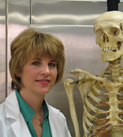
We are proud to announce that our own contributor and associate Dr. Elizabeth Murray, Ph.D., has been awarded the Anthropology Section's 2018 "T. Dale Stewart Award" at the American Academy of Forensic Sciences (AAFS) 71st Annual Scientific Meeting in late February.
Dr. Murray's involvement with AAFS has included chairing the Academy-wide annual meeting as well as committee-level service in long-term planning, Board of Trustees, and the Student Academy.
Well-known and respected as a forensic anthropologist, Dr. Murray teaches at the University of Mount St. Joseph courses in anatomy and physiology, gross anatomy, and forensic science for the Department of Biology. Her most recent book, published in 2019, is "The Dozier School for Boys: Forensics, Survivors, and a Painful Past."
Our congratulations to her for yet another incredible achievement in her illustrious career. We are glad to count her as a friend and as a contributor to "Medical Terminology Daily" and Clinical Anatomy Associates, Inc.