![]()
Medical Terminology Daily (MTD) is a blog sponsored by Clinical Anatomy Associates, Inc. as a service to the medical community. We post anatomical, medical or surgical terms, their meaning and usage, as well as biographical notes on anatomists, surgeons, and researchers through the ages. Be warned that some of the images used depict human anatomical specimens.
You are welcome to submit questions and suggestions using our "Contact Us" form. The information on this blog follows the terms on our "Privacy and Security Statement" and cannot be construed as medical guidance or instructions for treatment.
We have 710 guests and no members online
")
Marcia Crocker Noyes
(1869 – 1946)
Further to my comment on old books and research that started with an interesting bookplate (Ex-Libris). I continued my research and found that the person in charge of the Osler library bookplate was a fascinating individual that today maybe a ghost in the MedChi library and building in Baltimore... This is certainly an article that can be called "A Moment in History"
Marcia Crocker Noyes was the librarian at The Maryland State Medical Society from 1896 to 1946 and was a founding member of the Medical Library Association.[1][2][3]
Sir William Osler, MD. a famous Johns Hopkins surgeon was a noted bibliophile and had a large personal collection of books on various topics. When he became the President of MedChi in 1896, he was dismayed at the condition of the library and knew that with the right person and some stewardship, it could become a significant collection. Sir William asked his friend, Dr. Bernard Steiner, a physician and President of the Enoch Pratt Free Library in Baltimore for suggestions of a librarian, and Dr. Steiner recommended Marcia Crocker Noyes. A native of New York, and a graduate of Hunter College, Marcia had moved to Baltimore for a lengthy visit with her sister, and took a “temporary” position at the Pratt Library, which turned into three years. Although she had no medical experience or background, she was enthusiastic, and most importantly, she was willing to move into the apartment provided for the librarian, who needed to be available 24 hours a day.
The image in this article is Ms. Noyes on her first year on the job. Marcia developed a book classification system for medical books, based on the Index Medicus, and called it the Classification for Medical Literature. The system uses the alphabet with capital letters for the major divisions of medicine and lower-case ones for the sub-sections. The system was used for many years, but it's now dated and the Faculty's original shelving scheme was never changed. The card catalogs still reflect her classification and many of the cards are written in Marcia's back-slanting handwriting.
Marcia knew enough to ask the Faculty's members about medical questions, terminology and literature. She gradually won over the predominantly male membership and they became her greatest allies; Sir William at the start, and then for nearly 40 years, Dr. John Ruhräh, a wealthy pediatrician with no immediate family of his own. She made a point of attending almost every Faculty function, and in 1904, under guidelines from the American Medical Association, Marcia was made the Faculty Secretary. For much of her first 10 years, she was the Faculty's only full-time employee, only being assisted by Mr. Caution, the Faculty's janitor. Later in life Marcia would say that she hired him because of his name!
Within ten years, the library had outgrown its space, and plans, spearheaded by Marcia and Sir William before his move to Oxford, were made to build a headquarters building, mainly to house the library's growing collection of medical books and journals.
Marcia was instrumental in the design and building of the new headquarters. She travelled to Philadelphia, New York and Boston to look at their medical society buildings, and eventually, the Philadelphia architectural firm, Ellicott & Emmart was selected to design and build the new Faculty building. Every detail of the building held her imprimatur, from the graceful staircase, to the light-filled reading room, and all of the myriad details of the millwork, marble tesserae, and most of all, the four-story cast iron stacks. She was on-site, climbing up unfinished staircases, checking out the progress of the building, which was built in less than one year at a cost of $90,000.
Among the features of the new building was a fourth-floor apartment for her. She referred to it as the "first penthouse in Baltimore" and it had a garden and rooftop terrace. The library collection eventually grew to more than 65,000 volumes from medical and specialty societies around the world. Journals were traded back and forth, and physicians eagerly anticipated the arrival of each new issue. At the same time, Marcia was involved in the Medical Library Association as one of eight founding members. The MLA promotes medical libraries and the exchange of information. One of the earliest mandates of the MLA was the Exchange, a distribution and trade service for those who had duplicates or little-used books in their collections. Initially, the Exchange was run out of the Philadelphia medical society, but in 1900 it was moved to Baltimore and Marcia oversaw it. Several hundred periodicals and journals were received and sent each month, a huge amount of work for a tiny staff. In 1904, the Faculty had run out of room to manage the Exchange, so it was moved to the Medical Society of the Kings County (Brooklyn). But without Marcia's excellent administrative skills, it floundered and in 1908, the MLA asked Marcia to take charge once again.
In 1909, when the new Faculty building opened, there was enough room to run the Exchange and with the help of MLA Treasurer, noted bibliophile and close friend, Dr. John Ruhräh, it once again became successful. Additionally, Marcia and Dr. Ruhräh combined forces to revive the MLA's bulletin, which had all but ceased publication in 1908, taking the Exchange with it. This duo maintained editorial control from 1911 until 1926. In 1934, around the time of Dr. Ruhräh's death, Marcia became the first “unmedicated” professional to head the MLA. During her tenure, the MLA incorporated, the first seal was adopted, and the annual meeting was held in Baltimore. Marcia wanted to write the history of the MLA once she retired from full-time work at the Faculty, but her health was beginning to fail. She had back problems and had suffered a serious burn on her shoulder as a young woman, possibly from her time running a summer camp, Camp Seyon, for young ladies in the Adirondack Mountains. In 1946, a celebration was planned to honor Marcia's 50 years at the Faculty. But she was adamant that the physicians wait until November, the actual date of her 50 years. However, they knew she was gravely ill, and might not make it until then, so a huge party was held in April. More than 250 physicians attended the celebration, but the ones she was closest to in the early years, were long gone. She was presented with a suitcase, a sum of money to use for travelling, and her favorite painting of Dr. John Philip Smith, a founder of the Medical College in Winchester, Virginia. It was painted by Edward Caledon Smith, a Virginia painter who had been a student of the painter Thomas Sully.[4] She adored this painting and vowed, jokingly, to take it with her wherever she went.
The painting was not to stay with her for very long, for she died in November 1946, and left it to the Faculty in her will. Her funeral was held in the Faculty's Osler Hall, named for her dear friend. More than 60 physicians served as her pallbearers, and she was buried at Baltimore's Green Mount Cemetery. In 1948, the MLA decided to establish an award in the name of Marcia Crocker Noyes. It was for outstanding achievement in medical library field and was to be awarded every two years, or when a truly worthy candidate was submitted. In 2014, the Faculty began giving a bouquet of flowers to the winner of the award in Marcia's name, and in honor of her work. Much evidence exists for this tradition, as we know that the physicians, especially Drs. Osler and Ruhräh, frequently gave her bouquets of flowers. Marcia also cultivated flower gardens at the Faculty and decorated the rooms with her work.
Today, the MedChi building is open for tours and if the rumors are to be believed Ms. Marcia Crocker Noyes is still at work in her beloved library as the "resident ghost" [1][5]

NOTE: This article has been modified from the original Wikipedia article on Marcia Crocker Noyes. The article itself is well-written with interesting images of the subject. I would encourage you to visit it. The second insert is from book 00736 in my personal library and shows in pencil, the incredibly small handwriting of Marsha C. Noyes.
Sources:
1. "Marcia, Marcia, Marcia" MedChi Archives blog.
2. "Marcia C. Noyes, Medical Librarian" (PDF). Bulletin of the Medical Library Association. 35 (1): 108–109. 1947. PMC 194645
3. Smith, Bernie Todd (1974). "Marcia Crocker Noyes, Medical Librarian: The Shaping of a Career" (PDF). Bulletin of the Medical Library Association. 62 (3): 314–324. PMC 198800Freely accessible. PMID 4619344.
4. Edward Caledon BRUCE (1825-1901)"
5. Behind the scenes tour MedChiBuilding
"Clinical Anatomy Associates, Inc., and the contributors of "Medical Terminology Daily" wish to thank all individuals who donate their bodies and tissues for the advancement of education and research”.
Click here for more information


- Details

Change the text of the image
UPDATED: The [phrenoesophageal ligament] or phrenoesophageal membrane is part of a complex system that closes off the esophageal hiatus, one of the seven hiatuses in the respiratory diaphragm, preventing the herniation of abdominal structures into the thoracic mediastinum.
The connective tissue layer called the endoabdominopelvic fascia, which lines the inner aspect of the abdominopelvic cavity, is found as a "glue" between the respiratory diaphragm and the parietal peritoneum. At this point the endoabdominopelvic fascia is called the "infradiaphragmatic fascia".
When the infradiaphragmatic fasia gets to the edge of the esophageal hiatus, it splits into ascending and a descending components or limbs. These are the superior and inferior phrenoesophageal ligaments or phrenoesophageal membranes. These phrenoesophageal ligaments create a circular disc-like plug between the abdomen and the thorax. This "plug" is reinforced by a infradiaphragmatic fat pad found internal to the phrenoesophageal ligament.
The phrenoesophageal ligaments are reinforced externally. On their thoracic aspect by the endothoracic fascia, and on the abdominal side, by parietal peritoneum.
The ascending limb fuses superiorly with the esophageal fascia, which lines the external aspect of the longitudinal muscle of the esophagus, as the thoracic esophagus does not have a serosa layer. The descending limb fuses inferiorly with the esophageal fascial covering of the longitudinal ligament as it is covered by the peritoneum . Failure of the phrenoesophageal ligaments can predispose to esophageal hiatus hernia.
- Details
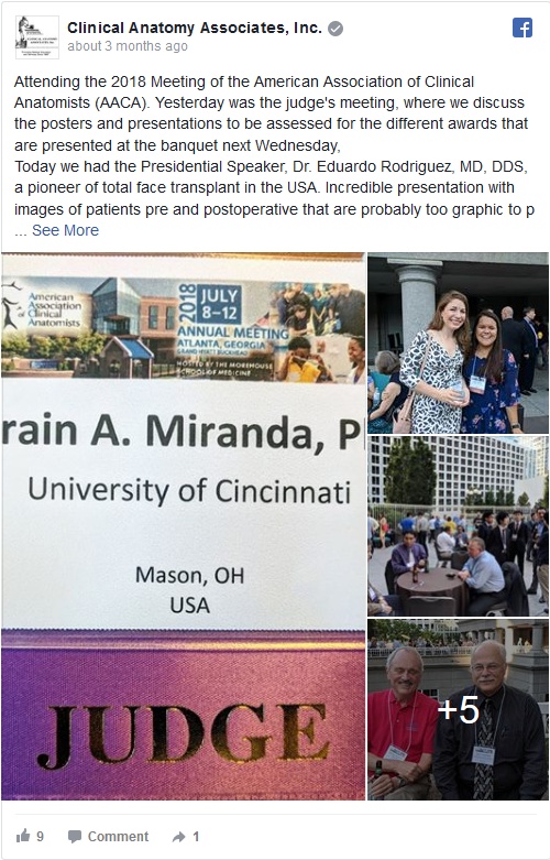
The following article is embedded from our Facebook page https://www.facebook.com/CAAInc.
This year the 2018 meeting of the American Association of Clinical Anatomists is being held in Atlanta, GA., at the Grand Hyatt Buckhead Hotel and Conference Center. The program is full of interesting topics and is already a hit with all the attendees. Looking forward to the program.
- Details

Esophageal hiatus hernia in situ.
The arrow points to stomach and greater
omentum herniating into the thorax
UPDATED: An esophageal hiatus hernia (also known as a hiatal hernia) is caused by a dilation of the esophageal hiatus and its component structures, the phrenoesophageal membranes (ligaments).
Since the intraabdominal pressure is higher than the intrathoracic pressure, abdominal contents -usually stomach and greater omentum- can herniate through the dilated esophageal hiatus into the mediastinum, the central region of the thoracic cavity. This presents as a hernia sac whose walls are formed by endothoracic fascia, phrenoesophageal membranes and parietal peritoneum.
There are two main types of esophageal hiatus hernias. Type I is known as a "sliding hiatal hernia" and is characterized by a complete ascension of the esophagogastric junction and abdominal esophagus into the thoracic hernia sac. This is usually accompanied by a typical "hourglass image" in a radiographic assessment, and also presents with gastroesophageal reflux disease (GERD). Type I esophageal hiatus hernias are more common.

Esophageal hiatus hernia, reduced.
The dotted line shows the edge of
the enlarged esophageal hiatus
Type II esophageal hiatus hernia is known as a "paraesophageal hernia" and represent about 5 - 15% of esophageal hiatus hernias. In this case, the esophagogastric junction maintains its anatomical position inferior to the respiratory diaphragm, but the fundus and body of the stomach, along with some greater omentum herniate alongside the esophagus into the mediastinal region of the thoracic cavity. Although there can be GERD, this type of hernia usually presents with little symptomatology, and when it does, symptoms are related to ischemia or partial to complete obstruction. There are variations of type II hernia, which are classified as Type III and IV. Type IV, although rare, will include other viscera in the hernia sac, including colon, spleen, or even small intestine.
The accompanying images above depict a Type I esophageal hiatus hernia. The superior image shows the hernia in situ where the stomach and greater omentum are still in the hernia sac. The inferior image shows the contents reduced and the abdominal esophagus being pulled into the abdominal cavity. The dotted line shows the dilated esophageal hiatus that needs to be repaired to prevent recurrence of the pathology.
Click on this link for additional information on esophageal hiatus hernia surgery.
The image below answers a question by Victoria Guy Ratcliffe, who asked via Facebook "What would it be if it feels like you've got a blockage right at the level of the heart? That's too high for a hiatal hernia, isn't it?" The image answers the question. It shows a dissection of the left side of the thorax. The anterior thoracic wall and the left lung have been removed. The heart is immediately superior and anterior to the esophageal hiatus, and the hernia sac of a Type I esophageal hiatus hernia is seen immediately posterior and in contact with the heart. Whether this means that you will "feel" the hernia, it is up for debate, as all these structures have visceral innervation. Most probably, a well-developed Type II esophageal hiatus hernia might interfere with swallowing at this level, causing the sensation she mentions. Thanks for the question, Tori.

For additional information:
"Approaches to the Diagnosis and Grading of Hiatal Hernia" Kahrilas et al Best Pract Res Clin Gastroenterol. 2008 ; 22(4): 601–616.
- Details
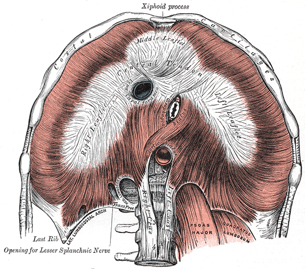
Respiratory diaphragm
The term [hiatus] derives from the Latin word [hiare], meaning to "gape" or to "yawn". In human anatomy this term is used to mean an "opening" or a "defect". It must be pointed out that in anatomy (and surgery) the term "defect" does not necessarily mean "defective". In most cases a "defect" is a normal opening in a structure, such as the esophageal hiatus. The plural form is either [hiatus] or [hiatuses].
In the case of the respiratory diaphragm, there are seven such openings, seven normal hiatuses. On top of this, you can find an abnormal opening caused by incomplete congenital closure of the dome of the diaphragm, a congenital diaphragmatic hernia (CDH), also known as Bochdalek's hernia, found in the posterior aspect of the respiratory diaphragm.
The seven hiatuses of the respiratory diaphragm are:
• Aortic hiatus
• Inferior vena cava hiatus
• Hiatuses (2) for the superior epigastric vessels, which are the inferior continuation of the internal thoracic (mammary) vessels. Also known as the hiatuses of Morgagni. A hernia in a newborn through this hiatus is also considered a CDH.
• Hiatuses (2) for the splanchnic nerves
Based on the above it is wrong (maybe not wrong, but incomplete) to say that a patient has a "hiatal hernia", as the term does not include which hiatus is involved. In fact the hernia of Morgagni is also a "hiatal hernia" as the hernia passes through a normal defect in the respiratory diaphragm. Come to think of it, it could also be a hernia in a hiatus somewhere else in the body, such as a hernia of Schwalbe, a type or pelvic diaphragm hernia.
Note: Thanks to DHREAMS of the Columbia University Medical Center for the link on CDH.
Sources:
1. "The Origin of Medical Terms" Skinner, HA 1970 Hafner Publishing Co.
2. "Medical Meanings - A Glossary of Word Origins" Haubrich, WD. ACP Philadelphia
3 "Tratado de Anatomia Humana" Testut et Latarjet 8 Ed. 1931 Salvat Editores, Spain
4. "Anatomy of the Human Body" Henry Gray 1918. Philadelphia: Lea & Febiger Image modified by CAA, Inc. Original image by Henry Vandyke Carter, MD., courtesy of bartleby.com
- Details
Esophagogastric junction
UPDATED: The esophageal hiatus is one of the seven hiatuses found in the respiratory diaphragm allowing passage of structures between the thorax and abdomen. As it name implies, the esophageal hiatus is the passageway for the esophagus. It also allows passage of the anterior and posterior vagus nerves, (CN X).
The hiatus is bound by two muscular crura, both of which arise from the right tendinous aortic crus. Since the intraabdominal pressure is higher than the intrathoracic pressure, there is a series of structures at the phrenoesophagogastric junction to close the esophageal hiatus.
The infradiaphragmatic parietal peritoneum reflects off the diaphragm towards the stomach to form its serosa layer (visceral peritoneum). At the same time the infradiaphragmatic fascia, also known as the endoabdominopelvic fascia, splits into two components or limbs. These are the superior and inferior phrenoesophageal ligaments or phrenoesophageal membranes. (the root [-phren-] means "diaphragm"). These phrenoesophageal ligaments create a disc-like plug between the abdomen and the thorax. This "plug" is reinforced by a circular infradiaphragmatic fat pad. The phrenoesophageal ligaments are reinforced on their thoracic aspect by the endothoracic fascia.
The lower esophagus has a dilation (evident in the image) called the "esophageal ampulla", in relation to this dilation the circular muscle layer of the esophagus slightly thickens creating the so-called "lower esophageal sphincter". This area is not a true anatomical sphincter, but rather is a functional sphincter.
The esophagogastric mucosal junction shows a marked transition in the shape of a wavy line. This is called the Z-line or the ora serrata. Extensions of the gastric mucosa and submucosa inferior to the ora serrata create a valve-like flap called the "gastroesophageal flap valve". When viewing this mucosal flap through and endoscope, it looks corrugated and flower-like, hence it is also called the "rosette".
The congenital or pathological dilation of the esophageal hiatus can predispose to esophageal hiatus hernia.
Sources:
1 "Tratado de Anatomia Humana" Testut et Latarjet 8 Ed. 1931 Salvat Editores, Spain
2. "Anatomy of the Human Body" Henry Gray 1918. Philadelphia: Lea & Febiger
Original image by Dr. E. Miranda
- Details
Esophagogastric junction
The esophagogastric junction is a complex anatomical region found at the esophageal hiatus of the respiratory diaphragm. It allows passage of he esophagus from the thorax into the abdomen.
The hiatus is bound by two muscular crura, both of which arise from the right tendinous aortic crus. Since the intraabdominal pressure is higher than the intrathoracic pressure, there is a series of structures at the phrenoesophagogastric junction to close the esophageal hiatus.
The infradiaphragmatic parietal peritoneum reflects off the diaphragm towards the stomach to form its serosa layer (visceral peritoneum). At the same time the infradiaphragmatic fascia, also known as the endoabdominopelvic fascia, splits into two components or limbs. These are the superior and inferior phrenoesophageal ligaments or phrenoesophageal membranes. (the root [-phren-] means "diaphragm"). These phrenoesophageal ligaments create a disc-like plug between the abdomen and the thorax. This "plug" is reinforced by a circular infradiaphragmatic fat pad. The phrenoesophageal ligaments are reinforced on their thoracic aspect by the endothoracic fascia.
The lower esophagus has a dilation (evident in the image) called the "esophageal ampulla", in relation to this dilation the circular muscle layer of the esophagus slightly thickens creating the so-called "lower esophageal sphincter". This area is not a true anatomical sphincter, but rather is a functional sphincter.
The esophagogastric mucosal junction shows a marked transition in the shape of a wavy line. This is called the Z-line or the ora serrata. Extensions of the gastric mucosa and submucosa inferior to the ora serrata create a valve-like flap called the "gastroesophageal flap valve". When viewing this mucosal flap through and endoscope, it looks corrugated and flower-like, hence it is also called the "rosette".
The congenital or pathological dilation of the esophageal hiatus can predispose to esophageal hiatus hernia.
Sources:
1 "Tratado de Anatomia Humana" Testut et Latarjet 8 Ed. 1931 Salvat Editores, Spain
2. "Anatomy of the Human Body" Henry Gray 1918. Philadelphia: Lea & Febiger
Original image by Dr. E. Miranda