![]()
Medical Terminology Daily (MTD) is a blog sponsored by Clinical Anatomy Associates, Inc. as a service to the medical community. We post anatomical, medical or surgical terms, their meaning and usage, as well as biographical notes on anatomists, surgeons, and researchers through the ages. Be warned that some of the images used depict human anatomical specimens.
You are welcome to submit questions and suggestions using our "Contact Us" form. The information on this blog follows the terms on our "Privacy and Security Statement" and cannot be construed as medical guidance or instructions for treatment.
We have 1033 guests and no members online
")
Marcia Crocker Noyes
(1869 – 1946)
Further to my comment on old books and research that started with an interesting bookplate (Ex-Libris). I continued my research and found that the person in charge of the Osler library bookplate was a fascinating individual that today maybe a ghost in the MedChi library and building in Baltimore... This is certainly an article that can be called "A Moment in History"
Marcia Crocker Noyes was the librarian at The Maryland State Medical Society from 1896 to 1946 and was a founding member of the Medical Library Association.[1][2][3]
Sir William Osler, MD. a famous Johns Hopkins surgeon was a noted bibliophile and had a large personal collection of books on various topics. When he became the President of MedChi in 1896, he was dismayed at the condition of the library and knew that with the right person and some stewardship, it could become a significant collection. Sir William asked his friend, Dr. Bernard Steiner, a physician and President of the Enoch Pratt Free Library in Baltimore for suggestions of a librarian, and Dr. Steiner recommended Marcia Crocker Noyes. A native of New York, and a graduate of Hunter College, Marcia had moved to Baltimore for a lengthy visit with her sister, and took a “temporary” position at the Pratt Library, which turned into three years. Although she had no medical experience or background, she was enthusiastic, and most importantly, she was willing to move into the apartment provided for the librarian, who needed to be available 24 hours a day.
The image in this article is Ms. Noyes on her first year on the job. Marcia developed a book classification system for medical books, based on the Index Medicus, and called it the Classification for Medical Literature. The system uses the alphabet with capital letters for the major divisions of medicine and lower-case ones for the sub-sections. The system was used for many years, but it's now dated and the Faculty's original shelving scheme was never changed. The card catalogs still reflect her classification and many of the cards are written in Marcia's back-slanting handwriting.
Marcia knew enough to ask the Faculty's members about medical questions, terminology and literature. She gradually won over the predominantly male membership and they became her greatest allies; Sir William at the start, and then for nearly 40 years, Dr. John Ruhräh, a wealthy pediatrician with no immediate family of his own. She made a point of attending almost every Faculty function, and in 1904, under guidelines from the American Medical Association, Marcia was made the Faculty Secretary. For much of her first 10 years, she was the Faculty's only full-time employee, only being assisted by Mr. Caution, the Faculty's janitor. Later in life Marcia would say that she hired him because of his name!
Within ten years, the library had outgrown its space, and plans, spearheaded by Marcia and Sir William before his move to Oxford, were made to build a headquarters building, mainly to house the library's growing collection of medical books and journals.
Marcia was instrumental in the design and building of the new headquarters. She travelled to Philadelphia, New York and Boston to look at their medical society buildings, and eventually, the Philadelphia architectural firm, Ellicott & Emmart was selected to design and build the new Faculty building. Every detail of the building held her imprimatur, from the graceful staircase, to the light-filled reading room, and all of the myriad details of the millwork, marble tesserae, and most of all, the four-story cast iron stacks. She was on-site, climbing up unfinished staircases, checking out the progress of the building, which was built in less than one year at a cost of $90,000.
Among the features of the new building was a fourth-floor apartment for her. She referred to it as the "first penthouse in Baltimore" and it had a garden and rooftop terrace. The library collection eventually grew to more than 65,000 volumes from medical and specialty societies around the world. Journals were traded back and forth, and physicians eagerly anticipated the arrival of each new issue. At the same time, Marcia was involved in the Medical Library Association as one of eight founding members. The MLA promotes medical libraries and the exchange of information. One of the earliest mandates of the MLA was the Exchange, a distribution and trade service for those who had duplicates or little-used books in their collections. Initially, the Exchange was run out of the Philadelphia medical society, but in 1900 it was moved to Baltimore and Marcia oversaw it. Several hundred periodicals and journals were received and sent each month, a huge amount of work for a tiny staff. In 1904, the Faculty had run out of room to manage the Exchange, so it was moved to the Medical Society of the Kings County (Brooklyn). But without Marcia's excellent administrative skills, it floundered and in 1908, the MLA asked Marcia to take charge once again.
In 1909, when the new Faculty building opened, there was enough room to run the Exchange and with the help of MLA Treasurer, noted bibliophile and close friend, Dr. John Ruhräh, it once again became successful. Additionally, Marcia and Dr. Ruhräh combined forces to revive the MLA's bulletin, which had all but ceased publication in 1908, taking the Exchange with it. This duo maintained editorial control from 1911 until 1926. In 1934, around the time of Dr. Ruhräh's death, Marcia became the first “unmedicated” professional to head the MLA. During her tenure, the MLA incorporated, the first seal was adopted, and the annual meeting was held in Baltimore. Marcia wanted to write the history of the MLA once she retired from full-time work at the Faculty, but her health was beginning to fail. She had back problems and had suffered a serious burn on her shoulder as a young woman, possibly from her time running a summer camp, Camp Seyon, for young ladies in the Adirondack Mountains. In 1946, a celebration was planned to honor Marcia's 50 years at the Faculty. But she was adamant that the physicians wait until November, the actual date of her 50 years. However, they knew she was gravely ill, and might not make it until then, so a huge party was held in April. More than 250 physicians attended the celebration, but the ones she was closest to in the early years, were long gone. She was presented with a suitcase, a sum of money to use for travelling, and her favorite painting of Dr. John Philip Smith, a founder of the Medical College in Winchester, Virginia. It was painted by Edward Caledon Smith, a Virginia painter who had been a student of the painter Thomas Sully.[4] She adored this painting and vowed, jokingly, to take it with her wherever she went.
The painting was not to stay with her for very long, for she died in November 1946, and left it to the Faculty in her will. Her funeral was held in the Faculty's Osler Hall, named for her dear friend. More than 60 physicians served as her pallbearers, and she was buried at Baltimore's Green Mount Cemetery. In 1948, the MLA decided to establish an award in the name of Marcia Crocker Noyes. It was for outstanding achievement in medical library field and was to be awarded every two years, or when a truly worthy candidate was submitted. In 2014, the Faculty began giving a bouquet of flowers to the winner of the award in Marcia's name, and in honor of her work. Much evidence exists for this tradition, as we know that the physicians, especially Drs. Osler and Ruhräh, frequently gave her bouquets of flowers. Marcia also cultivated flower gardens at the Faculty and decorated the rooms with her work.
Today, the MedChi building is open for tours and if the rumors are to be believed Ms. Marcia Crocker Noyes is still at work in her beloved library as the "resident ghost" [1][5]

NOTE: This article has been modified from the original Wikipedia article on Marcia Crocker Noyes. The article itself is well-written with interesting images of the subject. I would encourage you to visit it. The second insert is from book 00736 in my personal library and shows in pencil, the incredibly small handwriting of Marsha C. Noyes.
Sources:
1. "Marcia, Marcia, Marcia" MedChi Archives blog.
2. "Marcia C. Noyes, Medical Librarian" (PDF). Bulletin of the Medical Library Association. 35 (1): 108–109. 1947. PMC 194645
3. Smith, Bernie Todd (1974). "Marcia Crocker Noyes, Medical Librarian: The Shaping of a Career" (PDF). Bulletin of the Medical Library Association. 62 (3): 314–324. PMC 198800Freely accessible. PMID 4619344.
4. Edward Caledon BRUCE (1825-1901)"
5. Behind the scenes tour MedChiBuilding
"Clinical Anatomy Associates, Inc., and the contributors of "Medical Terminology Daily" wish to thank all individuals who donate their bodies and tissues for the advancement of education and research”.
Click here for more information


- Details
- Hits: 87731

Click for a larger image
UPDATED: The "triangle of doom" is a name given to a roughly triangular area in the posterior aspect of the anterior wall of the lower abdominopelvic region. It is used by surgeons repairing an inguinofemoral hernia with a mesh and they want to avoid large vascular structures, namely the external iliac artery and vein. The "triangle of doom" will be highlighted when you hover your cursor over the image.
The so-called "triangle of doom" is a misnomer perpetuated by the first laparoscopic surgeons who observed the anatomy of the inguinofemoral region from the posterior aspect. It is neither a triangle (as it only has two boundaries), nor is it an eponym (no such person existed - that is why uppercase should not be used). It does indicate an area where it is extremely dangerous to place staples or sutures during laparoscopic hernia surgery.
The "triangle of doom" is an inverted "V" shaped area with its apex at the internal (deep) inguinal ring. The "triangle of doom" is bound laterally by the gonadal vessels, and medially by the vas deferens in the male, or the round ligament of the uterus in the female. Within the boundaries of this area you can find the external iliac artery and vein.
It should be pointed out that although the "triangle of doom" landmark does protect the surgeon from damaging the external iliac vessels, a portion of these vessels lie outside of this area. In fact, there are several other areas of concern for neurovascular damage when performing a laparoscopic herniorrhaphy.
Hover over the image in this article, and the triangle of doom will appear. Clicking on the image will show a larger image of the posteroinferior abdominal region. The image also depicts other structures of anatomical importance for laparoscopic herniorrhaphy:
• Arcuate line (b)
• Hesselbach's triangle (in yellow)
• Aberrant obturator artery (Corona Mortis) (a)
• Inferior (deep) epigastric artery (c)
Image property of: CAA.Inc. . Artist: M. Zuptich.
Clinical anatomy of the inguinofemoral hernias, as well as abdominal and perineal hernias are some of the lecture topics developed and delivered to the medical devices industry by Clinical Anatomy Associates, Inc. For more information Contact Us.
- Details
- Written by: Pascale Pollier-Green
This article is part of the series "A Moment in History" where we honor those who have contributed to the growth of medical knowledge in the areas of anatomy, medicine, surgery, and medical research.
If you arrived directly to this article, the first article in this three-page series can be read HERE
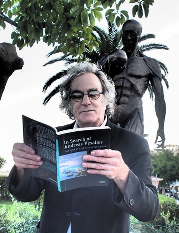
Bryan Green reading his poem in Theo Dirix's
book “ In Search of Andreas Vesalius”
Click on the image for a larger version
I would like to shine a light on my husband poet /sculptor Bryan Green , who wrote a poem on Vesalius that is published in Theo’s book; “In Search of Andreas Vesalius: The Quest for the Lost Grave” and gave a performance at the Fabrica Vitae exhibition opening.. Bryan has constantly worked behind the scenes editing many letters, articles, books, and leaflets, I couldn’t have done it all without his help and advice. He also made the long lorry journey to Zakynthos from Belgium with me and our friend James Gatehouse to deliver the monument.
Vesalius Continuum also marked the start of our touring exhibition “Fabrica Vitae” curated by Eleanor Crook, my sister Chantal Pollier, and myself. The exhibition toured all over Europe and the US with the help and support of Theo Dirix and Belgian Embassies world wide .
The conference and accompanying events could not have happened without financial funds and I hereby would like to thank all our sponsors: Professor Peter Abrahams with his infectious energy and professor Robert Jordan; St Georges University of Grenada, Ruth Richardson and Brian Hurwitz and Mark Gardiner for getting funding from the Wellcome trust, Marie Dauenheimer and the Vesalius Trust, BIOMAB, Ann van the Velde and The University of Antwerp, The AEIMS and MAA, William Nagels, warmly thank the local authorities and the mayor of Zakynthos, ARSIC, Theo Dirix, and Stephen Joffe, and a special thank you also Stephen for writing a beautiful foreword for our book In the Shadow of Vesalius.
You can imagine after such an exciting and wonderful adventure, which took quite a few years to organize, and a quite a few years to reminisce over, we decided we wanted to keep the momentum going and thus the Vesalius triennial was born.
In 2017 BIOMAB, in collaboration with Vesaliana, organized the first triennial in Zakynthos ‘Uniting Medicine with Poetry, History and Culture’
It seems like another world in which we made our plans for the 2nd edition of the Vesalius Triennial Congress, 4 months before the COVID-19 pandemic lock down. From the vain belief that COVID-19 would not hit most countries, to hopes that everything would have blown over by 13th November 2020 (the day when the next Vesalius Triennial Congress would take place in Antwerp) to realizing that we were going to have to take action, the scientific committee has transformed from one in which everyone knew their time-tried and perfected role, to one requiring invention in uncharted territory.
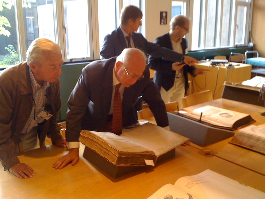
Professors Vivian Nutton and Omer Steeno
looking at a first edition of the Fabrica
Canceling the 2nd Vesalius Triennial was not a welcome prospect , since facilitating human communication is the corner stone of a scientific community. So we set sail for the vast virtual-reality realm. To discover just how far we could delve into virtual communication with a dedicated but small organising committee, was an eventful, insightful voyage. Sadly after long and careful consideration and several online meetings we finally decided to postpone all international congress keynote lectures and educational sessions until 2023.
However we would like to invite all the friends of Vesalius for a virtual book launch on Nov 13th we will soon post the event details on how to register for this event on social media, and on Vesalius continuum website
The book "In the shadow of Vesalius" can be ordered here: http://garant.be/shadow-of-vesalius/
Finally I would like to thank everyone who has been part of this adventure, special thank you to Professor Dr. Efrain Miranda ( Clinical Anatomy) for his continuous support, EBSA, Prof. Stefanos Geroulos, Vasia Hatzi (MEDinART), Pavlos Plessas, Nicos Varvianis, Maria Sidirokastriti-Kontoni & Fr. Panagiotis Kapodistrias, the many wonderful speakers, the local organisers, our Keynote speaker Professor Martin Kemp for his wonderful contribution, Eleni Andrianaki; ibis el greco , the wonderful delegates, the artists of the Fabrica Vitae exhibition, the museum and universities where we took our exhibition, a special thank you to Juris Salaks and Ieva Lebiete for hosting our exhibition at the Stradins museum and for all the help and support, Apostolis Sarris, Nikos Papadopoulos, Sylviane Déderix, Jan Driessen, Theo Dirix, Chr. Merkouri.and to the all the friends of Vesalius who like to keep his spirit alive.
Pascale Pollier-Green
Oct 2020
Personal note: I would like to thank Pascale Pollier-Green for authoring this series of articles and wish Professor Robrecht Van Hee the best success publishing this new book on the history and influence of Andreas Vesalius on anatomy, medicine, science, and the Arts. Dr. Miranda.
- Details
- Written by: Pascale Pollier-Green
This article is part of the series "A Moment in History" where we honor those who have contributed to the growth of medical knowledge in the areas of anatomy, medicine, surgery, and medical research.
If you arrived directly to this article, the first article in this three-page series can be read HERE
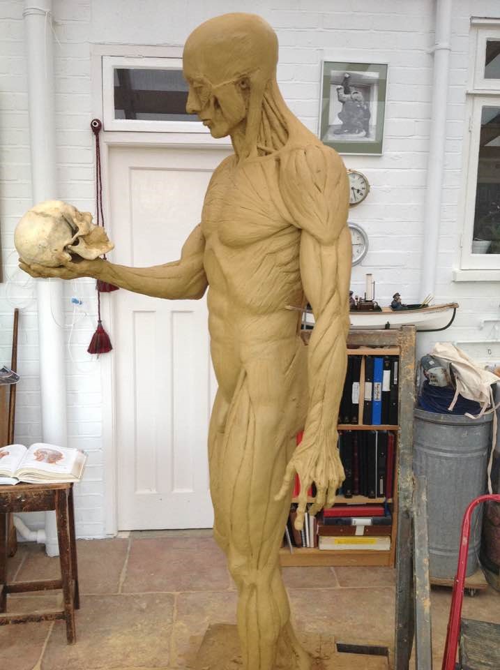
Sculpting the monument in Richard Neave’s studio
One of the most important conferences that BIOMAB initiated and co-organized was without doubt “Vesalius Continuum” which took place in 2014 on the island of Zakynthos . This joint AEIMS conference was organized in collaboration with Dr Mark Gardiner and the then Consul at the Belgian Embassy in Greece - Theo Dirix, and brought together many prominent scholars and Vesalius experts who presented papers on the life, work and death of Vesalius, and the influence of the Fabrica on modern anatomy, and on medical art and contemporary art (pdf file).
Without the organizing skills and drive and energy of Dr. Mark Gardiner and the countless meetings and emails with Theo Dirix, the conference Vesalius Continuum would not have been the huge world class event it became.
In 2013 Dr. Mark Gardiner and I first introduced ourselves to Professor Vivian Nutton who was giving a lecture at Warwick University about the incredible find of a hand written (by Vesalius himself !) edited version of the Fabrica’s second edition, which would have become the third edition, …had Vesalius not met his untimely death. Mark and I were blown away by this wonderfully exciting lecture and we asked Professor Nutton to be a speaker at our conference in 2014, which he accepted gladly. He was invited again in 2017 for the triennial and has now especially for this publication written an exciting account of Vesalius in England, 1544 to 1547.
Vesalius’s 500 th anniversary celebrations did not end with the organization of the conference, but became a collection of several events.
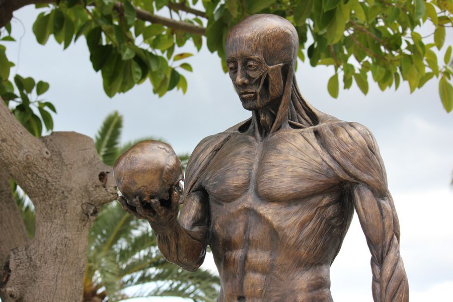
My colleague and dear friend forensic medical artist Richard Neave and I sculpted a bronze monument to commemorate Vesalius’s death on the Island of Zakynthos on 15th Oct 1564. This monument might never have been erected if it were not for the wonderful idea of Antwerp GP and president of Vesaliana Dr Marc de Roeck together with William Nagels, who devised a way of self-funding this large undertaking. We had countless meetings discussing the monument and agreed with Dr. de Roeck that by making a bronze facial reconstruction portrait (made by Richard Neave and myself) and selling 12 copies we would gather enough money together to make the monument, pay to have it cast in Bronze and drive the sculpture from Belgium to Zakynthos ready to have its grand inauguration at the start of the conference. This was all achieved successfully and the sculpture now stands on the Island of Zakynthos’s Vesalius square, facing the Ionian sea.
Dr. Marc De Roeck and Pascale Pollier
sailing to Zakynthos
In 2009 I had completed a facial reconstruction course at the academy of fine arts in Maastricht , the Netherlands, and as Vesalius had always been my big inspiration and the reason why I chose to become a medical artist, it was my dream to make a facial reconstruction of Vesalius. The dream soon turned into a passion, and the passion into an obsession to go in search of the grave of Vesalius and find his skull.
Ann suggested to Marc we sailed to Zakynthos with the MYC-Medical Yachting Club to start the quest for the grave. I will never forget the day that Marc gave me the ships wheel as we got closer to the island and I sailed into the harbor of Zakynthos! An amazing feeling!
After our first visit to “what we thought then was the grave site“ at Laganas, I wrote a letter to the Belgian embassy in Greece, asking for help with our quest, a year later when Theo Dirix took office as Consul he wrote back to me, my letter had ignited a flame in the heart of taphophile Theo Dirix.
Soon the quest became an official scientific research collaborative project between the Belgian School of Athens (Jan Driessen, Apostolis Sarris, Sylviane Déderix) and the Greek authorities, together with the invaluable research of Omer Steeno, Maurits Biesbrouk and Theodoor Godeeris, who all share their latest findings in this book. With this wonderful collaborative effort, even though we have not yet found the actual grave, we can truly claim that we have made some history.
Theo Dirix wrote 2 books on the quest of the grave and now will reveal some new insights with his article in the book “In the Shadow of Vesalius”.
This article continues HERE
- Details
- Written by: Pascale Pollier-Green
This article is part of the series "A Moment in History" where we honor those who have contributed to the growth of medical knowledge in the areas of anatomy, medicine, surgery, and medical research.
The following article was written by my good friend Pascale Pollier-Green and it recounts the long road that has taken her and other Vesaliana followers all over the world, and has resulted in the publication of the book "In the Shadow of Vesalius". Dr. Miranda
The concept behind the book “In the shadow of Vesalius” is probably best described by the opening address of the editor and one of the authors, Professor Robrecht Van Hee. Following here are a few excerpts from his transcript:

Pascale Pollier-Green, author of this article
“The quincentenary of Vesalius’s birthday in 2014 has been characterized by a flow of colloquia and publications on the Flemish anatomist, often presenting new insights concerning his life and death, as well as concerning his works and iconography.
This revival of interest has subsisted and induced new symposia and initiatives, resulting in new congress proceedings and publications, reflecting the search of an increasing number of scholars into the anatomical, social, and artistic influences of Vesalius and his opus magnum book "De Humani Corporis Fabrica Libri Septem" on 16th century and later scientific evolution.”
This book focuses on some of these anatomists, artists, publishers and other personalities, who in different ways remained in the shadow’ of Vesalius.
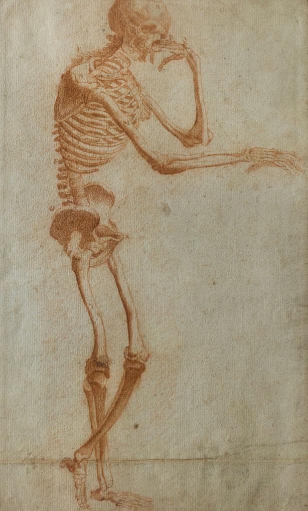
This is most pertinent in the case of Vesalius’s collaborator and artist Jan Steven van Calcar, whom Vesalius only mentioned as ‘his friend’, but was probably responsible for at least an important number of the plates and figures in the famous Fabrica and Epitome. This gradually gains momentum enhanced by a recently found drawing (figure 1) which is commented on in the book by Caiati and co-workers, and is believed to be a preliminary sketch by Jan Steven van Calcar of one of the most iconic drawings in the Fabrica, namely the so-called ‘Philosopher’. (see image)
It is in this light that I would like to take a few people out of the shadows and bring them into the spotlight, and by this, explaining how this book came about.
Click on the image for a larger version
I first was introduced to Bob Van Hee in 2006 by Ann Van de Velde. Professor Robrecht H. Van Hee who is a surgeon and medical historian, with over 160 publications to his name, has always had a special interest in Andreas Vesalius. In 2005 he promoted Vesalius for a television contest programme about “Who’s the most famous Belgian in history”.
In 2007 Ann and I were organizing the AEIMS conference “Confronting Mortality with Art and Science”. This conference brought together a group of artists, scientists and medical illustrators.
Bob van Hee and anatomist Francis Van Glabbeek both gave excellent lectures on Vesalius, Triverius and Philip Verheyen during a nocturnal visit to the Plantin Moretus museum in Antwerp.
It was also at this conference I invited Joanna Ebenstein to give a talk on her then new project” The morbid anatomy library blog” which has grown into the amazing art /science platform it is today.
The conference was such a success that we decided to set up BIOMAB, Biological and Medical Art in Belgium, with a teaching programme ARSIC ( art researches science international collaborations) .
BIOMAB would never have seen the light of day if it wasn’t for the energy, and drive and fearless enterprising spirit of hematologist and medical artist; Dr. Ann Van de Velde. Over the years we have organized many dissection drawing workshops, exhibitions, conferences, made films and books and have written many articles and given many lectures. Our article in the book “Anatomical Drawing From Cadavers – Limb by Limb Removed -- Brought Us Together” elucidates our collaborations and our intension and achievements.
Anatomist and surgeon Professor Francis Van Glabbeek, founding member and currently President of BIOMAB, and a passionate collector of antiquarian medical books, has been a driving force in continuing Vesalius’s legacy by bringing artists together with scientists, and, in the spirit of Vesalius, teaching anatomy from direct observation. I remember the first meeting with Francis with much fondness. He lovingly showed me a first edition of the Fabrica and spoke with so much passion and knowledge about the work and influence that Vesalius and the Fabrica still have on anatomy today.
This article continues HERE
- Details
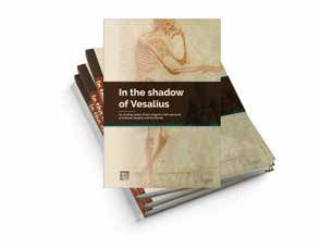
Clicking on this image will download a
PDF file with the book information
This is a new, as yet unpublished, book authored by several of my good friends who with me follow Vesaliana all over the world. There are too many of my friends to count, but I highly recommend this book. Dr. Miranda
In this richly illustrated book, a multitude of academic scholars present new findings concerning Andreas Vesalius Bruxellensis and his contemporaries. One of those discoveries includes a preparatory sketch by Jan Steven van Calcar for a drawing in Vesalius’ famous ‘Fabrica’, called ‘The Philosopher’.
Included are authentic letters, written by Vesalius to his friends Benedetto Varchi and Octavo Landi, are presented and translated for the first time and are thoroughly discussed, shedding new light on crucial periods of Vesalius’ life like his leave from academic Padua, exchanged for imperial service to Charles V, or his contribution in the treatment of Philips II’s son, crown prince Carlos in Spain.
Anatomical novelties, discovered by Vesalius’ friends and contemporaries, are equally broadly exposed, like Canani’s input in human arm musculature or Valverde’s ‘corrections’ of Vesalius’ Epitome. Valverde’s publication became one of the greatest ‘bestsellers’ in anatomy during the 16th and 17th century, thereby spreading the ‘Vesalian Revolution’ all over Europe. But also, the relationship between Vesalius and his Paduan room mate John Kay or Caius is explicated, as are a number of family descendants of Vesalius.
That Vesalius influenced artists in anatomical models and drawings is newly acknowledged in this book, as well as his influence on veterinary medicine.
In short, an inspiring new account of Vesalius’ extraordinary long-term influence on anatomy, science and art in general. One interesting point is that until now, many of the series of articles that composes the book where only available in Greek!
Jacqueline Vons (Emer. Professor of Latin and Medical History, University of Tours, France) comments "Centered around the figure of Andreas Vesalius (1514-1564), this book ‘In the Shadow of Vesalius’ offers a vast panorama of the anatomical knowledge in Europe during early Modern times.
Some new documents and private letters have been discovered and are finely analyzed, as are anatomical books written by contemporaries and successors of Vesalius. The studies gathered here show how the text and iconography’s diffusion of De Humani Corporis Fabrica and Epitome made Vesalius the first modern medical authority (auctoritas): his work was quoted, copied, discussed by all those who were interested in the anatomy of the human body."
Click here to download a PDF file with a description of the book, the Table of Contents, and the information to pre-order this book which will be launched on November 14, 2020. To order a book at a pre-launch discounted price, you can go directly to the publisher's website (http://garant.be/shadow-of-vesalius/).
Pascale recently wrote an article looking back at how this book came to be. Here is the story of "The Long Road to the book "In the Shadow of Vesalius"
- Details
Anatomical position
UPDATED:The term [proximal], from the Latin [proximus] meaning "next" and its counterpart [distal], from the Latin [distans] meaning "distant", have been poorly defined and this causes misunderstanding in the proper use of these terms. This is particularly true in the medical industry.
The classical definition of [proximal] are "nearest, closer to the origin, closer to the point of reference" and also "closer to the beginning", or "opposite of distal". [Distal] is, of course, the opposite. All of these definitions are lacking a consensus between the participants in a conversation. This lack of proper definition could potentially lead to problems in an interventional situation and a patient could be injured.
In our lectures and training materials we use a working definition1 as follows:
“Proximal has two meanings:
1- Closer to the point of attachment, where one end of the attached structure is free, and
2- Closer to the point of origin of flow of a fluid”
Distal is of course, opposite to proximal.
In the first acception of the word, a clear example is the attachment of the upper and lower extremities. Moving away from the shoulder or the hip joint is a distal movement. “The wrist joint is distal to the elbow joint”. The same is true for the Fallopian (uterine) tube, where the proximal attachment of the tube is to the uterus and the free distal end of the tube is its fimbriated end.
In the second acception of the word, in any anatomical structure, organ, or system where there is flow of a fluid (food, urine, bile, blood, etc.) it is accepted that normal flow (antegrade flow) goes from proximal to distal and that abnormal flow (retrograde flow) goes from distal to proximal.
1. Use of this definition is permitted, as long as CAA, Inc. is credited, or a link to this article is posted with it.
Image property of: CAA.Inc. Artist: Victoria G. Ratcliffe