![]()
Medical Terminology Daily (MTD) is a blog sponsored by Clinical Anatomy Associates, Inc. as a service to the medical community. We post anatomical, medical or surgical terms, their meaning and usage, as well as biographical notes on anatomists, surgeons, and researchers through the ages. Be warned that some of the images used depict human anatomical specimens.
You are welcome to submit questions and suggestions using our "Contact Us" form. The information on this blog follows the terms on our "Privacy and Security Statement" and cannot be construed as medical guidance or instructions for treatment.
We have 752 guests and no members online
")
Marcia Crocker Noyes
(1869 – 1946)
Further to my comment on old books and research that started with an interesting bookplate (Ex-Libris). I continued my research and found that the person in charge of the Osler library bookplate was a fascinating individual that today maybe a ghost in the MedChi library and building in Baltimore... This is certainly an article that can be called "A Moment in History"
Marcia Crocker Noyes was the librarian at The Maryland State Medical Society from 1896 to 1946 and was a founding member of the Medical Library Association.[1][2][3]
Sir William Osler, MD. a famous Johns Hopkins surgeon was a noted bibliophile and had a large personal collection of books on various topics. When he became the President of MedChi in 1896, he was dismayed at the condition of the library and knew that with the right person and some stewardship, it could become a significant collection. Sir William asked his friend, Dr. Bernard Steiner, a physician and President of the Enoch Pratt Free Library in Baltimore for suggestions of a librarian, and Dr. Steiner recommended Marcia Crocker Noyes. A native of New York, and a graduate of Hunter College, Marcia had moved to Baltimore for a lengthy visit with her sister, and took a “temporary” position at the Pratt Library, which turned into three years. Although she had no medical experience or background, she was enthusiastic, and most importantly, she was willing to move into the apartment provided for the librarian, who needed to be available 24 hours a day.
The image in this article is Ms. Noyes on her first year on the job. Marcia developed a book classification system for medical books, based on the Index Medicus, and called it the Classification for Medical Literature. The system uses the alphabet with capital letters for the major divisions of medicine and lower-case ones for the sub-sections. The system was used for many years, but it's now dated and the Faculty's original shelving scheme was never changed. The card catalogs still reflect her classification and many of the cards are written in Marcia's back-slanting handwriting.
Marcia knew enough to ask the Faculty's members about medical questions, terminology and literature. She gradually won over the predominantly male membership and they became her greatest allies; Sir William at the start, and then for nearly 40 years, Dr. John Ruhräh, a wealthy pediatrician with no immediate family of his own. She made a point of attending almost every Faculty function, and in 1904, under guidelines from the American Medical Association, Marcia was made the Faculty Secretary. For much of her first 10 years, she was the Faculty's only full-time employee, only being assisted by Mr. Caution, the Faculty's janitor. Later in life Marcia would say that she hired him because of his name!
Within ten years, the library had outgrown its space, and plans, spearheaded by Marcia and Sir William before his move to Oxford, were made to build a headquarters building, mainly to house the library's growing collection of medical books and journals.
Marcia was instrumental in the design and building of the new headquarters. She travelled to Philadelphia, New York and Boston to look at their medical society buildings, and eventually, the Philadelphia architectural firm, Ellicott & Emmart was selected to design and build the new Faculty building. Every detail of the building held her imprimatur, from the graceful staircase, to the light-filled reading room, and all of the myriad details of the millwork, marble tesserae, and most of all, the four-story cast iron stacks. She was on-site, climbing up unfinished staircases, checking out the progress of the building, which was built in less than one year at a cost of $90,000.
Among the features of the new building was a fourth-floor apartment for her. She referred to it as the "first penthouse in Baltimore" and it had a garden and rooftop terrace. The library collection eventually grew to more than 65,000 volumes from medical and specialty societies around the world. Journals were traded back and forth, and physicians eagerly anticipated the arrival of each new issue. At the same time, Marcia was involved in the Medical Library Association as one of eight founding members. The MLA promotes medical libraries and the exchange of information. One of the earliest mandates of the MLA was the Exchange, a distribution and trade service for those who had duplicates or little-used books in their collections. Initially, the Exchange was run out of the Philadelphia medical society, but in 1900 it was moved to Baltimore and Marcia oversaw it. Several hundred periodicals and journals were received and sent each month, a huge amount of work for a tiny staff. In 1904, the Faculty had run out of room to manage the Exchange, so it was moved to the Medical Society of the Kings County (Brooklyn). But without Marcia's excellent administrative skills, it floundered and in 1908, the MLA asked Marcia to take charge once again.
In 1909, when the new Faculty building opened, there was enough room to run the Exchange and with the help of MLA Treasurer, noted bibliophile and close friend, Dr. John Ruhräh, it once again became successful. Additionally, Marcia and Dr. Ruhräh combined forces to revive the MLA's bulletin, which had all but ceased publication in 1908, taking the Exchange with it. This duo maintained editorial control from 1911 until 1926. In 1934, around the time of Dr. Ruhräh's death, Marcia became the first “unmedicated” professional to head the MLA. During her tenure, the MLA incorporated, the first seal was adopted, and the annual meeting was held in Baltimore. Marcia wanted to write the history of the MLA once she retired from full-time work at the Faculty, but her health was beginning to fail. She had back problems and had suffered a serious burn on her shoulder as a young woman, possibly from her time running a summer camp, Camp Seyon, for young ladies in the Adirondack Mountains. In 1946, a celebration was planned to honor Marcia's 50 years at the Faculty. But she was adamant that the physicians wait until November, the actual date of her 50 years. However, they knew she was gravely ill, and might not make it until then, so a huge party was held in April. More than 250 physicians attended the celebration, but the ones she was closest to in the early years, were long gone. She was presented with a suitcase, a sum of money to use for travelling, and her favorite painting of Dr. John Philip Smith, a founder of the Medical College in Winchester, Virginia. It was painted by Edward Caledon Smith, a Virginia painter who had been a student of the painter Thomas Sully.[4] She adored this painting and vowed, jokingly, to take it with her wherever she went.
The painting was not to stay with her for very long, for she died in November 1946, and left it to the Faculty in her will. Her funeral was held in the Faculty's Osler Hall, named for her dear friend. More than 60 physicians served as her pallbearers, and she was buried at Baltimore's Green Mount Cemetery. In 1948, the MLA decided to establish an award in the name of Marcia Crocker Noyes. It was for outstanding achievement in medical library field and was to be awarded every two years, or when a truly worthy candidate was submitted. In 2014, the Faculty began giving a bouquet of flowers to the winner of the award in Marcia's name, and in honor of her work. Much evidence exists for this tradition, as we know that the physicians, especially Drs. Osler and Ruhräh, frequently gave her bouquets of flowers. Marcia also cultivated flower gardens at the Faculty and decorated the rooms with her work.
Today, the MedChi building is open for tours and if the rumors are to be believed Ms. Marcia Crocker Noyes is still at work in her beloved library as the "resident ghost" [1][5]

NOTE: This article has been modified from the original Wikipedia article on Marcia Crocker Noyes. The article itself is well-written with interesting images of the subject. I would encourage you to visit it. The second insert is from book 00736 in my personal library and shows in pencil, the incredibly small handwriting of Marsha C. Noyes.
Sources:
1. "Marcia, Marcia, Marcia" MedChi Archives blog.
2. "Marcia C. Noyes, Medical Librarian" (PDF). Bulletin of the Medical Library Association. 35 (1): 108–109. 1947. PMC 194645
3. Smith, Bernie Todd (1974). "Marcia Crocker Noyes, Medical Librarian: The Shaping of a Career" (PDF). Bulletin of the Medical Library Association. 62 (3): 314–324. PMC 198800Freely accessible. PMID 4619344.
4. Edward Caledon BRUCE (1825-1901)"
5. Behind the scenes tour MedChiBuilding
"Clinical Anatomy Associates, Inc., and the contributors of "Medical Terminology Daily" wish to thank all individuals who donate their bodies and tissues for the advancement of education and research”.
Click here for more information


- Details
- Written by: Prof. Claudio R. Molina, MSc
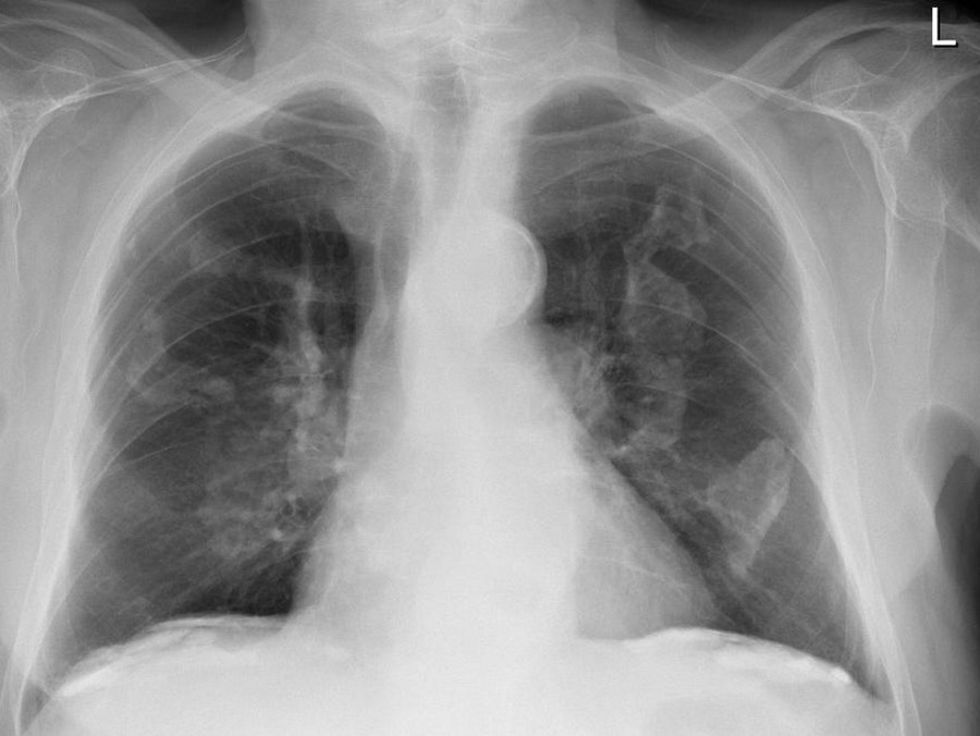
Holly leaf sign.
Click on the image for a larger depiction
The Holly is a tree/shrub of the genus Ilex , with perhaps the most well know being Ilex aquifolium. The plant has shiny prickly evergreen leaves and bright red berries. Cut branches of Holly are widely used as a traditionally Christmas decoration especially in wreaths and Christmas cards as illustrations. “The Holly and the Ivy” is a popular traditional English Christmas carol.
The [Holly leaf sign] refers to the appearance of calcified pleural plaques seen on chest radiographs. Pleural plaques are common in patients who have been exposed to asbestos, are asymptomatic and are most useful as a marker of asbestos exposure or asbestosis. They can be identified in 3-14% of dockyard workers and in 58% in insulation workers.
They are themselves not malignant, but patients with this plaques have a greater risk of mesothelioma and bronchogenic cancer than the general population and patients with exposed to asbestos but not pleural plaques.
The plaques arise in the parietal pleura and have predilection for the diaphragmatic dome and the undersurface of the lower posterolateral ribs. Rarely involve the visceral pleura but occasionally they are found in the fissures of the lungs.
On plain radiographic plaques appear as a geographic, usually calcified, opacities with irregular but well-defined edges. The irregular thickened nodular edges of the pleural plaques are likened to appearance of a Holly leaf, which has sharp spines along its margin.
Sources:
1. Jane R, Gulati A., Dwivedi R., Avula S., Curtis J., Abernethy L. (2013) We wish you a Merry X-Ray-mas: Christmas signs in radiology. BMJ 347:f7020 doi: 10.1136/bmj.f7020
2. Walker C., Takasugi J., Chung J., Reddy, G., Done S., Pipavath S., Schmidt R., Godwin J. (2012). Tumor-like Conditions of the Pleura. Radiographics 32:971–985.
3. Case radiograph courtesy of Dr Çağlayan Çakır, Radiopaedia.org. From the case rID: 22986
Figure below. Ilex aquifolium. Courtesy of A.Prof Frank Gaillard, Radiopaedia.org. From the case rID: 12398

Holly leaf sign.
Click on the image for a larger depiction
Article submitted by: Prof. Claudio R. Molina, MsC.
- Details
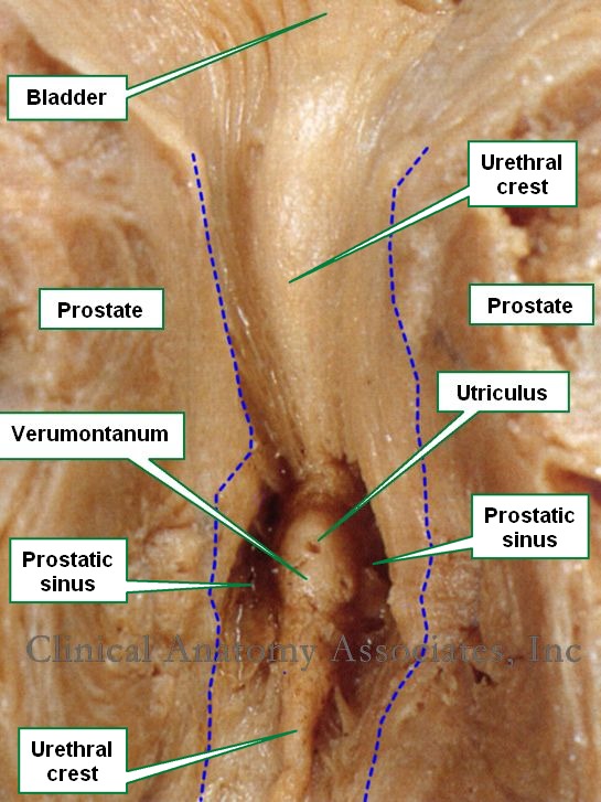
Anterior view of a section of the prostate gland. The blue
dotted line shows the edges of the prostatic urethra
[UPDATED] The [prostatic utricle], also known as "utriculus prostaticus" or "utriculus" is a small 6 mm small dead-end channel found in the male prostatic urethra.
The word [utriculus] is Latin and means "little sac"..
What is interesting about this structure is that it is the embryological remnant in the male of the Müllerian ducts that form the vagina and the uterus in the female. In fact, in some texts the prostatic utricle is referred to as "uterus masculinus". Some researchers differ and point to the fact that this structure may not be a Müllerian duct derivate.
The prostatic utricle is found inside the prostate, forming part of the posterior wall of the prostatic urethra. It is in the upper part of a small mound which is part of the prostatic crest. This mound is called the [colliculus seminalis] or [verumontanum], which is Latin and translates as the "mountain or mound of truth". On the verumontanum are the two slit-like openings of the ejaculatory ducts. Lateral to the verumontanum are the prostatic sinuses, depressions where the prostatic ducts are found.
Sources:
1. "The prostatic utricle is not a M?llerian duct remnant: immunohistochemical evidence for a distinct urogenital sinus origin" Shapiro E, Huang H, McFadden DE, et al. (2004) J Urol 172; 1753–1756
2. "Gray's Anatomy"38th British Ed. Churchill Livingstone 1995
3. "Tratado de Anatomia Humana" Testut et Latarjet 8 Ed. 1931 Salvat Editores, Spain
- Details
- Written by: Efrain A. Miranda, Ph.D.
This article is part of the series "A Moment in History" where we honor those who have contributed to the growth of medical knowledge in the areas of anatomy, medicine, surgery, and medical research.

Jane Todd Crawford - Daguerrotype
When writing the article “The Ephraim McDowell House and Museum” I realized that there are so many patients that by volunteering to a novel or sometimes experimental procedure or donating their bodies have been the catalyst of the advancement of medical science, surgery, and anatomy. Benigno says it so clearly in his paper explaining the physician/patient relation of McDowell and his patient: “Because of his innovative genius and finally honed surgical skills, Ephraim McDowell gave Jane Todd Crawford her life, and she, in return, gave him immortality”.
Few patients have influenced local history more than Jane Todd Crawford. In Kentucky there is a road named after her, a hospital bears her name in Greenville, KY, and there is even a formal "Jane Todd Crawford Day" on December 13!
By contrast, there are so many unknown patients whose names history has forgotten, and yet the fame of the physician continues through time in eponymic hospitals, educational institutions, named surgical procedures or maneuvers, surgical instruments, etc.
Some of the names and stories have survived, but many have not. In some cases, we know the name, but little else.
Dr. Henry Heimlich used his “Heimlich maneuver” for the first time to save his neighbor Patty Ris, in 2016, forty-two years after publishing it in 1974. The maneuver itself was used that same year (1974) to save the first person, Irene Bogachus, who was choking at a restaurant. Hundreds of thousands of people have been saved from death from choking by the proper use of this maneuver.
Dr. Christiaan Barnard, performed the first successful heart transplant on December 3, 1967. We know the name of the donor, 25 year-old Denise Darvall, and the recipient Lewis Washkansky.
Dr. Antoine Dubois and Dr. Dominique-Jean Larrey in France performed the first mastectomy on September 30, 1811. This was decades before the advent of anesthesia or aseptic technique. The patients was Fanny Burney, a famous novelist.
Dr. Edward Jenner developed the smallpox vaccine after working with a milkmaid, Sarah Nelmes. Jenner’s work saved the Americas from the smallpox epidemic through the work of Don Antonio de Gimbernat y Arbós and Don Francisco Javier de Balmis i Berenguer and his “Balmis Expedition”
The examples can continue, but who was the patient on the first Billroth procedure, who was the patient in the first Scopinaro procedure? Who was the patient on whom Dr. Eric Muhe performed the first laparoscopic cholecystectomy? Many are unknown yet they helped pave the way of the future.
The same can be said for the world of human anatomy. Today we honor the donors who will their bodies so that future physicians can study the intricacy of the human body, but we never know their names or their stories. Many a time I have stood at the side of a body while medical students dissect and study and wondered about their identities, the life they had, and what led them to give us their bodies as a wonderful gift to science and medicine.
There was a time (long ago) when the dissection of a human body was punished by the Church, or the times when the scarcity of bodies was such that some started to rob graves, or when the punishment for a crime was “death and a public anatomy”.
Some of these people we know, most of them we do not. Some have given their body willingly, others have not.
Joseph Paul Jernigan, a murderer, who after given the death penalty, donated his body to a now world-renown endeavor, the Visible Human Project.
The oldest known anatomical preparation is a skeleton mounted in Basel (Belgium) by Andreas Vesalius in 1543. The skeleton belongs to Jacob Karrer von Geweiler, a bigamist and attempted murderer who was beheaded for his crimes.
It is sad that we know the names of these criminals, and in some cases not that of their victims.
We do not know the names of many who, during the Nazi regime in WWII, were taken from concentration camps for medical experiments and as we understand, possibly murdered and dissected to illustrate now infamous anatomical atlases. Research is being done to discover their identities.
Times have changed and body donation has become accepted and praised by society. I am always touched by the words of Morgagni above the entrance to the dissection rooms at the University of Cincinnati: “hic locus est ubi mors gaudet succurrere vitae” meaning “in this place death rejoices helping the living”. The University of Cincinnati has an Annual Memorial Ceremony to honor the body donors and their families. A video of the ceremony can be watched here.
I cannot but end this article with the words that are found in the left side column of this blog and will always be there:
“Clinical Anatomy Associates, Inc., and the contributors of "Medical Terminology Daily" wish to thank all individuals who donate their bodies and tissues for the advancement of education and research”.
- Details
- Written by: Efrain A. Miranda, Ph.D.
This article is part of the series "A Moment in History" where we honor those who have contributed to the growth of medical knowledge in the areas of anatomy, medicine, surgery, and medical research.

Susan C. Potter
Image capture from a video
The title of this article is a reference to another article in this blog: “The unknown patient / donor” which honors all those who have anonymously donated their bodies to further the anatomical training of many in the medical field. They trusted that those who would use their bodies would do so ethically and with respect, but they did not know exactly how they were to be used or what was going to be done with their bodies.
Susan Potter was the exact opposite. She knew that her body was going to be coated with polyvinyl alcohol, frozen, cut into four pieces with a huge handsaw, and then it would be ground or milled into 27,000 slices of 63 microns each, which were to be photographed in exquisite detail.
She offered her body to science and spoke with Dr. Vic Spitzer, who had directed the Visible Human Project, the first digital cadaver in 1994. She agreed to the donation, but only after she had toured the facilities and only after she clearly understood what was going to her body and why.
The why is the most interesting part of her story. Susan had a very interesting medical history, including spinal surgery , double mastectomy, and a hip replacement. Normally her body would have been rejected, but doctors see this type of patients in their practices. Patients who are old, frail, with prior surgeries and a multitude of problems. This is why she was chosen
If images are needed, usually cadavers are scanned and imaged postmortem, but in her case, Susan underwent many imaging studies while she was alive. She was interviewed and filmed countless times so that her videos would be added to the digital cadaver that was going to be made of her, becoming de facto, a digital patient.
Susan donated her body in the year 2000 died of pneumonia in 2015. During those 15 years she became a friend of Dr. Spitzer, gave talks to medical students, and collaborated with this project.
National Geographic followed Susan for these 15 years and documented her life and death. You can read her story here or watch the video in this article. The development of the software continues. I am sure we will hear more from Susan Potter's contributions long after her death.
NOTE: My thanks to our contributor Pascalle Pollier for bringing Susan Potter to my attention. Dr. Miranda
“Clinical Anatomy Associates, Inc., and the contributors of "Medical Terminology Daily" wish to thank all individuals who donate their bodies and tissues for the advancement of education and research”.
- Details

Azygos system
The azygos venous system drains the posterior aspect of the thorax via the posterior intercostal veins It also connects the vascular territories of the superior vena cava and the inferior vena cava, and is the superior continuation of the lumbar veins. The azygos system was first described by Bartolomeo Eustachius (c1500 - 1574).
The name azygos comes from the Greek [ζεύγος] and means “unyoked” or better “asymmetrical”. This system is different on each side of the body, also having important anatomical variations.
The azygos vein (Lat: vena azygos major) is the larger vein of the azygos system and is found on the right side of the body. It begins at the level of the first or second lumbar vertebra as a continuation of the right ascending lumbar vein; sometimes by a branch from the right renal vein or from the inferior vena cava. It enters the thoracic cavity through the aortic hiatus of the respiratory diaphragm, and ascends along the right side of the vertebral column to level of the fourth thoracic vertebra, where it arches forward over the root of the right lung, at this point the vein is called the azygos arch, which terminates in the posterior aspect of the superior vena cava (SVC) just superior to the point where the SVC enters the pericardium.
In the thorax, the azygos vein is found to the right of the thoracic duct on the right side of the descending aorta; it lies upon the intercostal arteries and is partly covered by the parietal pleura.
The azygos vein receives the right subcostal vein, nine or ten right posterior intercostal veins, the hemiazygos vein, the accessory hemiazygos vein, the right superior intercostal vein, and several minor esophageal, mediastinal, and pericardial veins.
The left side of this system is more complex and presents with more anatomical variations. Its main component is the hemiazygos vein (Lat: vena azygos minor), also known as the left lower azygos vein. It is a continuation of the left ascending lumbar vein, and it sometimes may arise from the left renal vein and passes into the thorax usually through the left aortic crus of the respiratory diaphragm. It ascends to the level of the 7th or 8th thoracic vertebra where it crosses the midline posterior to the esophagus, descending aorta and thoracic duct to empty into the right-sided azygos vein. It receives the left subcostal vein and three to four lower posterior intercostal veins, and some esophageal and mediastinal veins.
The second component of the left azygos system is the accessory hemiazygos vein, also known as the left upper hemiazygos. This component varies in size depending on the third venous drainage component of the left posterior thoracic wall. This is the left superior intercostal vein (see attached diagram).
The accessory hemiazygos, similar to the hemiazygos vein will cross the midline posterior to the esophagus, descending aorta and thoracic duct to empty into the right-sided azygos vein. It may do so by a common vein or by a separate vein as shown in the attached diagram. If there is a common vein the hemiazygos is considered to be the inferior component and the hemiazygos is considered to be the superior component.
The left superior intercostal vein receives three or four posterior intercostal veins, and empties into the left brachiocephalic vein. In rare cases of absence of the hemiazygos vein, this left superior intercostal vein will extend as low as the fifth or sixth intercostal space.
Although not considered to be part of the azygos system, the drainage of the posterior thoracic wall is completed by the right and left supreme intercostal veins which empty the posterior aspect of the first intercostal space into the left and right brachiocephalic veins respectively.
The azygos system of veins constitute an important collateral venous circulation pathway which can be seen in action in cases of blockage of the superior or inferior vena cavæ.
Sources:
1. “Gray’s Anatomy” Henry Gray, 1918
2. "Tratado de Anatomia Humana" Testut et Latarjet 8th Ed. 1931 Salvat Editores, Spain
3. "Gray's Anatomy" 38th British Ed. Churchill Livingstone 1995
4. "Reconstructive Anatomy: A Method for the Study of Human Structure: Arnold, M WB Saunders1968
Image modified from the original from Arnold (4)
- Details
- Written by: Efrain A. Miranda, Ph.D.
This article is part of the series "A Moment in History" where we honor those who have contributed to the growth of medical knowledge in the areas of anatomy, medicine, surgery, and medical research.
Further to my comment on old books and research that started with an interesting bookplate (Ex-Libris). I continued my research and found that the person in charge of the Osler library bookplate was a fascinating individual that today maybe a ghost in the MedChi library and building in Baltimore... This is certainly an article that can be called "A Moment in History"

Marcia Crocker Noyes
Marcia Crocker Noyes was the librarian at The Maryland State Medical Society from 1896 to 1946 and was a founding member of the Medical Library Association.[1][2][3]
Sir William Osler, MD. a famous Johns Hopkins surgeon was a noted bibliophile and had a large personal collection of books on various topics. When he became the President of MedChi in 1896, he was dismayed at the condition of the library and knew that with the right person and some stewardship, it could become a significant collection. Sir William asked his friend, Dr. Bernard Steiner, a physician and President of the Enoch Pratt Free Library in Baltimore for suggestions of a librarian, and Dr. Steiner recommended Marcia Crocker Noyes. A native of New York, and a graduate of Hunter College, Marcia had moved to Baltimore for a lengthy visit with her sister, and took a “temporary” position at the Pratt Library, which turned into three years. Although she had no medical experience or background, she was enthusiastic, and most importantly, she was willing to move into the apartment provided for the librarian, who needed to be available 24 hours a day.
The image in this article is Ms. Noyes on her first year on the job. Marcia developed a book classification system for medical books, based on the Index Medicus, and called it the Classification for Medical Literature. The system uses the alphabet with capital letters for the major divisions of medicine and lower-case ones for the sub-sections. The system was used for many years, but it's now dated and the Faculty's original shelving scheme was never changed. The card catalogs still reflect her classification and many of the cards are written in Marcia's back-slanting handwriting.
Marcia knew enough to ask the Faculty's members about medical questions, terminology and literature. She gradually won over the predominantly male membership and they became her greatest allies; Sir William at the start, and then for nearly 40 years, Dr. John Ruhräh, a wealthy pediatrician with no immediate family of his own. She made a point of attending almost every Faculty function, and in 1904, under guidelines from the American Medical Association, Marcia was made the Faculty Secretary. For much of her first 10 years, she was the Faculty's only full-time employee, only being assisted by Mr. Caution, the Faculty's janitor. Later in life Marcia would say that she hired him because of his name!
Within ten years, the library had outgrown its space, and plans, spearheaded by Marcia and Sir William before his move to Oxford, were made to build a headquarters building, mainly to house the library's growing collection of medical books and journals.
Marcia was instrumental in the design and building of the new headquarters. She travelled to Philadelphia, New York and Boston to look at their medical society buildings, and eventually, the Philadelphia architectural firm, Ellicott & Emmart was selected to design and build the new Faculty building. Every detail of the building held her imprimatur, from the graceful staircase, to the light-filled reading room, and all of the myriad details of the millwork, marble tesserae, and most of all, the four-story cast iron stacks. She was on-site, climbing up unfinished staircases, checking out the progress of the building, which was built in less than one year at a cost of $90,000.
Among the features of the new building was a fourth-floor apartment for her. She referred to it as the "first penthouse in Baltimore" and it had a garden and rooftop terrace. The library collection eventually grew to more than 65,000 volumes from medical and specialty societies around the world. Journals were traded back and forth, and physicians eagerly anticipated the arrival of each new issue. At the same time, Marcia was involved in the Medical Library Association as one of eight founding members. The MLA promotes medical libraries and the exchange of information. One of the earliest mandates of the MLA was the Exchange, a distribution and trade service for those who had duplicates or little-used books in their collections. Initially, the Exchange was run out of the Philadelphia medical society, but in 1900 it was moved to Baltimore and Marcia oversaw it. Several hundred periodicals and journals were received and sent each month, a huge amount of work for a tiny staff. In 1904, the Faculty had run out of room to manage the Exchange, so it was moved to the Medical Society of the Kings County (Brooklyn). But without Marcia's excellent administrative skills, it floundered and in 1908, the MLA asked Marcia to take charge once again.
 In 1909, when the new Faculty building opened, there was enough room to run the Exchange and with the help of MLA Treasurer, noted bibliophile and close friend, Dr. John Ruhräh, it once again became successful. Additionally, Marcia and Dr. Ruhräh combined forces to revive the MLA's bulletin, which had all but ceased publication in 1908, taking the Exchange with it. This duo maintained editorial control from 1911 until 1926. In 1934, around the time of Dr. Ruhräh's death, Marcia became the first “unmedicated” professional to head the MLA. During her tenure, the MLA incorporated, the first seal was adopted, and the annual meeting was held in Baltimore. Marcia wanted to write the history of the MLA once she retired from full-time work at the Faculty, but her health was beginning to fail. She had back problems and had suffered a serious burn on her shoulder as a young woman, possibly from her time running a summer camp, Camp Seyon, for young ladies in the Adirondack Mountains. In 1946, a celebration was planned to honor Marcia's 50 years at the Faculty. But she was adamant that the physicians wait until November, the actual date of her 50 years. However, they knew she was gravely ill, and might not make it until then, so a huge party was held in April. More than 250 physicians attended the celebration, but the ones she was closest to in the early years, were long gone. She was presented with a suitcase, a sum of money to use for travelling, and her favorite painting of Dr. John Philip Smith, a founder of the Medical College in Winchester, Virginia. It was painted by Edward Caledon Smith, a Virginia painter who had been a student of the painter Thomas Sully.[4] She adored this painting and vowed, jokingly, to take it with her wherever she went.
The painting was not to stay with her for very long, for she died in November 1946, and left it to the Faculty in her will. Her funeral was held in the Faculty's Osler Hall, named for her dear friend. More than 60 physicians served as her pallbearers, and she was buried at Baltimore's Green Mount Cemetery. In 1948, the MLA decided to establish an award in the name of Marcia Crocker Noyes. It was for outstanding achievement in medical library field and was to be awarded every two years, or when a truly worthy candidate was submitted. In 2014, the Faculty began giving a bouquet of flowers to the winner of the award in Marcia's name, and in honor of her work. Much evidence exists for this tradition, as we know that the physicians, especially Drs. Osler and Ruhräh, frequently gave her bouquets of flowers. Marcia also cultivated flower gardens at the Faculty and decorated the rooms with her work.
Today, the MedChi building is open for tours and if the rumors are to be believed Ms. Marcia Crocker Noyes is still at work in her beloved library as the "resident ghost" [1][5]
NOTE: This article has been modified from the original Wikipedia article on Marcia Crocker Noyes. The article itself is well-written with interesting images of the subject. I would encourage you to visit it. The second insert is from book ML-0736 in my personal library and shows in pencil, the incredibly small handwriting of Marsha C. Noyes.
Sources:
1. "Marcia, Marcia, Marcia" MedChi Archives blog.
2. "Marcia C. Noyes, Medical Librarian" (PDF). Bulletin of the Medical Library Association. 35 (1): 108–109. 1947. PMC 194645
3. Smith, Bernie Todd (1974). "Marcia Crocker Noyes, Medical Librarian: The Shaping of a Career" (PDF). Bulletin of the Medical Library Association. 62 (3): 314–324. PMC 198800Freely accessible. PMID 4619344.
4. Edward Caledon BRUCE (1825-1901)"
5. Behind the scenes tour MedChiBuilding