![]()
Medical Terminology Daily (MTD) is a blog sponsored by Clinical Anatomy Associates, Inc. as a service to the medical community. We post anatomical, medical or surgical terms, their meaning and usage, as well as biographical notes on anatomists, surgeons, and researchers through the ages. Be warned that some of the images used depict human anatomical specimens.
You are welcome to submit questions and suggestions using our "Contact Us" form. The information on this blog follows the terms on our "Privacy and Security Statement" and cannot be construed as medical guidance or instructions for treatment.
We have 1420 guests and no members online
")
Marcia Crocker Noyes
(1869 – 1946)
Further to my comment on old books and research that started with an interesting bookplate (Ex-Libris). I continued my research and found that the person in charge of the Osler library bookplate was a fascinating individual that today maybe a ghost in the MedChi library and building in Baltimore... This is certainly an article that can be called "A Moment in History"
Marcia Crocker Noyes was the librarian at The Maryland State Medical Society from 1896 to 1946 and was a founding member of the Medical Library Association.[1][2][3]
Sir William Osler, MD. a famous Johns Hopkins surgeon was a noted bibliophile and had a large personal collection of books on various topics. When he became the President of MedChi in 1896, he was dismayed at the condition of the library and knew that with the right person and some stewardship, it could become a significant collection. Sir William asked his friend, Dr. Bernard Steiner, a physician and President of the Enoch Pratt Free Library in Baltimore for suggestions of a librarian, and Dr. Steiner recommended Marcia Crocker Noyes. A native of New York, and a graduate of Hunter College, Marcia had moved to Baltimore for a lengthy visit with her sister, and took a “temporary” position at the Pratt Library, which turned into three years. Although she had no medical experience or background, she was enthusiastic, and most importantly, she was willing to move into the apartment provided for the librarian, who needed to be available 24 hours a day.
The image in this article is Ms. Noyes on her first year on the job. Marcia developed a book classification system for medical books, based on the Index Medicus, and called it the Classification for Medical Literature. The system uses the alphabet with capital letters for the major divisions of medicine and lower-case ones for the sub-sections. The system was used for many years, but it's now dated and the Faculty's original shelving scheme was never changed. The card catalogs still reflect her classification and many of the cards are written in Marcia's back-slanting handwriting.
Marcia knew enough to ask the Faculty's members about medical questions, terminology and literature. She gradually won over the predominantly male membership and they became her greatest allies; Sir William at the start, and then for nearly 40 years, Dr. John Ruhräh, a wealthy pediatrician with no immediate family of his own. She made a point of attending almost every Faculty function, and in 1904, under guidelines from the American Medical Association, Marcia was made the Faculty Secretary. For much of her first 10 years, she was the Faculty's only full-time employee, only being assisted by Mr. Caution, the Faculty's janitor. Later in life Marcia would say that she hired him because of his name!
Within ten years, the library had outgrown its space, and plans, spearheaded by Marcia and Sir William before his move to Oxford, were made to build a headquarters building, mainly to house the library's growing collection of medical books and journals.
Marcia was instrumental in the design and building of the new headquarters. She travelled to Philadelphia, New York and Boston to look at their medical society buildings, and eventually, the Philadelphia architectural firm, Ellicott & Emmart was selected to design and build the new Faculty building. Every detail of the building held her imprimatur, from the graceful staircase, to the light-filled reading room, and all of the myriad details of the millwork, marble tesserae, and most of all, the four-story cast iron stacks. She was on-site, climbing up unfinished staircases, checking out the progress of the building, which was built in less than one year at a cost of $90,000.
Among the features of the new building was a fourth-floor apartment for her. She referred to it as the "first penthouse in Baltimore" and it had a garden and rooftop terrace. The library collection eventually grew to more than 65,000 volumes from medical and specialty societies around the world. Journals were traded back and forth, and physicians eagerly anticipated the arrival of each new issue. At the same time, Marcia was involved in the Medical Library Association as one of eight founding members. The MLA promotes medical libraries and the exchange of information. One of the earliest mandates of the MLA was the Exchange, a distribution and trade service for those who had duplicates or little-used books in their collections. Initially, the Exchange was run out of the Philadelphia medical society, but in 1900 it was moved to Baltimore and Marcia oversaw it. Several hundred periodicals and journals were received and sent each month, a huge amount of work for a tiny staff. In 1904, the Faculty had run out of room to manage the Exchange, so it was moved to the Medical Society of the Kings County (Brooklyn). But without Marcia's excellent administrative skills, it floundered and in 1908, the MLA asked Marcia to take charge once again.
In 1909, when the new Faculty building opened, there was enough room to run the Exchange and with the help of MLA Treasurer, noted bibliophile and close friend, Dr. John Ruhräh, it once again became successful. Additionally, Marcia and Dr. Ruhräh combined forces to revive the MLA's bulletin, which had all but ceased publication in 1908, taking the Exchange with it. This duo maintained editorial control from 1911 until 1926. In 1934, around the time of Dr. Ruhräh's death, Marcia became the first “unmedicated” professional to head the MLA. During her tenure, the MLA incorporated, the first seal was adopted, and the annual meeting was held in Baltimore. Marcia wanted to write the history of the MLA once she retired from full-time work at the Faculty, but her health was beginning to fail. She had back problems and had suffered a serious burn on her shoulder as a young woman, possibly from her time running a summer camp, Camp Seyon, for young ladies in the Adirondack Mountains. In 1946, a celebration was planned to honor Marcia's 50 years at the Faculty. But she was adamant that the physicians wait until November, the actual date of her 50 years. However, they knew she was gravely ill, and might not make it until then, so a huge party was held in April. More than 250 physicians attended the celebration, but the ones she was closest to in the early years, were long gone. She was presented with a suitcase, a sum of money to use for travelling, and her favorite painting of Dr. John Philip Smith, a founder of the Medical College in Winchester, Virginia. It was painted by Edward Caledon Smith, a Virginia painter who had been a student of the painter Thomas Sully.[4] She adored this painting and vowed, jokingly, to take it with her wherever she went.
The painting was not to stay with her for very long, for she died in November 1946, and left it to the Faculty in her will. Her funeral was held in the Faculty's Osler Hall, named for her dear friend. More than 60 physicians served as her pallbearers, and she was buried at Baltimore's Green Mount Cemetery. In 1948, the MLA decided to establish an award in the name of Marcia Crocker Noyes. It was for outstanding achievement in medical library field and was to be awarded every two years, or when a truly worthy candidate was submitted. In 2014, the Faculty began giving a bouquet of flowers to the winner of the award in Marcia's name, and in honor of her work. Much evidence exists for this tradition, as we know that the physicians, especially Drs. Osler and Ruhräh, frequently gave her bouquets of flowers. Marcia also cultivated flower gardens at the Faculty and decorated the rooms with her work.
Today, the MedChi building is open for tours and if the rumors are to be believed Ms. Marcia Crocker Noyes is still at work in her beloved library as the "resident ghost" [1][5]

NOTE: This article has been modified from the original Wikipedia article on Marcia Crocker Noyes. The article itself is well-written with interesting images of the subject. I would encourage you to visit it. The second insert is from book 00736 in my personal library and shows in pencil, the incredibly small handwriting of Marsha C. Noyes.
Sources:
1. "Marcia, Marcia, Marcia" MedChi Archives blog.
2. "Marcia C. Noyes, Medical Librarian" (PDF). Bulletin of the Medical Library Association. 35 (1): 108–109. 1947. PMC 194645
3. Smith, Bernie Todd (1974). "Marcia Crocker Noyes, Medical Librarian: The Shaping of a Career" (PDF). Bulletin of the Medical Library Association. 62 (3): 314–324. PMC 198800Freely accessible. PMID 4619344.
4. Edward Caledon BRUCE (1825-1901)"
5. Behind the scenes tour MedChiBuilding
"Clinical Anatomy Associates, Inc., and the contributors of "Medical Terminology Daily" wish to thank all individuals who donate their bodies and tissues for the advancement of education and research”.
Click here for more information


- Details
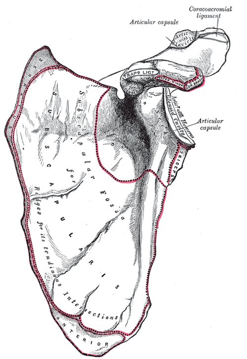
Anterior view of the left scapula
UPDATED: The scapula, known vernacularly as the "shoulder blade", is a flat, triangular bone that forms the posterior portion of the shoulder girdle. It is described with two surfaces, three borders, and three angles. The scapula attaches to the clavicle by way of the acromioclavicular joint and ligaments. Seventeen muscles attach to the scapula and are discussed in a different article.
This bone actually has two names depending on the language used. In English we use the word [scapula] which has a Latin origin, while in some Latin-based languages the word [omóplato] (Spanish, in this case) has a Greek origin!
[Scapula] originates from the Latin [scapula] (scapula), meaning “shoulder”, also “the back”. The later derivation into “shoulder blade” in English has no known history, except perhaps for the primitive use of animal scapulae as a blade or a spatula in daily chores.
In Greek the term [ωμοπλάτη] (omopláti) was used to name the scapula. The origin of the word was the combination of the terms [ώμος] (ómos) meaning “shoulder”, and the word [πλάτη] (pláti) meaning “back”. One of the seventeen muscles that attach to the [scapula] is the [omohyoid], where the root term [-omo-] indicates its relation to the scapula.
It was Andreas Vesalius who popularized the name [scapula] selecting it from the many names this bone had at the time (1543), and is today the the accepted “Nomina Anatomica” term.
There is still discrepancy on the name of the bone in other languages.
• English: scapula
• Spanish: omóplato, also escápula
• Italian: scapula
• French: omoplate
• Romanian: omoplat
• Portuguese: escápula
In German, they used the word [schulterblatt] which means "shoulder blade".
The anatomical description of this bone continues in this article
Sources:
1. "Tratado de Anatomia Humana" Testut et Latarjet 8 Ed. 1931 Salvat Editores, Spain
2. "Gray's Anatomy" 38th British Ed. Churchill Livingstone 1995
3. “The Origin of Medical Terms” Skinner HA 1970 Hafner Publishing Co.
4. "Terminologia Anatomica: International Anatomical Terminology (FCAT)" Thieme, 1998
Image in Public Domain, by Henry Vandyke Carter - Gray's Anatomy
Note: The links to Google Translate include an icon that will allow you to hear the Greek or Latin pronunciation of the word.
- Details
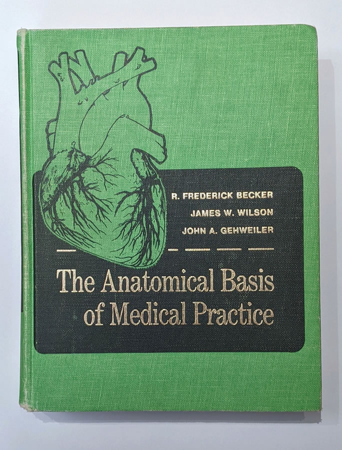
The Anatomical Basis of Medical Practice - Book Cover
Click on the image for a larger version
The title of this article paraphrases the title of a paper authored by Edward Halperin, MD, MA., and published in “Academic Medicine,” the Journal of the Association of American Medical Colleges in February 2009. More information in the "Sources" section of this article. I have also used other sources to complete this "strange tale."
"The Anatomical Basis of Medical Practice" was published in 1971 by Williams & Wilkins. A large and heavy book, it is one of the many anatomy books written for medical students. This one however, stirred a controversy because of the "tongue-in-cheek" writing style of the authors and mostly because of the photographs of female nude models in seductive poses pretending to depict surface anatomy. The pictures were taken by a famous photographer, Peter Gowland.
Initial reviews were positive, as stated in the British Journal of Surgery, where the book is praised saying: "one only wishes that such a book was available when one was a student in the dissecting room."
Other reviews were more negative, as Dr. Edward A. Edwards in 1972 states: "...I feel compelled to say that the numerous photographs of comely young women, while enticing, no not well demonstrate the muscles and bony points the legends suggest as their purpose." He also says: "The book is marred by a waggishness expressed in facetiousness and in long digressions...."
Soon negative reactions to the book took place, many of them from anatomy professors who saw the illustrations as "out of place" and "not needed". Interestingly only a small percentage of the total images were of the "Playboy" style, while the rest of the images, photographs and sketches are of high educational quality.
Estelle R. Ramey, Emeritus Professor of Physiology and Biophysics at the Georgetown University School of Medicine, in a letter to the Association of Women in Science stated “In effect, the entire book is a calculated insult to women and men alike. It demeans the whole profession of medicine and is openly contemptuous of middle-aged women whose breasts are not so round and may even be rotting with cancer.” The AWIS as a whole threatened to boycott Williams and Wilkins Publishing, and as a result, the book was pulled off the market.
The story gets more interesting. Because of public pressure, angry letters to the publisher, and boycotts, the publisher agreed to stop all promotion, marketing, and sales of the book. Many assumed that they pulled the book off the market when, apparently because of low sales, they just let the initial run sell out by December 1972 and never republished it.
I do agree that the book transgresses the lines of decorum and respect for women that exist today. At the time of its publication the feminine movement was starting, and I think the authors tried (and failed) to have a humorous approach to what is at best, a difficult subject.
The book itself is a great book on anatomy and superficial anatomy, notwithstanding the images and comments. Since it was published and because of its scarcity, it has become a rare book, and as Dr. Halperin states “a minor collector’s item.” When published, this book sold for US$23.00. In 2009, at the time Dr. Halperin published his paper, the book was valued between US$ 89.00 and US$ 300.00; today (2025,) the book is valued between US$ 575.00 and US$1,000.00 in Abebooks.com.
"The Anatomical Basis of Medical Practice" by Becker, R; Frederick; Wilson, James W; and Gehweiler, John, A. is one of the many books in my library and it has become known as “the green book” among my colleagues because of its bright green cloth hardcover. The image in this article is from the book in my library.
Sources:
1. “The Pornographic Anatomy Book? The Curious Tale of The Anatomical Basis of Medical Practice" Halperin, E. Academic Medicine: February 2009 - Volume 84 - Issue 2 - p 278-283
2. "The anatomical basis of medical practice." By R. Frederick Becker, Ph.D., James W. Wilson, Ph.D., M.D., and John A. Gehweiler, M.D. 11 × 8¼ in. Pp. 907 + xv. Illustrated. 1971. Edinburgh: Churchill Livingstone.
3: "Five centuries of gender bias in anatomy" Elisabeth Brander — December 9, 2021
4. " The Anatomical Basis of Medical Practice (NSFW)" September 10th, 2012
5. "The Anatomical Basis of Medical Practice" by Becker, R; Frederick; Wilson, James W; and Gehweiler, John, A. 1971. The Williams & Wilkins Company
6. "The Anatomical Basis of Medical Practice" Book Review by Edwards, E. Ach. Surg. 1972
- Details
This article is part of the series "A Moment in History" where we honor those who have contributed to the growth of medical knowledge in the areas of anatomy, medicine, surgery, and medical research.
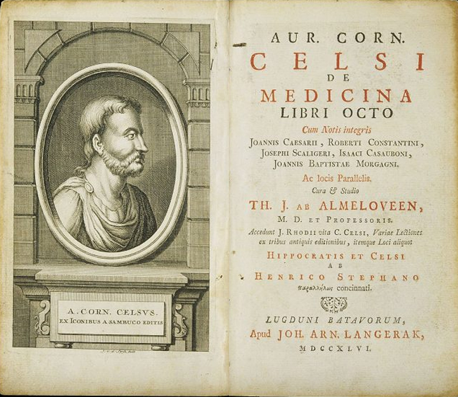
Celsus and Title Page De Medicina Libri Octo”
Click on the image for a larger version
Aulus Cornelius Celsus (25 BC - 45 AD) was Roman, probably born in the south of France. A controversial character, many dispute that he was even a physician or that he actually did what is mentioned in his opus magnum “De Medicina Libri Octo.” According to some, he was what today would be considered a compiler and editor of larger books where each chapter is written by a different author. Others present Celsus as a learned scholar of noble origin, a famous physician, either way, he influenced Medicine and Surgery for centuries. Some of his canons and medical terms he coined are in use today. such as omentum, cancer, vertebra, scalpel, etc.
He is the first to describe the four pillars of inflammation: dolor (pain), rubor (redness), calor (fever), and tumor (swelling) , and in passing criticizes Erasistratus on being wrong about inflammation. In his book he states “Notæ vero inflammationis sunt quatuor, rubor, et tumor, cum calore et dolore. Quo magis erravit Erasistratus, qui febre nullam sine hac esse dixir.” Translates as: “Note that there are four signs of inflammation: redness and swelling, with heat and pain. So much was Erasistratus mistaken, when he declared there was no fever without it.” Celsus popularized the use of the term “cancer”. although it was first used by Galen of Pergamon.
Not much is known about his life or work. Quintilian mentions a work of Celsus on rhetoric, but that work is lost. Quintilian, who apparently was not a fan of Celsus, writes: “…when even Cornelius Celsus, a man of a moderate share of genius, has not only composed treatises on all these arts, but has also left precepts of the military art, agriculture, and medicine.” His quote is what leads Alexander of Padua to state that his book “De Medicina, Libri Octo” is but a small part of a larger treatise that was called “The Arts.” Celsus is known by his book “De Medicina, Libri Octo,” (On Medicine, eight books). This book is sometimes presented as “De Re Medicina, Libri Octo.” Apparently, this book was part of a larger work by Celsus that has disappeared from History. This medical book was handwritten and was lost to history until one copy was discovered in the Vatican Library and released for copying as manuscripts. Of interest is the fact that the book found was titled "Aulii Cornelii Celsi liber sextus. idemque medicinæ primus," making it understood that these were six books, not eight! This was one of the first medical books to be printed upon the invention of the printing press by Gutenberg.
The first book begins with the principles for the preservation of health; the second is dedicated to the symptoms of diseases; the third is describes treatment of diseases which affect the whole organism and the fourth is focused on the diseases where the cause is found only in certain parts of the body, such as in head, lungs etc. The fifth book is dedicated to pharmacology. The sixth, seventh, and the eighth book deal with surgery, wounds, and fractures.
His book describes the importance of cleanliness of a wound and the use of topical treatment that today would be considered antiseptics. His contributions to surgery are many including anatomy of the eye, the description of ophthalmic diseases, methods of preventive treatment and surgery including eyelid surgery. He is probably the first to describe cataract surgery.
The eighth book, almost entirely dedicated to dislocations and fractures, provides an extensive description of head injuries such as extradural hematomas, lesions distant from the impact point, and intracranial damage in cases with no overlying fractures. He also provided the first description of brain swelling exceeding the level of the skull, described several surgical indications and craniotomy techniques, recommended treatment for depressed fractures (which had been previously considered untreatable), and detailed the surgical instruments employed.
De Medicina was among the first medical books to be printed (Florence, 1478), and more than 50 editions have appeared; it was required reading in most medical schools to the present century. It is the principal historical authority for the doctrinal medical teachings of Roman antiquity. The surgical section, which even Joseph Lister studied in the 19th century, is perhaps the best part of the treatise.
W. G. Spencer, the most recent translator of his works, supports an older, minority view that the details of medical procedure, the experienced judgment shown in the selection of treatment, and the frequent use of the first person reveal an author with an intimate acquaintance of clinical medicine who must have been himself a practitioner of the medical arts.
Because of this book and the clarity of the disease descriptions and treatments both pharmacological and surgical. Celsus has been lauded as one of the Fathers of Medicine and Surgery. His influence has stood the test of time and was read well into the 1800’s.
Personal note: This article was motivated by the recent acquisition of a 1713 copy of De Medicina, that is now part of my library. The book belonged to Dr. Harry Kaufmann Hines, a renown radiologist who lived and worked in Cincinnati, OH. The book was a gift to him from his sister-in-law Erma Wright Tateman neé Sharkey in August 1961. Dr. Miranda
Sources:
1. “The Contribution of Aulus Cornelius Celsus (25 B.C.–50 A.D.) to Eyelid Surgery” Davide Lazzeri, Tommaso Agostini, Michele Figus, Marco Nardi, Giuseppe Spinelli, Marcello Pantaloni & Stefano Lazzeri (2012), Orbit, 31:3, 162-167
2. “Aulus Cornelius Celsus and the Head Injuries” GiuseppeTalamonti Giuseppe D'Aliberti, MarcoCenzato World Neurosurgery Volume 133, January 2020, Pages 127-134
3. “A. Cornelius Celsus of medicine. In eight books.” Translated, with notes critical and explanatory, by James Greive, M.D. 1756
For anyone interested in reading an online copy of this book, you can find it here. For the translation by Greive click here.
- Details

UPDATED: After a long hiatus, I am happy to report that this website has been updated to a newer version of Joomla!, and still hosted by GoDaddy. The "look" of the website has not changed, but the software is faster and with a higher level of security. This work was done by my good friend from Greece, Christopher Mavros, who is an expert at designing web templates, web sites, and Joomla! extensions. This also meant that my library catalog was left unfinished for a long time and this required lots of additional work.
First, the library needed new and updated bookplates, which were printed by BookplateInk, a USA based company that I strongly recommend. In case anyone wonders, the famous anatomists that are depicted in the bookplate are Harvey, Vesalius, Spigelius, and Albinus. Second, I needed software that could create the code to publish the titles of the library online. This work was done by my daughter Evelyn, to whom I am deeply grateful.
Recataloging the library is an ongoing effort. This will also require repairing and rebinding some of the older and damaged books. Due to this, during the next weeks and months this listing will change, but for a bibliophile this is not work, but pleasure.
As many of you know I am a collector of old and antique medical and anatomy books and I also have several copies of some of these books in different languages.
The oldest book in the library dates from 1696 and is "Opera Chirurgica Anotomica Conformata al Moto Circolare Del Sangue, & Altre Inuenziono De'piu Moderni. Aggivuntovi Un Trattato Della Peste" by Paolo Barbette. I recently acquired the 1713 book by Aulus Cornelius Celsus "De Medicina, Libri Octo".
Another book I am very proud of is not old, but still a rare book. It is "The Fabric of the Human Body, An Annotated Translation of the 1543 and 1555 Editions of “De Humani Corporis Fabrica Libri Septem", by Drs. Garrison and Hast This monumental translation work took twenty years and only 948 copies of this book were published in 2014. Completely sold out, it is today a rare book!
There are many other books that are important to me, either by its rarity (for those who know.... "The Green Book") or its controversy, as are the books by Pernkopf and Stefano Mancini.
You are welcome to peruse my library catalog, but be forewarned; I am not selling any of my books. Should you want to find a copy, you can use Abebooks.com as a great source for old and new books.
- Details
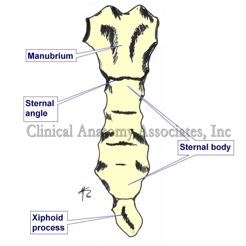
Click on the image for a larger version.
UPDATED: The term [xiphoid] is Greek in origin [ξίφος] and points to the similarity [-oid] of the sternum to a short straight sword used by the Greek. The term xiphoid refers to the lower cartilaginous inferior tip of the sternum. It presents with anatomical variations that include a central opening, or large processes that look like a disc, or even a bifid xiphoid process.
It has been named the xiphisternum, the metasternum, and the ensiform process. It is the last cartilage to ossify in the human, and it is used as a landmark for laparoscopic surgery. i.e. a subxiphoid port is a location for a laparoscopic port in the abdomen.
As a side note, another term used for [sternum] is the Latin word [gladius], referring to the short Roman straight sword of the gladiators. This term is no longer in use, but if it were, the xiphoid appendix would be the tip of the sword.
Think about this: why is a certain plant called a "gladiolus"?
Image property of:CAA.Inc.. Artist:David M. Klein
- Details
This article is part of the series "A Moment in History" where we honor those who have contributed to the growth of medical knowledge in the areas of anatomy, medicine, surgery, and medical research.

Andreas Vesalius Bruxellensis (1514- 1564). A Flemish anatomist and surgeon, Andreas Vesalius was born on December 31, 1514 in Brussels, Belgium. He is considered to be the father of the science of Anatomy. Up until his studies and publications human anatomy studies consisted only on the confirmation of the old doctrines of Galen of Pergamon (129AD - 200AD). Anatomy professors would read to the students from Galen's work and a demonstrator would point in a body to the area being described, if a body was used at all. The reasoning was that there was no need to dissect since all that was needed to know was already written in Galen's books. Vesalius, Fallopius, and others started the change by describing what they actually saw in a dissection as opposed to what was supposed to be there.
Vesalius had a notorious career, both as an anatomist and as a surgeon. His revolutionary book "De Humani Corporis Fabrica: Libri Septem" was published in May 26, 1543. One of the most famous anatomical images is his plate 22 of the book, called sometimes "The Hamlet". You can see this image if you hover over Vesalius' only known portrait which accompanies this article. Sir William Osler said of this book "... it is the greatest book ever printed, from which modern medicine dates"
After the original 1543 printing, the Fabrica was reprinted in 1555. It was re-reprinted and translated in many languages, although many of these printings were low-quality copies with no respect for copyright or authorship.
The story of the wood blocks with the carved images used for the original printing extends into the 20th century. In 1934 these original wood blocks were used to print 617 copies of the book "Iconaes Anatomica". This book is rare and no more can be printed because, sadly, during a 1943 WWII bombing raid over Munich all the wood blocks were burnt.
One interesting aspect of the book was the landscape panorama in some of his most famous woodcuts which was only "discovered" until 1903.
Vesalius was controversial in life and he still is in death. We know that he died on his way back from a pilgrimage to Jerusalem, but how he died, and exactly where he died is lost in controversy. We do know he was alive when he set foot on the port of Zakynthos in the island of the same name in Greece. He is said to have suddenly collapsed and die at the gates of the city, presumably as a consequence of scurvy. Records show that he was interred in the cemetery of the Church of Santa Maria delle Grazie, but the city and the church were destroyed by an earthquake and Vesalius' grave lost to history. Modern researchers are looking into finding the lost grave and have identified the location of the cemetery. This story has not ended yet.
For a detailed biography of Andreas Vesalius CLICK HERE.
Personal note: To commemorate Andrea Vesalius' 500th birthday in 2014, there were many scientific meetings throughout the world, one of them was the "Vesalius Continuum" anatomical meeting on the island of Zakynthos, Greece on September 4-8, 2014. This is the island where Vesalius died in 1564. I had the opportunity to attend and there are several articles in this website on the presence of Andreas Vesalius on Zakynthos island. During 2015 I also attended a symposium on "Vesalius and the Invention of the Modern Body" at the St. Louis University. At this symposium I had the honor of meeting of Drs. Garrison and Hast, authors of the "New Fabrica". For other articles on Andreas Vesalius, click here. Dr. Miranda