![]()
Medical Terminology Daily (MTD) is a blog sponsored by Clinical Anatomy Associates, Inc. as a service to the medical community. We post anatomical, medical or surgical terms, their meaning and usage, as well as biographical notes on anatomists, surgeons, and researchers through the ages. Be warned that some of the images used depict human anatomical specimens.
You are welcome to submit questions and suggestions using our "Contact Us" form. The information on this blog follows the terms on our "Privacy and Security Statement" and cannot be construed as medical guidance or instructions for treatment.
We have 834 guests and no members online
")
Marcia Crocker Noyes
(1869 – 1946)
Further to my comment on old books and research that started with an interesting bookplate (Ex-Libris). I continued my research and found that the person in charge of the Osler library bookplate was a fascinating individual that today maybe a ghost in the MedChi library and building in Baltimore... This is certainly an article that can be called "A Moment in History"
Marcia Crocker Noyes was the librarian at The Maryland State Medical Society from 1896 to 1946 and was a founding member of the Medical Library Association.[1][2][3]
Sir William Osler, MD. a famous Johns Hopkins surgeon was a noted bibliophile and had a large personal collection of books on various topics. When he became the President of MedChi in 1896, he was dismayed at the condition of the library and knew that with the right person and some stewardship, it could become a significant collection. Sir William asked his friend, Dr. Bernard Steiner, a physician and President of the Enoch Pratt Free Library in Baltimore for suggestions of a librarian, and Dr. Steiner recommended Marcia Crocker Noyes. A native of New York, and a graduate of Hunter College, Marcia had moved to Baltimore for a lengthy visit with her sister, and took a “temporary” position at the Pratt Library, which turned into three years. Although she had no medical experience or background, she was enthusiastic, and most importantly, she was willing to move into the apartment provided for the librarian, who needed to be available 24 hours a day.
The image in this article is Ms. Noyes on her first year on the job. Marcia developed a book classification system for medical books, based on the Index Medicus, and called it the Classification for Medical Literature. The system uses the alphabet with capital letters for the major divisions of medicine and lower-case ones for the sub-sections. The system was used for many years, but it's now dated and the Faculty's original shelving scheme was never changed. The card catalogs still reflect her classification and many of the cards are written in Marcia's back-slanting handwriting.
Marcia knew enough to ask the Faculty's members about medical questions, terminology and literature. She gradually won over the predominantly male membership and they became her greatest allies; Sir William at the start, and then for nearly 40 years, Dr. John Ruhräh, a wealthy pediatrician with no immediate family of his own. She made a point of attending almost every Faculty function, and in 1904, under guidelines from the American Medical Association, Marcia was made the Faculty Secretary. For much of her first 10 years, she was the Faculty's only full-time employee, only being assisted by Mr. Caution, the Faculty's janitor. Later in life Marcia would say that she hired him because of his name!
Within ten years, the library had outgrown its space, and plans, spearheaded by Marcia and Sir William before his move to Oxford, were made to build a headquarters building, mainly to house the library's growing collection of medical books and journals.
Marcia was instrumental in the design and building of the new headquarters. She travelled to Philadelphia, New York and Boston to look at their medical society buildings, and eventually, the Philadelphia architectural firm, Ellicott & Emmart was selected to design and build the new Faculty building. Every detail of the building held her imprimatur, from the graceful staircase, to the light-filled reading room, and all of the myriad details of the millwork, marble tesserae, and most of all, the four-story cast iron stacks. She was on-site, climbing up unfinished staircases, checking out the progress of the building, which was built in less than one year at a cost of $90,000.
Among the features of the new building was a fourth-floor apartment for her. She referred to it as the "first penthouse in Baltimore" and it had a garden and rooftop terrace. The library collection eventually grew to more than 65,000 volumes from medical and specialty societies around the world. Journals were traded back and forth, and physicians eagerly anticipated the arrival of each new issue. At the same time, Marcia was involved in the Medical Library Association as one of eight founding members. The MLA promotes medical libraries and the exchange of information. One of the earliest mandates of the MLA was the Exchange, a distribution and trade service for those who had duplicates or little-used books in their collections. Initially, the Exchange was run out of the Philadelphia medical society, but in 1900 it was moved to Baltimore and Marcia oversaw it. Several hundred periodicals and journals were received and sent each month, a huge amount of work for a tiny staff. In 1904, the Faculty had run out of room to manage the Exchange, so it was moved to the Medical Society of the Kings County (Brooklyn). But without Marcia's excellent administrative skills, it floundered and in 1908, the MLA asked Marcia to take charge once again.
In 1909, when the new Faculty building opened, there was enough room to run the Exchange and with the help of MLA Treasurer, noted bibliophile and close friend, Dr. John Ruhräh, it once again became successful. Additionally, Marcia and Dr. Ruhräh combined forces to revive the MLA's bulletin, which had all but ceased publication in 1908, taking the Exchange with it. This duo maintained editorial control from 1911 until 1926. In 1934, around the time of Dr. Ruhräh's death, Marcia became the first “unmedicated” professional to head the MLA. During her tenure, the MLA incorporated, the first seal was adopted, and the annual meeting was held in Baltimore. Marcia wanted to write the history of the MLA once she retired from full-time work at the Faculty, but her health was beginning to fail. She had back problems and had suffered a serious burn on her shoulder as a young woman, possibly from her time running a summer camp, Camp Seyon, for young ladies in the Adirondack Mountains. In 1946, a celebration was planned to honor Marcia's 50 years at the Faculty. But she was adamant that the physicians wait until November, the actual date of her 50 years. However, they knew she was gravely ill, and might not make it until then, so a huge party was held in April. More than 250 physicians attended the celebration, but the ones she was closest to in the early years, were long gone. She was presented with a suitcase, a sum of money to use for travelling, and her favorite painting of Dr. John Philip Smith, a founder of the Medical College in Winchester, Virginia. It was painted by Edward Caledon Smith, a Virginia painter who had been a student of the painter Thomas Sully.[4] She adored this painting and vowed, jokingly, to take it with her wherever she went.
The painting was not to stay with her for very long, for she died in November 1946, and left it to the Faculty in her will. Her funeral was held in the Faculty's Osler Hall, named for her dear friend. More than 60 physicians served as her pallbearers, and she was buried at Baltimore's Green Mount Cemetery. In 1948, the MLA decided to establish an award in the name of Marcia Crocker Noyes. It was for outstanding achievement in medical library field and was to be awarded every two years, or when a truly worthy candidate was submitted. In 2014, the Faculty began giving a bouquet of flowers to the winner of the award in Marcia's name, and in honor of her work. Much evidence exists for this tradition, as we know that the physicians, especially Drs. Osler and Ruhräh, frequently gave her bouquets of flowers. Marcia also cultivated flower gardens at the Faculty and decorated the rooms with her work.
Today, the MedChi building is open for tours and if the rumors are to be believed Ms. Marcia Crocker Noyes is still at work in her beloved library as the "resident ghost" [1][5]

NOTE: This article has been modified from the original Wikipedia article on Marcia Crocker Noyes. The article itself is well-written with interesting images of the subject. I would encourage you to visit it. The second insert is from book 00736 in my personal library and shows in pencil, the incredibly small handwriting of Marsha C. Noyes.
Sources:
1. "Marcia, Marcia, Marcia" MedChi Archives blog.
2. "Marcia C. Noyes, Medical Librarian" (PDF). Bulletin of the Medical Library Association. 35 (1): 108–109. 1947. PMC 194645
3. Smith, Bernie Todd (1974). "Marcia Crocker Noyes, Medical Librarian: The Shaping of a Career" (PDF). Bulletin of the Medical Library Association. 62 (3): 314–324. PMC 198800Freely accessible. PMID 4619344.
4. Edward Caledon BRUCE (1825-1901)"
5. Behind the scenes tour MedChiBuilding
"Clinical Anatomy Associates, Inc., and the contributors of "Medical Terminology Daily" wish to thank all individuals who donate their bodies and tissues for the advancement of education and research”.
Click here for more information


- Details
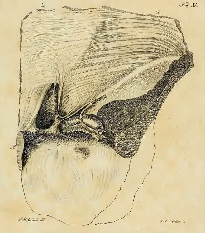
Hesselbach's triangle
Hesselbach’s triangle is a triangular region in the lower posterior aspect of the anterior abdominal wall (see yellow inset in the image). It is bound medially by the lateral border of the rectus abdominis muscle, superolaterally by the inferior (deep) epigastric vessels (label “C”) and by the inguinal ligament inferolaterally.
Hesselbach’s triangle is described as the area where a direct inguinal hernia will extrude from posterior to anterior, to protrude directly (hence the name) through the external (superficial) inguinal ring.
Franz Kaspar Hesselbach (1759-1816) was a German surgeon and anatomist who described inguinofemoral hernias in detail, publishing several books on the subject. His name is attached to several regions and structures:
• Hesselbach’s triangle, described in this article
• Hesselbach’s fascia. Known as the cribriform fascia, this perforated fascia covers the saphenous opening in the superior femoral region.
• Hesselbach’s ligament. Also known as the interfoveolar ligament, this is a thickening of the transversalis fascia in relation to the inferior (deep) epigastric vessels.
If you click on the picture, an original image by Hesselbach will appear. This image shows a defect in Hesselbach’s triangle, setting the stage for a direct inguinal hernia, as well as the interfoveolar ligament. Incidentally, Hesselbach's triangle as described today is not the area described originally by Dr. Hesselbach, where the lower border of the triangle was Cooper's ligament.
Initial image property of:CAA.Inc.. Artist:M. Zuptich. Secondary image by F.K. Hesselbach.
Clinical anatomy of the inguinofemoral hernias, as well as abdominal and perineal hernias are some of the lecture topics developed and delivered to the medical devices industry by Clinical Anatomy Associates, Inc. For more information Contact Us.
- Details
The suffix [-plasia] originates from the Greek word [plasis] or [plassein], meaning "to form", or "to develop". In medical terminology the suffix [-plasia] is used to mean "development". Here are some applications of this term:
- Aplasia: the prefix [a-] means "absence of" or "no", therefore "no development", or "no formation"
- Dysplasia: the prefix [dys-] means "abnormal" - abnormal development
- Hyperplasia: the prefix [hyper-] means "excessive" - excessive development
- Neoplasia: the prefix [neo-] means "new", therefore "a new development" or a "new formation". The term [neoplasia] is used to denote a malignant condition, a cancer tumor.
- Details

Click for a larger image
UPDATED: The eponymic [Thebesian valve], or valve of the coronary sinus is a fold of endocardial tissue situated at the exit ostium of the coronary sinus. Named after Adam Christian Thebesius, the morphology of the valve presents with anatomical variations that go from total absence (see image here) to endocardial folds that cover up to 65% of the coronary sinus opening (see image in this article). The variations of the Thebesian valve include fenestrations, cribiriform valves, and the presence of pectineal cardiac muscle.
The Thebesian valve is important because it can be an obstruction to the passage of a catheter performing retrograde cardioplegia in electrophysiological studies, catheter ablation, and percutaneous mitral valve repair.
Thanks to Dr. Karuna Katti for allowing us the use of the image in this article. The image shows the interior of the right atrium with the inferior vena cava removed to demonstrate the Thebesian valve. In this case, the Thebesian valve covers >65% of the coronary sinus ostium. A catheter is being passed from the superior vena cava into the coronary sinus ostium.
The image shown a human heart with the right atrium and ventricle opened. VC, inferior vena cava; SVC, superior vena cava; TV, Thebesian valve.
Source:"The Thebesian Valve: Gatekeeper to the Coronary Sinus"Karuna Katti and Nikhil Prakash PatilClin Anat 25:379–385 (2012)
- Details

Click for a larger image
[Dysmorphism] is a medical term formed by the prefix [dys-] meaning "abnormal", the root term [-morph-], arising from the Greek word [μορφος] (morfos), meaning "form" or "shape", and the suffix [-ism], also of Greek origin, in this case meaning "condition". A condition of abnormal shape, or "misshapen".
The term dysmorphism is used to denote an anatomical anomaly, usually superficial, that goes beyond the objective (and sometimes subjective) boundaries of normalcy. An example of these are craniofacial dysmorphisms associated with specific congenital disorders, such as Crouzon syndrome; birth defects such as hemifacial microsomia, or as in the case of the image on this article, Mevalonate Kinase Deficiency, a metabolic disease.
Some craniofacial dysmorphisms can affect articulation and speech, such as cleft palate, cleft lip (harelip), prognathism, and retrognathism.
Dysmorphism is also used in some mental disorders where the patient has an abnormal self-image, seeing his/her body or body parts as abnormal, when they are not. This is known as Body Dysmorphic Disorder (BDD), and can be associated with obsessive-compulsive behavior, anxiety, or depression. In some cases BDD can be associated or found in eating disorders such as Anorexia Nervosa, or bulimia.
The image shows a small child with mevalonic aciduria, a deficiency of mevalonate kinase deficiency. This is a rare genetic autosomal recessive metabolic disorder.
Thanks to Maria E. Gallegos, Chair of the Speech Pathology School, Iberoamerican University, Santiago Chile, for suggesting this article. Dr. Miranda
Image Source: Haas D, Hoffmann GF. Mevalonate kinase deficiencies: from mevalonic aciduria to hyperimmunoglobulinemia D syndrome. Orphanet J Rare Dis. 1, 13. 2006. PMID 16722536. DOI:10.1186/1750-1172-1-13 Image in the public domain
- Details

Coccyx, anterior view
The word [coccyx] arises from the Greek term [κούκος] (pronounced koúkos)and means "cuckoo". It is the name of the lower segment of the spinal column, and was named by Herophilus of Alexandria (325-255BC) because of a resemblance of this structure to the bill of the cuckoo bird. Vesalius also used the same analogy. There is another structure of the body named after the beak of a bird, do you know it? If not, click here.
The coccyx (vernacularly known as "tailbone") is usually represented by four rudimentary vertebrae, although the number varies between 3 to 5 vertebrae. There have been reported cases of "human tails" but these do not have a bony structure and are usually related to congenital abnormalities such as spina bifida.
The coccyx has a well-formed superior component, which usually presents with two cornua (horns) which serve as part of a rudimentary zygapophyseal (facet) joint. The lower coccygeal vertebra is usually a small bony node.
The coccyx has an anterior sacrococcygeal ligament, which is continued with the anoccygeal raphe, a ligamentous structure that serves as a posterior attachment for muscular components of the pelvic diaphragm, and helps anchor the anal canal. The coccygeus muscle, the posterior component of the pelvic diaphragm and part of the sacrospinous ligament also attach to the anterolateral aspect of the coccyx.
Coccygeal pain is referred to as coccydynia.
Sources:
1. "The Origin of Medical Terms" Skinner, HA 1970 Hafner Publishing Co.
2. "Medical Meanings - A Glossary of Word Origins" Haubrich, WD. ACP Philadelphia
3. "Dorlands's Illustrated Medical Dictionary" 26th Ed. W.B. Saunders 1994
5 "Tratado de Anatomia Humana" Testut et Latarjet 8 Ed. 1931 Salvat Editores, Spain
6. "Anatomy of the Human Body" Henry Gray 1918. Philadelphia: Lea & Febiger
Image modified by CAA, Inc. Original image courtesy of bartleby.com
Note: Google Translate includes the symbol (?). Clicking on it will allow you to hear the pronunciation of the word.
- Details

Click for a larger image
The [coracoid process] is a curved bony projection that attaches to the superior aspect of the neck of the scapula . At its origin it has a broad base continuous with a segment that projects slightly superomedially, it then continues anterolaterally with a narrower distal segment.
The pectoralis minor and coracobrachialis muscles attach to the coracoid process as well as the tendon of the short head of the biceps brachii muscle. The trapezoid and conoid ligaments, which also attach to the clavicle, attach to the coracoid process.
The root term [-corac-] originates from the Greek word [κοράκι] (koríki), meaning “raven” or “crow. The suffix [-oid], also of Greek origin, means “similar to". The word [coracoid] was coined because of the similarity of the coracoid process to a raven’s beak.
Trivia question: What other part of the body is named after a bird’s beak? For the answer, click here. What other parts of the body are named after birds? For the answer, click here.
The image shows the anterior view of the left scapula. Modified from image in Public Domain, by Henry Vandyke Carter - Gray's Anatomy
Source:
“Gray’s Anatomy” Henry Gray, 1918