![]()
Medical Terminology Daily (MTD) is a blog sponsored by Clinical Anatomy Associates, Inc. as a service to the medical community. We post anatomical, medical or surgical terms, their meaning and usage, as well as biographical notes on anatomists, surgeons, and researchers through the ages. Be warned that some of the images used depict human anatomical specimens.
You are welcome to submit questions and suggestions using our "Contact Us" form. The information on this blog follows the terms on our "Privacy and Security Statement" and cannot be construed as medical guidance or instructions for treatment.
We have 1437 guests and no members online
")
Marcia Crocker Noyes
(1869 – 1946)
Further to my comment on old books and research that started with an interesting bookplate (Ex-Libris). I continued my research and found that the person in charge of the Osler library bookplate was a fascinating individual that today maybe a ghost in the MedChi library and building in Baltimore... This is certainly an article that can be called "A Moment in History"
Marcia Crocker Noyes was the librarian at The Maryland State Medical Society from 1896 to 1946 and was a founding member of the Medical Library Association.[1][2][3]
Sir William Osler, MD. a famous Johns Hopkins surgeon was a noted bibliophile and had a large personal collection of books on various topics. When he became the President of MedChi in 1896, he was dismayed at the condition of the library and knew that with the right person and some stewardship, it could become a significant collection. Sir William asked his friend, Dr. Bernard Steiner, a physician and President of the Enoch Pratt Free Library in Baltimore for suggestions of a librarian, and Dr. Steiner recommended Marcia Crocker Noyes. A native of New York, and a graduate of Hunter College, Marcia had moved to Baltimore for a lengthy visit with her sister, and took a “temporary” position at the Pratt Library, which turned into three years. Although she had no medical experience or background, she was enthusiastic, and most importantly, she was willing to move into the apartment provided for the librarian, who needed to be available 24 hours a day.
The image in this article is Ms. Noyes on her first year on the job. Marcia developed a book classification system for medical books, based on the Index Medicus, and called it the Classification for Medical Literature. The system uses the alphabet with capital letters for the major divisions of medicine and lower-case ones for the sub-sections. The system was used for many years, but it's now dated and the Faculty's original shelving scheme was never changed. The card catalogs still reflect her classification and many of the cards are written in Marcia's back-slanting handwriting.
Marcia knew enough to ask the Faculty's members about medical questions, terminology and literature. She gradually won over the predominantly male membership and they became her greatest allies; Sir William at the start, and then for nearly 40 years, Dr. John Ruhräh, a wealthy pediatrician with no immediate family of his own. She made a point of attending almost every Faculty function, and in 1904, under guidelines from the American Medical Association, Marcia was made the Faculty Secretary. For much of her first 10 years, she was the Faculty's only full-time employee, only being assisted by Mr. Caution, the Faculty's janitor. Later in life Marcia would say that she hired him because of his name!
Within ten years, the library had outgrown its space, and plans, spearheaded by Marcia and Sir William before his move to Oxford, were made to build a headquarters building, mainly to house the library's growing collection of medical books and journals.
Marcia was instrumental in the design and building of the new headquarters. She travelled to Philadelphia, New York and Boston to look at their medical society buildings, and eventually, the Philadelphia architectural firm, Ellicott & Emmart was selected to design and build the new Faculty building. Every detail of the building held her imprimatur, from the graceful staircase, to the light-filled reading room, and all of the myriad details of the millwork, marble tesserae, and most of all, the four-story cast iron stacks. She was on-site, climbing up unfinished staircases, checking out the progress of the building, which was built in less than one year at a cost of $90,000.
Among the features of the new building was a fourth-floor apartment for her. She referred to it as the "first penthouse in Baltimore" and it had a garden and rooftop terrace. The library collection eventually grew to more than 65,000 volumes from medical and specialty societies around the world. Journals were traded back and forth, and physicians eagerly anticipated the arrival of each new issue. At the same time, Marcia was involved in the Medical Library Association as one of eight founding members. The MLA promotes medical libraries and the exchange of information. One of the earliest mandates of the MLA was the Exchange, a distribution and trade service for those who had duplicates or little-used books in their collections. Initially, the Exchange was run out of the Philadelphia medical society, but in 1900 it was moved to Baltimore and Marcia oversaw it. Several hundred periodicals and journals were received and sent each month, a huge amount of work for a tiny staff. In 1904, the Faculty had run out of room to manage the Exchange, so it was moved to the Medical Society of the Kings County (Brooklyn). But without Marcia's excellent administrative skills, it floundered and in 1908, the MLA asked Marcia to take charge once again.
In 1909, when the new Faculty building opened, there was enough room to run the Exchange and with the help of MLA Treasurer, noted bibliophile and close friend, Dr. John Ruhräh, it once again became successful. Additionally, Marcia and Dr. Ruhräh combined forces to revive the MLA's bulletin, which had all but ceased publication in 1908, taking the Exchange with it. This duo maintained editorial control from 1911 until 1926. In 1934, around the time of Dr. Ruhräh's death, Marcia became the first “unmedicated” professional to head the MLA. During her tenure, the MLA incorporated, the first seal was adopted, and the annual meeting was held in Baltimore. Marcia wanted to write the history of the MLA once she retired from full-time work at the Faculty, but her health was beginning to fail. She had back problems and had suffered a serious burn on her shoulder as a young woman, possibly from her time running a summer camp, Camp Seyon, for young ladies in the Adirondack Mountains. In 1946, a celebration was planned to honor Marcia's 50 years at the Faculty. But she was adamant that the physicians wait until November, the actual date of her 50 years. However, they knew she was gravely ill, and might not make it until then, so a huge party was held in April. More than 250 physicians attended the celebration, but the ones she was closest to in the early years, were long gone. She was presented with a suitcase, a sum of money to use for travelling, and her favorite painting of Dr. John Philip Smith, a founder of the Medical College in Winchester, Virginia. It was painted by Edward Caledon Smith, a Virginia painter who had been a student of the painter Thomas Sully.[4] She adored this painting and vowed, jokingly, to take it with her wherever she went.
The painting was not to stay with her for very long, for she died in November 1946, and left it to the Faculty in her will. Her funeral was held in the Faculty's Osler Hall, named for her dear friend. More than 60 physicians served as her pallbearers, and she was buried at Baltimore's Green Mount Cemetery. In 1948, the MLA decided to establish an award in the name of Marcia Crocker Noyes. It was for outstanding achievement in medical library field and was to be awarded every two years, or when a truly worthy candidate was submitted. In 2014, the Faculty began giving a bouquet of flowers to the winner of the award in Marcia's name, and in honor of her work. Much evidence exists for this tradition, as we know that the physicians, especially Drs. Osler and Ruhräh, frequently gave her bouquets of flowers. Marcia also cultivated flower gardens at the Faculty and decorated the rooms with her work.
Today, the MedChi building is open for tours and if the rumors are to be believed Ms. Marcia Crocker Noyes is still at work in her beloved library as the "resident ghost" [1][5]

NOTE: This article has been modified from the original Wikipedia article on Marcia Crocker Noyes. The article itself is well-written with interesting images of the subject. I would encourage you to visit it. The second insert is from book 00736 in my personal library and shows in pencil, the incredibly small handwriting of Marsha C. Noyes.
Sources:
1. "Marcia, Marcia, Marcia" MedChi Archives blog.
2. "Marcia C. Noyes, Medical Librarian" (PDF). Bulletin of the Medical Library Association. 35 (1): 108–109. 1947. PMC 194645
3. Smith, Bernie Todd (1974). "Marcia Crocker Noyes, Medical Librarian: The Shaping of a Career" (PDF). Bulletin of the Medical Library Association. 62 (3): 314–324. PMC 198800Freely accessible. PMID 4619344.
4. Edward Caledon BRUCE (1825-1901)"
5. Behind the scenes tour MedChiBuilding
"Clinical Anatomy Associates, Inc., and the contributors of "Medical Terminology Daily" wish to thank all individuals who donate their bodies and tissues for the advancement of education and research”.
Click here for more information


- Details
This article is part of the series "A Moment in History" where we honor those who have contributed to the growth of medical knowledge in the areas of anatomy, medicine, surgery, and medical research.
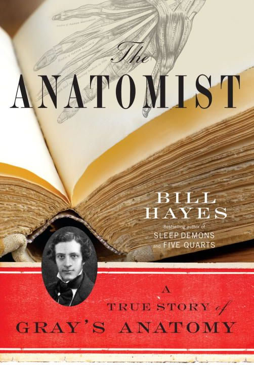
Book cover
As you know, I am interested in the history of Science, Medicine, and specially, Human Anatomy. Because of that and as part of this website we added a series called "A Moment in History". The objective was to create a series of articles to honor those individuals who have contributed to the growth of medical knowledge in the areas of anatomy, medicine, surgery, and medical research including individuals who have contributed in different ways, but still added their life work to the advancement of medical knowledge.
One of the individuals who piqued my interest was Henry Gray, FRS author of one of the great teaching books on anatomy ever written, “Gray’s Anatomy”. This book was first published in England in 1858, later published in the USA from 1862 to 1990. The English Edition is still published and is now in its 42nd Edition.
I started to look deeper into his life which, I learned with surprise, was not only very short, but obscure. Researching into Henry Gray’s life, I came about this book: “The Anatomist – A True Story of Gray’s Anatomy” by William (Bill) Hayes.
Bill Hayes started looking into Henry Gray’s life with the intent of writing a biography. He rapidly run into a wall. Besides a well-detailed list of academic titles and positions, degrees awarded, and scientific papers that he published. As the author states “what I had gathered about him would amount to little more than a Curriculum Vitae”. So, he starts a journey to uncover more data about Gray’s life and his well-known book.
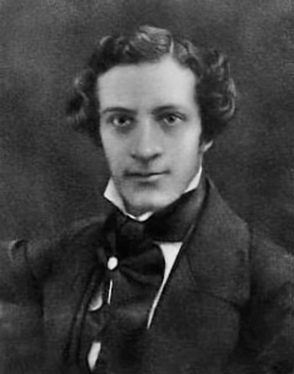
Henry Gray, FRS

Henry Vandyke Carter, MD
Part of his journey was to understand the importance of anatomy as a component of the medical curriculum. Bill Hayes was accepted as a participant in the anatomy laboratory for physical therapy students and later visited medical students in the anatomy lab. Just his observations and comments throughout the book on these activities is worth reading this book.
What is interesting is the lack of personal information on Henry Gray. To this date, we do not have information about his birth, either location or date. There is more information on his family that on the subject. Most agree that Henry Gray was born in 1827, but some propose 1825. Either way, there is no firm data. What we do know is that he died on June 12, 1861, having contracted smallpox most probably from his nephew Charles Gray. Henry Gray was 36 years old.
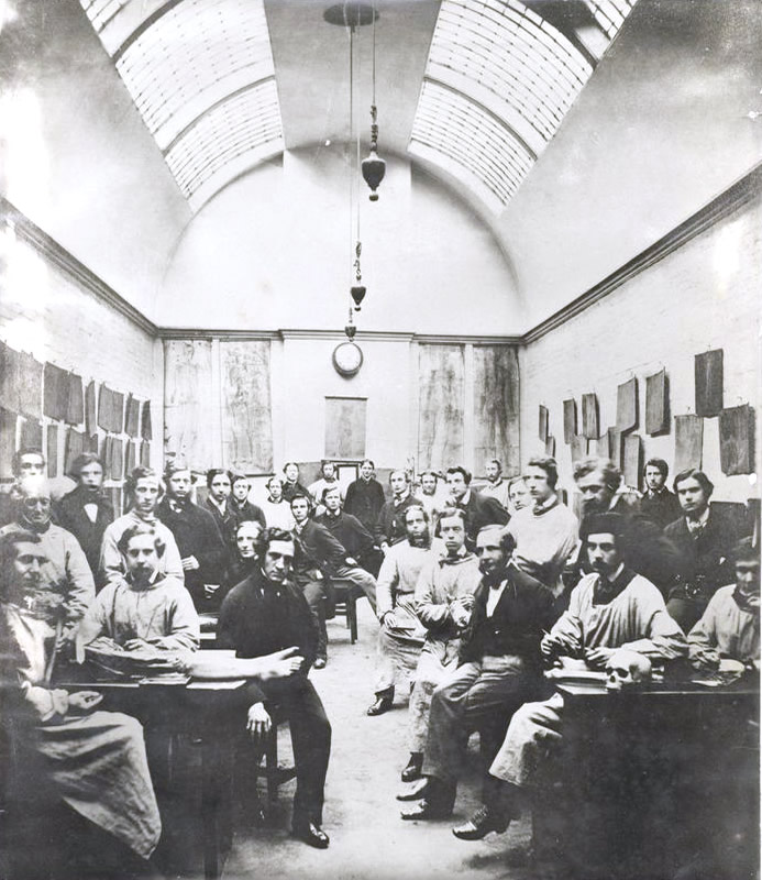
Gray and students
Here is one of the few photographs that exists of Henry Gray. In this particular 1870 picture by Joseph Langhorn, Gray is in the anatomy laboratory (forefront, third from the left) at St. George’s Hospital with his students. At the time it was customary for medical students to pose in the lab with bones and cadavers. This is a practice not in use today and disappeared circa 1930. For anyone interested in this now considered gruesome custom, I would recommend the book "Dissection" by John H. Warner and James M. Edmonson.
In his research, the author discovers Henry Gray’s collaborator and illustrator of the book: Henry Vandyke Carter, MD. Where Gray is obscure and with no personal information, Henry Carter writes a diary daily, and for some time he actually writes another diary that he calls “Reflections” on more personal and religious topics. Carter is a troubled, complicated individual blessed with incredible anatomical knowledge and drawing capabilities that can be seen throughout the book.
It is because of these diaries that we know Henry Carter and we can glimpse (almost at a distance) the character of Henry Gray, but it is not enough to elucidate his biography. In some ways it is like looking at an individual through a veil. You see him, but it is nebulous.
Much of the book is concentrated on Henry Carter, his life, his depression bouts, his self doubts and the work that he did illustrating Gray’s book. In some ways he is behind the scenes, and even though he did much of the dissection work and illustration, Henry Carter is mostly unknown to the anatomical world, as in may cases the medical illustrators are, with some notable exceptions.
Henry Carter is paid only 150 pounds for his work and even before getting paid he pays for a ticket to India where he accomplishes his objectives in life and in academia. Henry Carter eventually retires and comes back to England where he dies in 1897.
Bill Hayes and his partner visited rare book libraries at different universities and eventually go to London to visit areas and locations where Henry Gray lived and worked. There is a sense of accomplishment, but also a sense that we can almost touch the life of Henry Gray but fall short of seeing him.
The book ending is poignant, dealing with personal matters and thoughts on life and death. Eventually the author is very clear that the study of anatomy, although on dead subjects who donated their bodies to the universities is actually an activity that helps us understand life.
Bill Hayes is an awarded author of seven books, a frequent contributor to the New York Times and a photographer. More information about him and his activities at https://www.billhayes.com
Personal note: This is a book that I personally recommend and proud to add to my personal library. My one observation is that the author should have probably name the book “The Anatomists - A True Story of Gray’s Anatomy” as it is the story of the life of the two Henrys: Gray and Carter. In fact, I believe that Gray’s Anatomy could have been called “Gray’s and Carter’s Anatomy”, had history been slightly different – Dr. Miranda.
Sources:
1. “Henry Gray and Henry Vandyke Carter: Creators of a Famous Textbook" Roberts, S. J Med Biog 2000 8: 206-212
2. "Henry Gray, Anatomist: An Appreciation" Boland, F Am J Med Sci 1908 1827-1924
3. "The Anatomist: A True Story of Gray’s Anatomy" Hayes, B. Random House PG 2007
- Details
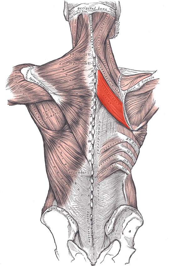
Rhomboid major muscle
The rhomboid major muscle is found in the posterior aspect of the thorax between the spine and scapula. This muscle originates from the spinous processes of the thoracic vertebrae T2 to T5 as well as the supraspinous ligament at these levels by way of an aponeurosis that varies in width. It inserts on the medial border of the scapula, from about the level of the scapular spine to the inferior angle of the scapula by way of short tendinous fibers.
It adducts, elevates, and inferiorly rotates the scapula. It also helps to maintain the scapula flush with the posterior aspect of the thoracic cage. It is innervated by the dorsal scapular nerve (C4, C5), a branch of the brachial plexus.
The Terminologia Anatomica name for the muscle is musculus rhomboideus major.
The rhomboid major is one of the 17 muscles that attach to the scapula.
Sources:
1. “Gray’s Anatomy” Henry Gray, 1918
2. "Tratado de Anatomia Humana" Testut et Latarjet 8th Ed. 1931 Salvat Editores, Spain
3. "Gray's Anatomy" 38th British Ed. Churchill Livingstone 1995
4. “An Illustrated Atlas of the Skeletal Muscles” Bowden, B. 4th Ed. Morton Publishing. 2015
5. "Morris' Human Anatomy" Pearce, J. (1942) Blakiston Co. Philadlephia USA
6. "Trail Guide to The Body" 4th. Ed. Biel, A. Books of Discovery. 2010
Image modified from the original by Henry VanDyke Carter, MD. in the book "Grays's Anatomy" by Henry Gray FRS. Public domain
- Details
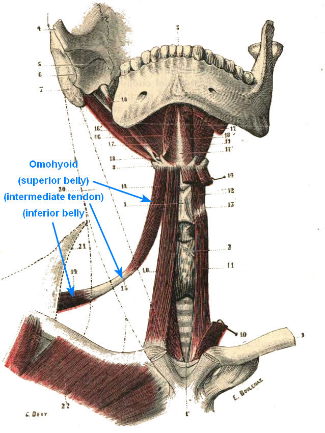
Omohyoid muscle.
Click on the image for a larger depiction
The omohyoid also known as the musculus omohyoideus is the most lateral of the infrahyoid muscles, the others being the sternohyoid, sternothyroid, and thyroid muscles.The omohyoid is a long muscle formed by two muscular bellies (superior and inferior) and an intermediate tendon.
The inferior belly originates from the superior margin of the scapula, near the superior transverse ligament of the scapula. Sometimes, as a variation, the tendon of the inferior belly originates directly from this ligament. The inferior belly passes medially, posteriorly, and slightly superiorly towards the intermediate tendon. The medial aspect of this belly is deep to the trapezius muscle.
The intermediate tendon is held in place by a strong extension of the middle layer of the cervical fascia. This extension has firm attachments to the first rib and the posterior surface of the clavicle.
Along with the other infrahyoid muscles, the omohyoid muscle depresses the hyoid bone. Each belly receives a separate branch that arises from the ansa cervicalis (C1, C2, and C3).
Note: The image in this article was taken from the book "Tratado de Anatomía Humana" by Testut and Latarjet. The illustration was made by George Devy, a famous French painter who specialized in anatomical illustrations (? -1902) and the engraving was made by a Swiss artist, Edmond Boulenaz (1859-1905). This image is in the Public Domain and has been edited by us.
Sources:
1. “Gray’s Anatomy” Henry Gray, 1918
2. "Tratado de Anatomia Humana" Testut et Latarjet 8th Ed. 1931 Salvat Editores, Spain
3. "Gray's Anatomy" 42nd British Ed. Churchill Livingstone 2021
4. “An Illustrated Atlas of the Skeletal Muscles” Bowden, B. 4th Ed. Morton Publishing. 2015
5. "Trail Guide to The Body" 4th. Ed. Biel, A. Books of Discovery. 2010
6. "Petite histoire de l'iconographie anatomique" Huard, P; Imbalt-Huart, MJ Communication présentée à la Société Française d'Histoire de la Médecine, le30 septembre 1972.
7. https://www.anatomicalterms.info
- Details
- Written by: Efrain A. Miranda, Ph.D.

Brachialis muscle.
Click on the image for a larger depiction
UPDATED: The brachialis muscle is a skeletal muscle attached proximally to the anterior surface of the humerus and distally to the coronoid process and tuberosity of the ulna. It is one of the three muscles in the anterior compartment of the arm (flexor compartment), the other two being the biceps brachii and the coracobrachialis.
It is a strong flexor of the elbow found deep to the biceps brachii. Because it does not attach to the radius, the brachialis muscle does not participate in the pronation and supination of the forearm.
The brachialis is supplied by branches of the brachial artery and by the recurrent radial artery.
The innervation of the brachialis muscle is a point to be discussed. Most modern books of anatomy state that this muscle is innervated by the musculocutaneous nerve (C5, C6, and C7). Older and more detailed books state that this muscle has a dual innervation. A 2011 research paper published in Spanish (see Sources #6) describes this dual innervation. The proximal portion of the muscles is indeed innervated by the musculocutaneous nerve, but the distal portion (in 90% of the cases) is innervated by muscular branches that arise off the radial nerve. The radial nerve (C5, C6, C7, C8 & T1) is a branch of the brachial plexus.
Following is an excerpt from the "Trail Guide to the Body" by Andrew Biel: "Ironically, (because it is deep to the biceps) the brachialis girth only helps the biceps brachii to bulge further from the arm, making the brachialis the biceps' "best friend"
Personal note: The research paper that describes the double innervation of the brachialis muscle was done at my alma mater, the University of Chile, and the authors' listing includes two of the contributors to this blog, Professors Claudio Molina and Cristian Uribe. Dr. Miranda
Sources:
1. “Gray’s Anatomy” Henry Gray, 1918
2. "Tratado de Anatomia Humana" Testut et Latarjet 8th Ed. 1931 Salvat Editores, Spain
3. "Gray's Anatomy" 42nd British Ed. Churchill Livingstone 2021
4. “An Illustrated Atlas of the Skeletal Muscles” Bowden, B. 4th Ed. Morton Publishing. 2015
5. "Trail Guide to The Body" 4th. Ed. Biel, A. Books of Discovery. 2010
6. "Doble Innervacion del Musculo Brachial en la Poblacion Chilena" Claudio Molina; Cristián Uribe; Álvaro Heras; Cristián Astorga;Jorge Lemus & Alberto Rodríguez. Int. J. Morphol, 2011. 29(4):1207-1211. A PDF copy of this paper is available here.
Note: The side image modified from the original by Anatomography, CC BY-SA 2.1 JP <https://creativecommons.org/licenses/by-sa/2.1/jp/deed.en>, via Wikimedia Commons following Creative Commons attributes.
- Details

Biceps brachii muscle.
Click on the image for a larger depiction
The musculus biceps brachii is a long muscle found in the anterior, aspect of the arm and is one of the three muscles contained in the anterior compartment (flexor compartment) of the arm, the other two being the brachialis and coracobrachialis muscles. It is composed by two muscular heads, one long (lateral) , and one short (medial) that originate superiorly from separate tendons that attach to the scapula. These two heads join to form a single long, oval-shaped belly with a single tendon that crosses the elbow joint and attaches to the radius.
The short tendon of the biceps brachii passes anteromedial to the shoulder joint and attaches to the coracoid process of the scapula by way of a tendon that mixes with the tendon of the coracobrachialis muscle.
The long cylindrical tendon of the biceps brachii is found in the intertubercular (bicipital) groove (Lat: sulcus intertubercularis) of the humerus, and passes between the greater and lesser tubercles of the humerus, entering the articular cavity of shoulder joint, and continues superiorly to insert in the supraglenoid tubercle of the scapula.
The distal, common tendon of the biceps brachii courses inferiorly and attaches to the radial (bicipital) tuberosity of the radius. There is a well-defined bursa between the radial tuberosity and the biceps brachii tendon that allows for movement of the tendon.
Also, a flat, fascial extension of the tendon, known as the bicipital aponeurosis extends inferomedially to blend with the antebrachial aponeurosis that covers the epitrochlear muscles of the forearm (pronator teres, flexor carpi radialis muscles). The brachial artery passes between the tendon of the biceps brachii and the bicipital aponeurosis in the anterior aspect of the elbow joint.
The biceps brachii crosses both the shoulder and the elbow join. As such, its functions will depend on which joint is fixed and which one is not. This muscle flexes the elbow, supinates the forearm, and flexes the shoulder.
It is innervated by the musculocutaneous nerve (C5,D6) which is a branch of the brachial plexus. It receives arterial supply by way of muscular branches that arise from the brachial artery.
The name of the muscle literally means "two heads" as the prefix "bi" means "two" and the Latin term "-ceps" means "head".
Note: Because the long and the short head of the biceps brachii attach to different locations of the scapula, some authors and Internet websites say that there are 18 muscles that attach to the scapula. I do not agree, as the biceps brachii is a single muscle that happens to have to separate attachments to the scapula. It would be different if this article was titled "Name the 18 separate muscular attachment points of the scapula". Dr. Miranda
The image is modified from the original via Wikimedia. Public domain. Animated image below by Wikimedia Commons - Anatomography [CC BY-SA 2.1 following Creative Commons attributes.
 Sources:
1. “Gray’s Anatomy” Henry Gray, 1918
2. "Tratado de Anatomia Humana" Testut et Latarjet 8th Ed. 1931 Salvat Editores, Spain
3. "Gray's Anatomy" 42nd British Ed. Churchill Livingstone 2021
4. “An Illustrated Atlas of the Skeletal Muscles” Bowden, B. 4th Ed. Morton Publishing. 2015
5. "Trail Guide to The Body" 4th. Ed. Biel, A. Books of Discovery. 2010
- Details
- Written by: Efrain A. Miranda, Ph.D.
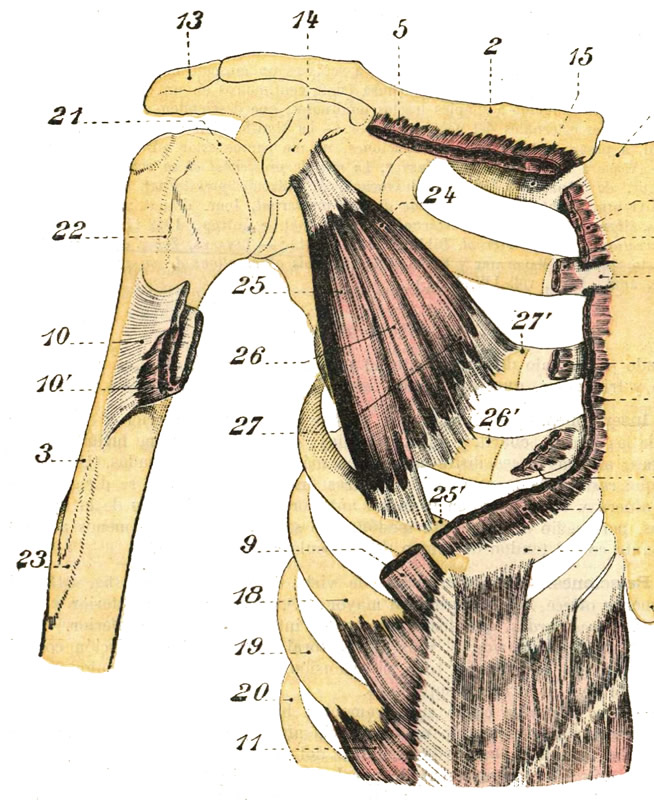
Pectoralis minor muscle (26)
Click on the image for a larger depiction
The pectoralis minor muscle is a small triangular muscle found deep to the pectoralis major in the anterior aspect of the thorax.
This muscle originates from three fleshy bellies that insert into the superior border and anterior surface of the third, fourth and fifth ribs. The muscle fibers converge superolaterally to insert into the inferomedial aspect of the coracoid process, of the scapula, where the tendon of the pectoralis minor intermingles and fuses with the tendon of the coracobrachialis muscle.
The pectoralis minor lies immediately anterior and covers some of the structures of the axillary region, the axillary artery and vein and some of the components of the brachial plexus. In fact, the pectoralis minor muscle is the landmark that divides the axillary artery into its three components: proximal (between the first rib and the medial border of the pectoralis minor). middle (deep to the pectoralis minor), and distal (between the lateral border of the pectoralis major and the inferior border of the teres major muscle). Thus defined the pectoralis major forms part of the anterior wall of the axilla.
In conjunction with other muscles, the pectoralis minor helps to maintain the scapular and shoulder joint in position. If the scapula is fixed, the pectoralis major assists to elevate the anterior thoracic wall during forced inhalation. The pectoralis minor also works as a depressor of the scapula and shoulder joint, abducts the scapula, and rotates the scapula.
The pectoralis minor is innervated by the medial pectoral nerve (C8.T1), a branch of the brachial plexus. Some of the fibers of the medial pectoral nerve perforate the pectoralis minor to provide nerve supply to a portion of the pectoralis major. The pectoralis minor is one of the 17 muscles that attach to the scapula.
Sources:
1. “Gray’s Anatomy” Henry Gray, 1918
2. "Tratado de Anatomia Humana" Testut et Latarjet 8th Ed. 1931 Salvat Editores, Spain
3. "Gray's Anatomy" 42nd British Ed. Churchill Livingstone 2021
4. “An Illustrated Atlas of the Skeletal Muscles” Bowden, B. 4th Ed. Morton Publishing. 2015
5. "Trail Guide to The Body" 4th. Ed. Biel, A. Books of Discovery. 2010