![]()
Medical Terminology Daily (MTD) is a blog sponsored by Clinical Anatomy Associates, Inc. as a service to the medical community. We post anatomical, medical or surgical terms, their meaning and usage, as well as biographical notes on anatomists, surgeons, and researchers through the ages. Be warned that some of the images used depict human anatomical specimens.
You are welcome to submit questions and suggestions using our "Contact Us" form. The information on this blog follows the terms on our "Privacy and Security Statement" and cannot be construed as medical guidance or instructions for treatment.
We have 274 guests and no members online
")
Marcia Crocker Noyes
(1869 – 1946)
Further to my comment on old books and research that started with an interesting bookplate (Ex-Libris). I continued my research and found that the person in charge of the Osler library bookplate was a fascinating individual that today maybe a ghost in the MedChi library and building in Baltimore... This is certainly an article that can be called "A Moment in History"
Marcia Crocker Noyes was the librarian at The Maryland State Medical Society from 1896 to 1946 and was a founding member of the Medical Library Association.[1][2][3]
Sir William Osler, MD. a famous Johns Hopkins surgeon was a noted bibliophile and had a large personal collection of books on various topics. When he became the President of MedChi in 1896, he was dismayed at the condition of the library and knew that with the right person and some stewardship, it could become a significant collection. Sir William asked his friend, Dr. Bernard Steiner, a physician and President of the Enoch Pratt Free Library in Baltimore for suggestions of a librarian, and Dr. Steiner recommended Marcia Crocker Noyes. A native of New York, and a graduate of Hunter College, Marcia had moved to Baltimore for a lengthy visit with her sister, and took a “temporary” position at the Pratt Library, which turned into three years. Although she had no medical experience or background, she was enthusiastic, and most importantly, she was willing to move into the apartment provided for the librarian, who needed to be available 24 hours a day.
The image in this article is Ms. Noyes on her first year on the job. Marcia developed a book classification system for medical books, based on the Index Medicus, and called it the Classification for Medical Literature. The system uses the alphabet with capital letters for the major divisions of medicine and lower-case ones for the sub-sections. The system was used for many years, but it's now dated and the Faculty's original shelving scheme was never changed. The card catalogs still reflect her classification and many of the cards are written in Marcia's back-slanting handwriting.
Marcia knew enough to ask the Faculty's members about medical questions, terminology and literature. She gradually won over the predominantly male membership and they became her greatest allies; Sir William at the start, and then for nearly 40 years, Dr. John Ruhräh, a wealthy pediatrician with no immediate family of his own. She made a point of attending almost every Faculty function, and in 1904, under guidelines from the American Medical Association, Marcia was made the Faculty Secretary. For much of her first 10 years, she was the Faculty's only full-time employee, only being assisted by Mr. Caution, the Faculty's janitor. Later in life Marcia would say that she hired him because of his name!
Within ten years, the library had outgrown its space, and plans, spearheaded by Marcia and Sir William before his move to Oxford, were made to build a headquarters building, mainly to house the library's growing collection of medical books and journals.
Marcia was instrumental in the design and building of the new headquarters. She travelled to Philadelphia, New York and Boston to look at their medical society buildings, and eventually, the Philadelphia architectural firm, Ellicott & Emmart was selected to design and build the new Faculty building. Every detail of the building held her imprimatur, from the graceful staircase, to the light-filled reading room, and all of the myriad details of the millwork, marble tesserae, and most of all, the four-story cast iron stacks. She was on-site, climbing up unfinished staircases, checking out the progress of the building, which was built in less than one year at a cost of $90,000.
Among the features of the new building was a fourth-floor apartment for her. She referred to it as the "first penthouse in Baltimore" and it had a garden and rooftop terrace. The library collection eventually grew to more than 65,000 volumes from medical and specialty societies around the world. Journals were traded back and forth, and physicians eagerly anticipated the arrival of each new issue. At the same time, Marcia was involved in the Medical Library Association as one of eight founding members. The MLA promotes medical libraries and the exchange of information. One of the earliest mandates of the MLA was the Exchange, a distribution and trade service for those who had duplicates or little-used books in their collections. Initially, the Exchange was run out of the Philadelphia medical society, but in 1900 it was moved to Baltimore and Marcia oversaw it. Several hundred periodicals and journals were received and sent each month, a huge amount of work for a tiny staff. In 1904, the Faculty had run out of room to manage the Exchange, so it was moved to the Medical Society of the Kings County (Brooklyn). But without Marcia's excellent administrative skills, it floundered and in 1908, the MLA asked Marcia to take charge once again.
In 1909, when the new Faculty building opened, there was enough room to run the Exchange and with the help of MLA Treasurer, noted bibliophile and close friend, Dr. John Ruhräh, it once again became successful. Additionally, Marcia and Dr. Ruhräh combined forces to revive the MLA's bulletin, which had all but ceased publication in 1908, taking the Exchange with it. This duo maintained editorial control from 1911 until 1926. In 1934, around the time of Dr. Ruhräh's death, Marcia became the first “unmedicated” professional to head the MLA. During her tenure, the MLA incorporated, the first seal was adopted, and the annual meeting was held in Baltimore. Marcia wanted to write the history of the MLA once she retired from full-time work at the Faculty, but her health was beginning to fail. She had back problems and had suffered a serious burn on her shoulder as a young woman, possibly from her time running a summer camp, Camp Seyon, for young ladies in the Adirondack Mountains. In 1946, a celebration was planned to honor Marcia's 50 years at the Faculty. But she was adamant that the physicians wait until November, the actual date of her 50 years. However, they knew she was gravely ill, and might not make it until then, so a huge party was held in April. More than 250 physicians attended the celebration, but the ones she was closest to in the early years, were long gone. She was presented with a suitcase, a sum of money to use for travelling, and her favorite painting of Dr. John Philip Smith, a founder of the Medical College in Winchester, Virginia. It was painted by Edward Caledon Smith, a Virginia painter who had been a student of the painter Thomas Sully.[4] She adored this painting and vowed, jokingly, to take it with her wherever she went.
The painting was not to stay with her for very long, for she died in November 1946, and left it to the Faculty in her will. Her funeral was held in the Faculty's Osler Hall, named for her dear friend. More than 60 physicians served as her pallbearers, and she was buried at Baltimore's Green Mount Cemetery. In 1948, the MLA decided to establish an award in the name of Marcia Crocker Noyes. It was for outstanding achievement in medical library field and was to be awarded every two years, or when a truly worthy candidate was submitted. In 2014, the Faculty began giving a bouquet of flowers to the winner of the award in Marcia's name, and in honor of her work. Much evidence exists for this tradition, as we know that the physicians, especially Drs. Osler and Ruhräh, frequently gave her bouquets of flowers. Marcia also cultivated flower gardens at the Faculty and decorated the rooms with her work.
Today, the MedChi building is open for tours and if the rumors are to be believed Ms. Marcia Crocker Noyes is still at work in her beloved library as the "resident ghost" [1][5]

NOTE: This article has been modified from the original Wikipedia article on Marcia Crocker Noyes. The article itself is well-written with interesting images of the subject. I would encourage you to visit it. The second insert is from book 00736 in my personal library and shows in pencil, the incredibly small handwriting of Marsha C. Noyes.
Sources:
1. "Marcia, Marcia, Marcia" MedChi Archives blog.
2. "Marcia C. Noyes, Medical Librarian" (PDF). Bulletin of the Medical Library Association. 35 (1): 108–109. 1947. PMC 194645
3. Smith, Bernie Todd (1974). "Marcia Crocker Noyes, Medical Librarian: The Shaping of a Career" (PDF). Bulletin of the Medical Library Association. 62 (3): 314–324. PMC 198800Freely accessible. PMID 4619344.
4. Edward Caledon BRUCE (1825-1901)"
5. Behind the scenes tour MedChiBuilding
"Clinical Anatomy Associates, Inc., and the contributors of "Medical Terminology Daily" wish to thank all individuals who donate their bodies and tissues for the advancement of education and research”.
Click here for more information


- Details
- Written by: Prof. Claudio R. Molina, MSc
Click for a larger image
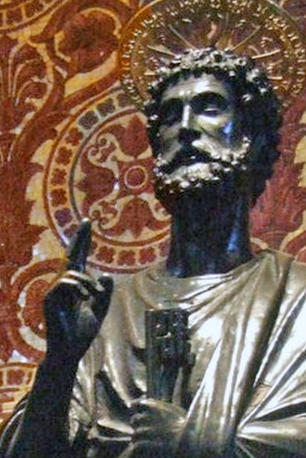
Also referred to as “benediction hand”, “Preacher`s hand”, “Papal Hand”, “Pope Blessing hand”, etc. This sign is linked to an ulnar nerve neuropathy, and there is a long time controversy about the name of this sign as well as its etiology, where some authors contend that there may be involvement of the median nerve.
Scholars, books and Internet sites are not clear about it. If there is an ulnar nerve neuropathy the person attempting to open his hand would be left with the second and third digits in extension, while the fourth and fifth digits would be flexed at the interphalangeal joints but extended at the metacarpophalangeal joints due the loss of the function of the interossei muscles and lumbrical muscles of the fourth and fifth digits.
The key for this sign is based in on the blessing act which is performed with an open hand and not with a fist. This is the reason why an ulnar neuropathy and not a median nerve neuropathy is the undelaying cause of the Papal Benediction Sign.
Presumably this term was the result of an injury of Saint Peter’s (the first Pope) ulnar nerve which caused him to bless using the “Preacher’s hand”. It stands to reason that everyone copied him as we can see in the lustrations and the art of the Catholic Church.
Article written by: Prof. Claudio R. Molina, MsC.
Sources:
1. Futterman, B. (2015). Analysis of the Papal Benediction Sign: The ulnar neuropathy of St. Peter. Clinical Anatomy (New York, N.Y.), 28(6), 696–701. Click here for the article
2. "From Vulcan Salute To Papal Blessing, Ulnar Nerve Damage Caused Original Benediction Sign | Box | NYIT". Nyit.edu. N.p., 2017. Web. 14 Jan. 2017
Image by By Mattana [Public domain], via Wikimedia Common: Click here for the link to the original image
- Details
Click for a larger image
We would like to welcome Professor Claudio R. Molina. MsC. as a contributor to Medical Terminology Daily.
Prof. Molina s a Physical Therapist, has a Masters degree in Biomedical Sciences, and is a Professor in the Human Anatomy Department of the Medical School at the Finis Terrae University in Santiago, Chile.
He is also a Postgraduate teacher for students of the “Anatomical Bases of Normal Imaging” diploma program at the Medical school of the same university.
Clinical Anatomy Associates, Inc is proud to have Dr. Cortés as a contributor to "Medical Terminology Daily" and as a consultant to our team.
- Details

Intended initially as a humorous view of anatomy, the "Anatomical Laws of Miranda" have a very serious objective. They support the fact that in every interventional case the operator should be extremely aware of the potential anatomical variations present. They also point to the fact that in real life, human anatomy does not look exactly like the anatomy books, models or prosections, and practice in the art of dissection and constant study are needed to ensure the proper identification of anatomical structures in surgery.
These laws are not original, they have been partially expressed at one time or another by several anatomists and surgeons, including Dr. Aaron Ruhalter and Dr. Robert Acland. What I have done is put them officially together and create the corollaries.
The anatomical laws of Miranda
- 1. The only constant in anatomy is variation
- 2. Nothing in the human body is really colored... or labeled
- 3. No anatomical structure has the moral obligation to be where they are supposed to be
There are corollaries to these laws and visitors to this web site are invited to provide us with their thoughts and addenda to the anatomical laws.
Corollaries:
1a. In the case of the so-called "anatomical constants"...law number 1 also applies
2.a. Black and white anatomy books are sometimes better to study than color atlases
2.b. Arteries are not red, nerves are not yellow, and lymphatic vessels are definitely not green!
2c. Nothing looks exactly like the anatomy books, computer simulations, or models. Food for thought for those medical schools that are eliminating dissection from their medical curricula.
3.a. This leads to that dreadful "Oooops!" sometimes heard in surgery
3.b. This also leads to the comment "It HAS to be around here!", which is dreadful if the "here" is a patient in surgery.
- Details
- Written by: Theo Dirix
This article is part of the series "A Moment in History" where we honor those who have contributed to the growth of medical knowledge in the areas of anatomy, medicine, surgery, and medical research.

Theo Dirix, Author and Taphophile
It is a truism that commemorations generate more attention for those being celebrated: since the quincentenary of 2014, the bibliography of the Flemish anatomist Andreas Vesalius (1514-1564) exceeds 3000 entries, and counting (1). Has the moment also come when hoaxes in his biography, some that were refuted over fifty or a hundred years ago, finally cease to circulate? (2) The Quest for his Lost Grave is entering a second crucial phase, but will we ever find his remains and learn the cause of his death?
There is a consensus of opinion that his early work "De Humani Corporis Fabrica Libri Septem" marks the transition to empiric research. His academic career and his advancement to the position of family physician at the court of Charles V, Emperor of the Holy Roman Empire, and at that of his successor Philip II, are well documented. His last months, days and moments become clearer too but obstinate pranks survive. Indeed, there is absolutely no proof that he ever ran into the otherwise so well documented Inquisition (3).
Recently rediscovered letters are evidence that Vesalius left Spain as a pious pilgrim: a laissez-passer by Philip II, notes from the Spanish Embassy in Venice and even the letter of thanks written by the Custodian of the Holy Places in Jerusalem, which Vesalius was to hand over to Philip II (4) The latter unequivocally refutes the other prank that a shipwreck during his return was the cause of his death.
In the running up to the quincentenary, medical artist, artisan and curator, Pascale Pollier has launched a romantic quest for his grave. Keen to make his facial reconstruction, she went looking for his cranium. When the Embassy of Belgium in Athens incorporated her project in its public diplomacy, the Quest had become cross-disciplinary.
First some contradictions about his final resting place had to be cleared up. Prominent Vesalius biographers, Omer Steeno, Maurits Biesbrouck and Theodoor Goddeeris have provided the research that convincingly points to the catholic church of Santa Maria delle Grazie in Zakynthos. Unfortunately the church, constructed in 1488, disappeared under the rubble of a major earthquake in 1953. The trio also documented the fact that several eyewitnesses had visited his sepulchre and copied the epitaph, Christoph Fürer von Haimendorf being the first in August 1565. In May 1566 Reiner Solenander quotes a merchant from Nuremberg who had been travelling with Vesalius. Is he the goldsmith or jeweller, mentioned in other literature? The grave was also seen in 1586 by Jean Zuallart and Filippo Pigafetta. As early as 1574 Johannes Sambucus states that Vesalius was buried in Zakynthos and in 1603 he added the name of the church: “D.[omus] Mariae” (5).
Once the spot had been defined, the research team, now calling itself Vesalius Continuum (6), turned to archaeologists: Prof. Jan Driessen, Université Catholique de Louvain (UCL) and Director of the Belgian School in Athens, EBSA, and Apostolos Sarris, Deputy Director of the Institute for Mediterranean Studies - Foundation for Research and Technology, Hellas (IMS-FORTH).
In 2014, Dr. Sylviane Déderix (UCL/IMS-FORTH) checked the presumed location of the church through the spatial analysis of a Geographical Information System (GIS). Her comparison of historical maps with modern cartographic data shows that the ruins are to be found on the northwest corner of the intersection of Kolyva Street and Kolokotroni Street, partly below the asphalt and partly under private property.
During construction works on that exact spot, funerary slabs have already been excavated, and provide yet further proof that there was a cemetery at this location. A geophysical approach to the further examination of anomalies under the surface is imperative. With the necessary official permission and funding, a team of researchers could collect and process data through non-destructive methods such as ground penetrating radar (GPR) and electrical resistivity tomography (ERT). If this was to prove conclusive, a third phase of small-scale excavations in search of remains may follow.
One of the unearthed funerary slabs dates from the sixteenth century: it belonged to a certain Bevilaqua who was given the position of Public Physician in 1593. Vesalius is not the only traveller who has been buried there. Other high profile guests may be Bishop Balthassar, Maria Remondini (1698-1777) and the French philhellene and author of acclaimed travel books, Pierre-Augustin Guys who was buried in the church on 27 September 1799.
It is obvious that if human remains were exhumed genetic identification is a must. Vesalius Continuum turned to Dr. Maarten Larmuseau of the Laboratory of Forensic Genetics and Molecular Archaeology of the KULeuven. He is a Specialist in the genetic identification of old-DNA and will compare potential mitochondrial DNA and/or Y-chromosomes of remains in the Santa Maria delle Grazie with those of living relatives who are in direct maternal or pattern line. In the case of Vesalius, his direct descendants, and those of his wife, cannot contribute to the identification, but maternal relatives of his mother, Elisabeth Crabbé, can.
This romantic quest for the lost bones of the father of modern anatomy, which has turned into a cross-disciplinary search, ostensibly does not end in death, but rather in curiosity, understanding, beauty, love, passion, life (7).
You too can join in the adventure by contributing to the crowd funding campaign to sponsor the next step in the archaeological campaign: www.gofundme.com/VesaliusContinuum
Note: This article was originally published in Theo Dirix's blog. Published here with his permission. Theo Dirix is a Vesaliana contributor to Medical Terminology Daily.
Sources:
1. Maurits Biesbrouck upgraded Dr. Harvey Cushing’s list of publications on Vesalius to more than 3000 records: http://www.andreasvesalius.be , accessed 8 January 2017.
2. DIRIX, Theo: Andreas Vesalius and his hoaxes, con variazioni, in: Vesalius, Journal of the International Society of the History of Medicine, Vol. XXII, nr. 1, June 2016, Special Issue, Proceedings of A Tribute to Andreas Vesalius, Padua, Italy - December 2015, pp. 103 - 111.
3. The source is post-mortem gossip spread in January 1565 by the French diplomat, Hubertus Languetus, in a note of 24 lines opening with: “rumour has it”. See: BIESBROUCK, Maurits, Theodoor GODDEERIS, Omer STEENO. ‘Post Mortem’ Andreae Vesalii (1514-1564), Deel I. De laatste reis van Andreas Vesalius en de omstandigheden van zijn dood), in: A.Vesalius, nr. 3 september 2015, Alfagen, Leuven, pp 154-161.
4. In total four letters have been discovered by José Baron Fernandez in the archives of Simancas, described and published since 1965, brought back to light by Steeno, Biesbrouck and Goddeeris.
5. Primary sources about the epitaphs are shown in:https://vimeo.com/album/4256560/video/190461188, accessed 15/01/2017
6. Within the initial ad hoc organising committee of the Vesalius Continuum Conference in September 2014 in Zakynthos, medical artist Pascale Pollier and the author, then Consul at the Embassy of Belgium in Athens, formed the Search team.
7. Closing lines of the “Conclusion, to be continued” in: DIRIX, Theo, In Search of Andreas Vesalius, The Quest for the Lost Grave, LannooCampus, Leuven, 2014, p.140.
- Details
- Written by: Fernanda Cortes, DDS, MSc

Click for a larger image
The temporal fascia (Lat:Fascia Temporalis) is thick and strong muscular (deep) fascia that covers the external surface of the temporal muscle.
It originates on a curved line formed by the posterosuperior part of the zygomatic bone, the temporal line of the frontal bone, the upper temporal line of the temporal bone and the area between both upper and lower temporal lines. It is divided in two laminae: superficial and deep which have insertion on the zygomatic arch. The deep portion provides insertion to the temporal muscle (1,2). The superficial layer is part of the epicraneal aponeurosis (3).
The two layers of the temporal fasica have separate arterial and venous blodd supply.
Article written by: Maria F. Cortés, DDS, MSc.
Images from:
Fig 1. Public domain, by Henry Vandyke Carter, MD - Gray's Anatomy, 1918
Sources:
1. “Anatomía humana” V.2. Latarjet- Ruiz Liard, 4ª ed. 6ª reimp. 2008 Médica Panamericana, Buenos Aires, Argentina.
2. “Anatomía humana: descriptiva, topográfica y funcional. Tomo 1. Cabeza y Cuello, Rouviere H – Delmas A, 11° ed. 2005 MASSON, S.A., Barcelona, Spain.
3. "Anatomy of the temporalis fascia" Wormald PJ, Alun-Jones T. J Laryngol Otol. 1991 Jul;105(7):522-4.
- Details
- Written by: Fernanda Cortes, DDS, MSc
Click for a larger image
The temporal muscle (Lat:Temporalis) is a bilateral muscle located on the side of the head. It belongs to a subgroup of head muscles called Masticatory Muscles, named after their function elevating the mandible to produce the mandible movements (1,2). Masticatory muscles are four per side: Temporalis, Masseter, Pterygoideus medialis and Pterygoideus lateralis (1,2).
The temporalis muscle is a fan-shaped muscle which occupies the temporal fossa from which its fascicles (fibers) converge to the coronoid process of the mandible. Classic description for this muscle recognizes three main muscular bodies (anterior, midle, and posterior) originated from the temporal fossa up to the lower temporalis line and the temporalis fascia, fascicles which descend through the inner part de the zygomatic arch converging to be inserted on the coronoid process of the mandible, its temporalis crest and anterior margin of the mandibular branch through thick tendons (1,2).
Click for a larger image
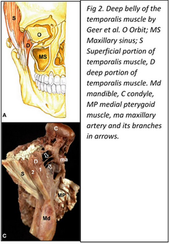
In 1996 Dunn et al. (3) reported the discovery of a so far unknown masticatory muscle called the “sphenomandibularis”, originated from the greater wing of the sphenoid bone medial to the temporalis muscle and descends on an oblique (lateral and slightly posterior) fashion reaching distally the coronoid process of the mandible. This muscular portion has been recognized as the “deep belly of the temporalis muscle” and has been described by several authors since then (4,5,6,8). The importance that has been given to this particular bundle lies on the fact that its medial insertion can reach a close relationship to the foramen rotundum, place of emergency from the cranium of the maxillary nerve, which has been hypothesized, could lead to eventual alteration of this nerve if it got trapped by this part of the muscle (6, 7).
The temporalis muscle receives innervation fundamentally from branches of the mandibular nerve: Deep temporal nerve (N. temporalis profundus)through its anterior middle and posterior branches.
The temporalis muscle is covered by a thick fascia layer: the temporalis fascia.
Article written by: Maria F. Cortés, DDS, MSc.
Images from:
Fig 1. Public domain, by Henry Vandyke Carter, MD - Gray's Anatomy, 1918
Fig 2. Geers C, Nyssen-Behets C, Cosnard G, Lengelé B. The deep belly of the temporalis muscle: an anatomical, histological and MRI study. Surg Radiol Anat. 2005 Aug;27(3):184-91. Epub 2005 Apr 9
Sources:
1. “Anatomía humana” V.2. Latarjet- Ruiz Liard, 4ª ed. 6ª reimp. 2008 Médica Panamericana, Buenos Aires, Argentina.
2. “Anatomía humana: descriptiva, topográfica y funcional. Tomo 1. Cabeza y Cuello, Rouviere H – Delmas A, 11° ed. 2005 MASSON, S.A., Barcelona, Spain.
3. Dunn GF, Hack GD, Robinson WL, Koritzer RT. Anatomical observation of a craniomandibular muscle originating from the skull base: the sphenomandibularis. Cranio. 1996 Apr;14(2):97-103; discussion 104-5.
4. Shimokawa T, Akita K, Soma K, Sato T. Innervation analysis of the small muscle bundles attached to the temporalis: truly new muscles or merely derivatives of the temporalis? Surg Radiol Anat. 1998;20(5):329-34.
5. Akita K, Shimokawa T, Sato T. Aberrant muscle between the temporalis and the lateral pterygoid muscles: M. pterygoideus proprius (Henle). Clin Anat. 2001 Jul;14(4):288-91.
6. Schön Ybarra MA, Bauer B. Medial portion of M. Temporalis and its potential involvement in facial pain. Clin Anat. 2001;14(1):25-30.
7. Fuentes E, Llanos S, Gómez R, Llanos P, Llanos F, Cortés-Sylvester MF, Solaria P, Melian A, Asfura J, Santos M, Zamorano E. Discovery of deep temporalis muscle belly close to maxillary nerve in a patient with trigeminal neuralgia: hypothesis of muscular compression and case report treated by Botox® Onabotulinum toxin tipe-A. Chirurgia 2016 June;29(3):99-102
8. Geers C, Nyssen-Behets C, Cosnard G, Lengelé B. The deep belly of the temporalis muscle: an anatomical, histological and MRI study. Surg Radiol Anat. 2005 Aug;27(3):184-91. Epub 2005 Apr 9.