![]()
Medical Terminology Daily (MTD) is a blog sponsored by Clinical Anatomy Associates, Inc. as a service to the medical community. We post anatomical, medical or surgical terms, their meaning and usage, as well as biographical notes on anatomists, surgeons, and researchers through the ages. Be warned that some of the images used depict human anatomical specimens.
You are welcome to submit questions and suggestions using our "Contact Us" form. The information on this blog follows the terms on our "Privacy and Security Statement" and cannot be construed as medical guidance or instructions for treatment.
We have 346 guests and no members online
")
Marcia Crocker Noyes
(1869 – 1946)
Further to my comment on old books and research that started with an interesting bookplate (Ex-Libris). I continued my research and found that the person in charge of the Osler library bookplate was a fascinating individual that today maybe a ghost in the MedChi library and building in Baltimore... This is certainly an article that can be called "A Moment in History"
Marcia Crocker Noyes was the librarian at The Maryland State Medical Society from 1896 to 1946 and was a founding member of the Medical Library Association.[1][2][3]
Sir William Osler, MD. a famous Johns Hopkins surgeon was a noted bibliophile and had a large personal collection of books on various topics. When he became the President of MedChi in 1896, he was dismayed at the condition of the library and knew that with the right person and some stewardship, it could become a significant collection. Sir William asked his friend, Dr. Bernard Steiner, a physician and President of the Enoch Pratt Free Library in Baltimore for suggestions of a librarian, and Dr. Steiner recommended Marcia Crocker Noyes. A native of New York, and a graduate of Hunter College, Marcia had moved to Baltimore for a lengthy visit with her sister, and took a “temporary” position at the Pratt Library, which turned into three years. Although she had no medical experience or background, she was enthusiastic, and most importantly, she was willing to move into the apartment provided for the librarian, who needed to be available 24 hours a day.
The image in this article is Ms. Noyes on her first year on the job. Marcia developed a book classification system for medical books, based on the Index Medicus, and called it the Classification for Medical Literature. The system uses the alphabet with capital letters for the major divisions of medicine and lower-case ones for the sub-sections. The system was used for many years, but it's now dated and the Faculty's original shelving scheme was never changed. The card catalogs still reflect her classification and many of the cards are written in Marcia's back-slanting handwriting.
Marcia knew enough to ask the Faculty's members about medical questions, terminology and literature. She gradually won over the predominantly male membership and they became her greatest allies; Sir William at the start, and then for nearly 40 years, Dr. John Ruhräh, a wealthy pediatrician with no immediate family of his own. She made a point of attending almost every Faculty function, and in 1904, under guidelines from the American Medical Association, Marcia was made the Faculty Secretary. For much of her first 10 years, she was the Faculty's only full-time employee, only being assisted by Mr. Caution, the Faculty's janitor. Later in life Marcia would say that she hired him because of his name!
Within ten years, the library had outgrown its space, and plans, spearheaded by Marcia and Sir William before his move to Oxford, were made to build a headquarters building, mainly to house the library's growing collection of medical books and journals.
Marcia was instrumental in the design and building of the new headquarters. She travelled to Philadelphia, New York and Boston to look at their medical society buildings, and eventually, the Philadelphia architectural firm, Ellicott & Emmart was selected to design and build the new Faculty building. Every detail of the building held her imprimatur, from the graceful staircase, to the light-filled reading room, and all of the myriad details of the millwork, marble tesserae, and most of all, the four-story cast iron stacks. She was on-site, climbing up unfinished staircases, checking out the progress of the building, which was built in less than one year at a cost of $90,000.
Among the features of the new building was a fourth-floor apartment for her. She referred to it as the "first penthouse in Baltimore" and it had a garden and rooftop terrace. The library collection eventually grew to more than 65,000 volumes from medical and specialty societies around the world. Journals were traded back and forth, and physicians eagerly anticipated the arrival of each new issue. At the same time, Marcia was involved in the Medical Library Association as one of eight founding members. The MLA promotes medical libraries and the exchange of information. One of the earliest mandates of the MLA was the Exchange, a distribution and trade service for those who had duplicates or little-used books in their collections. Initially, the Exchange was run out of the Philadelphia medical society, but in 1900 it was moved to Baltimore and Marcia oversaw it. Several hundred periodicals and journals were received and sent each month, a huge amount of work for a tiny staff. In 1904, the Faculty had run out of room to manage the Exchange, so it was moved to the Medical Society of the Kings County (Brooklyn). But without Marcia's excellent administrative skills, it floundered and in 1908, the MLA asked Marcia to take charge once again.
In 1909, when the new Faculty building opened, there was enough room to run the Exchange and with the help of MLA Treasurer, noted bibliophile and close friend, Dr. John Ruhräh, it once again became successful. Additionally, Marcia and Dr. Ruhräh combined forces to revive the MLA's bulletin, which had all but ceased publication in 1908, taking the Exchange with it. This duo maintained editorial control from 1911 until 1926. In 1934, around the time of Dr. Ruhräh's death, Marcia became the first “unmedicated” professional to head the MLA. During her tenure, the MLA incorporated, the first seal was adopted, and the annual meeting was held in Baltimore. Marcia wanted to write the history of the MLA once she retired from full-time work at the Faculty, but her health was beginning to fail. She had back problems and had suffered a serious burn on her shoulder as a young woman, possibly from her time running a summer camp, Camp Seyon, for young ladies in the Adirondack Mountains. In 1946, a celebration was planned to honor Marcia's 50 years at the Faculty. But she was adamant that the physicians wait until November, the actual date of her 50 years. However, they knew she was gravely ill, and might not make it until then, so a huge party was held in April. More than 250 physicians attended the celebration, but the ones she was closest to in the early years, were long gone. She was presented with a suitcase, a sum of money to use for travelling, and her favorite painting of Dr. John Philip Smith, a founder of the Medical College in Winchester, Virginia. It was painted by Edward Caledon Smith, a Virginia painter who had been a student of the painter Thomas Sully.[4] She adored this painting and vowed, jokingly, to take it with her wherever she went.
The painting was not to stay with her for very long, for she died in November 1946, and left it to the Faculty in her will. Her funeral was held in the Faculty's Osler Hall, named for her dear friend. More than 60 physicians served as her pallbearers, and she was buried at Baltimore's Green Mount Cemetery. In 1948, the MLA decided to establish an award in the name of Marcia Crocker Noyes. It was for outstanding achievement in medical library field and was to be awarded every two years, or when a truly worthy candidate was submitted. In 2014, the Faculty began giving a bouquet of flowers to the winner of the award in Marcia's name, and in honor of her work. Much evidence exists for this tradition, as we know that the physicians, especially Drs. Osler and Ruhräh, frequently gave her bouquets of flowers. Marcia also cultivated flower gardens at the Faculty and decorated the rooms with her work.
Today, the MedChi building is open for tours and if the rumors are to be believed Ms. Marcia Crocker Noyes is still at work in her beloved library as the "resident ghost" [1][5]

NOTE: This article has been modified from the original Wikipedia article on Marcia Crocker Noyes. The article itself is well-written with interesting images of the subject. I would encourage you to visit it. The second insert is from book 00736 in my personal library and shows in pencil, the incredibly small handwriting of Marsha C. Noyes.
Sources:
1. "Marcia, Marcia, Marcia" MedChi Archives blog.
2. "Marcia C. Noyes, Medical Librarian" (PDF). Bulletin of the Medical Library Association. 35 (1): 108–109. 1947. PMC 194645
3. Smith, Bernie Todd (1974). "Marcia Crocker Noyes, Medical Librarian: The Shaping of a Career" (PDF). Bulletin of the Medical Library Association. 62 (3): 314–324. PMC 198800Freely accessible. PMID 4619344.
4. Edward Caledon BRUCE (1825-1901)"
5. Behind the scenes tour MedChiBuilding
"Clinical Anatomy Associates, Inc., and the contributors of "Medical Terminology Daily" wish to thank all individuals who donate their bodies and tissues for the advancement of education and research”.
Click here for more information


- Details
HAPPY VALENTINE'S DAY!
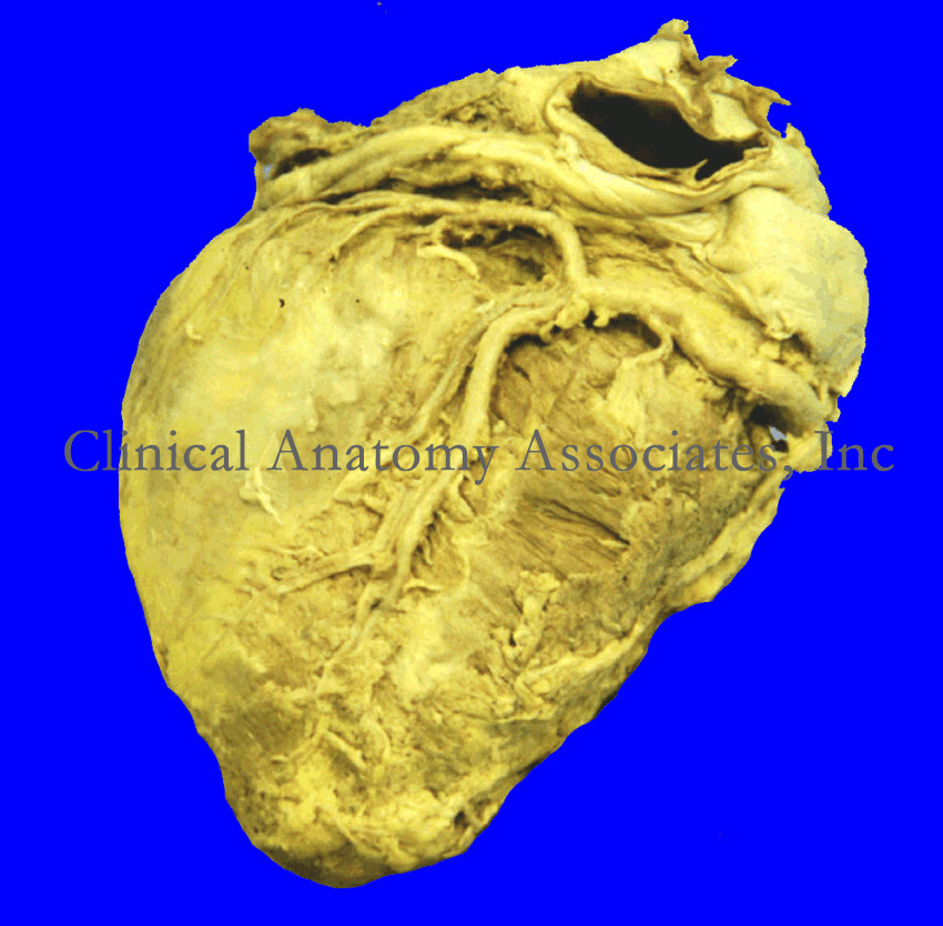
Click for a larger image
For years I tried to understand from where did the classical Valentine's Day heart shape(❤) came. It was not until I was observing a human heart from the posteroinferior aspect, the diaphragmatic surface of the heart that I realized I was looking at it! You see, in the early days of anatomy, the heart was considered to be the ventricles only. This lead to consider the chambers at the "entrance" to the heart to be called "atria". This image of the heart is only valid if you make abstraction of the atria and look at the ventricles only.
Of course, there is a view of the heart, a cross section where you can see all four chambers of the heart. This four-chambered view is called by some the "Valentine's view".
I like my interpretation better. At least now you know that when you draw a [❤] you are being anatomically correct! It is only the ventricles of the heart in a posteroinferior view. Happy Valentine's Day! Dr. Miranda
Image property of:CAA.Inc.Photographer:David M. Klein
- Details
Click for a larger image
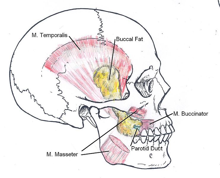
The [buccal fat pad] is dense, fatty trigone-shaped pad that is located in the cheek. It is formed by several connective-tissue encapsulated smaller fat pads. It covers partially the posterior aspect of the buccinator muscle, and is found deep to the anterior portion of the masseter muscle. Also known as “Bichat’s fat pad”, it was first described by Marie-François Xavier Bichat in 1802. It is also known as the suctorial fat pad and it helps in the suction process for breast feeding in infants, although because of its location it is also said to help in the gliding motion of the masticatory and facial expression muscles. The buccal fat pad is well developed in newborns and is not as evident in most adults.
Its anatomical description varies according to the authors, but it has a main body and three extensions, namely the anteromalar (anterior), pterygomaxillary (pterygoid), and temporal (posterotemporal) extensions. The blood supply to the buccal fat pad is by way of the anterior deep temporal, buccal, and posterior superior alveolar arteries.
Excessive development of this fat pad can lead to cosmetic surgery to eliminate, or at least reduce its size. This procedure is known in many countries as a “bichectomy”, Bichatectomy” of “cheek reduction surgery”, in some cases this procedure can be performed intraorally.
The buccal fat pad can also be used in maxillofacial reconstructive surgery, as well as the repair of skull base defects. When dissecting the buccal fat pad, care must be taken because of the relation of this structure with the parotid duct, the parotid gland, and branches of the facial nerve
Sources:
1. “Anatomy of the buccal fat pad and its clinical significance” Jackson, IT Plastic and Reconstructive Surgery, 06/1999, Volume 103, Issue 7
2. "A review of the gross anatomy, functions, pathology, and clinical uses of the buccal fat pad" Yousuf, S et al Surg Radiol Anat (2010) 32:427–436
3. "The Endonasal Endoscopic Harvest and Anatomy of the Buccal Fat Pad Flap for Closure of Skull Base Defects" Markey, J et al The Laryngoscope 125: 2247-2252
4. "Bichectomy or Bichatectomy - A Small and Simple Intraoral Surgical Procedure with Great Facial Results" Eber Luis de L S. Adv Dent & Oral Health. 2015; 1(1): 555555. DOI: 10.19080/ADOH.2015.01.555555.
5. "Tratado de Anatomia Humana" Testut et Latarjet 8th Ed. 1931 Salvat Editores, Spain
Image: By Otto Placik (Own work) [CC BY-SA 3.0 (http://creativecommons.org/licenses/by-sa/3.0) or GFDL (http://www.gnu.org/copyleft/fdl.html)], via Wikimedia Commons] Click here for the link to the original image
- Details
The term [quadratus] is Latin and means “square”, it is also the root of the Spanish word “cuadrado”.
It is used to several structures in the body that are of course, square, or at least square-like:
• Quadratus lumborum muscle
• Quadratus femoris muscle
• Pronator quadratus muscle
These muscles will be described in separate articles
Sources: 1. “Understanding Anatomical Terms” Mehta, LA, et al. Clin Anat 9:330-336 (1996)
- Details
The term [suctorial] is based on the Latin [sugere] meaning “sucking” and evolves to modern Latin as the term [suctorius]. It is the base for the English verb “to suck”, the Spanish term [succionar], and the French [succion].
It is used to describe the small fat pad that covers the external and posterior aspect of the buccinator muscle. Also known as Bichat’s fat pads, these pads are said to help the suction activity of the baby. As they are not needed later in life, they tend to reduce in size.
Sources:
1. “Anatomy of the buccal fat pad and its clinical significance” Jackson, IT Plastic and Reconstructive Surgery, 06/1999, Volume 103, Issue 7
2. “Gray’s Anatomy” Henry Gray, 1918
3. “Understanding Anatomical Terms” Mehta, LA, et al. Clin Anat 9:330-336 (1996)
- Details
Click for a larger image
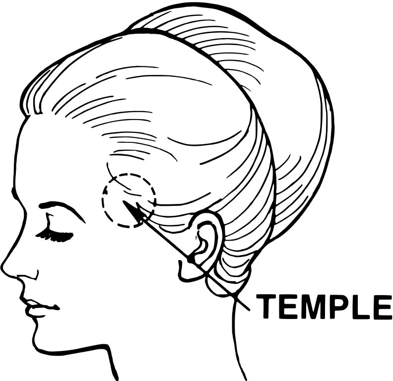
The etymology (origin) of the term [temporal] is Latin and derives from [tempus and temporis] meaning "time". It is said that the name was given to the area of the head where the hair initially starts to become gray and whitish, marking the "passage of time". Many philosophists and scholars have said that "life is only temporal".
Anatomically, the [temple] is the area of the temporal region. The word "temple" as used in anatomy has a separate etymology from the word temple, used as "place of worship". Both come from Latin, but the word for the place of worship comes from [templum] meaning a "shrine" or a "place of worship".
Use of the term [temporal] is found in:
• Temporal bone
• Temporal muscle
• Temporal fascia
Image: By Pearson Scott Foresman [Public domain], via Wikimedia Commons
Click here for the link to the original image
- Details
This article is part of the series "A Moment in History" where we honor those who have contributed to the growth of medical knowledge in the areas of anatomy, medicine, surgery, and medical research.

Thomas Willis
UPDATED: Thomas Willis (1621-1675). An English physician and anatomist, Willis was born on his parents' farm in Great Bedwyn, Wiltshire, where his father held the stewardship of the Manor. He was a kinsman of the Willys baronets of Fen Ditton, Cambridgeshire. He graduated M.A. from Christ Church, Oxford in 1642. In the Civil War years he was a royalist, and was dispossessed of the family farm at North Hinksey by Parliamentary forces. In the 1640's Willis was one of the royal physicians to Charles I of England. He obtained his medical degree in 1646.
Thomas Willis might well be one of the greatest physicians of the 17th century.He is one of the founders of the Royal Society of London. He is remembered by his many publications, especially "Cerebri Anatome: Cui accessit Nervorum Descriptio et Usu", where he describes the arterial anastomoses at the base of the brain. This work is also the first detailed description of the vasculature of the brain. Willis described nine cranial nerves.
He is considered as the father of Neurology as a discipline. He used the term "neurology" for the first time in 1664. He described several neurological conditions
The Arterial Circle of Willis is a famous eponymous structure found at the base of the brain. It represents an anastomotic roundabout that connects the right and left sides as well as the carotid and vertebral arterial territories that supply the brain. Named after Thomas Willis, this structure was known well before him, but it was Willis who described its function. You will be redirected to a detailed description of this structure if you click here.
Sources:
1. "The legendary contributions of Thomas Willis (1621-1675): the arterial circle and beyond" Rengachary SS et al J Neurosurg. 2008 Oct;109(4):765-75
2. "Thomas Willis, a pioneer in translational research in anatomy (on the 350th anniversary of Cerebri anatome)" Arraez-AybarJournal of Anatomy, 03/2015, Volume 226, Issue 3
3. " The naming of the cranial nerves: A historical review" Davis, M Clinical Anatomy, 01/2014, Volume 27, Issue 1
4. "Observations on the history of the circle of Willis". Meyer A, Hieros, R.Med Hist 6:119–130, 1962
Original image in the public domain courtesy of the National Library of Medicine.