![]()
Medical Terminology Daily (MTD) is a blog sponsored by Clinical Anatomy Associates, Inc. as a service to the medical community. We post anatomical, medical or surgical terms, their meaning and usage, as well as biographical notes on anatomists, surgeons, and researchers through the ages. Be warned that some of the images used depict human anatomical specimens.
You are welcome to submit questions and suggestions using our "Contact Us" form. The information on this blog follows the terms on our "Privacy and Security Statement" and cannot be construed as medical guidance or instructions for treatment.
We have 759 guests online
")
Marcia Crocker Noyes
(1869 – 1946)
Further to my comment on old books and research that started with an interesting bookplate (Ex-Libris). I continued my research and found that the person in charge of the Osler library bookplate was a fascinating individual that today maybe a ghost in the MedChi library and building in Baltimore... This is certainly an article that can be called "A Moment in History"
Marcia Crocker Noyes was the librarian at The Maryland State Medical Society from 1896 to 1946 and was a founding member of the Medical Library Association.[1][2][3]
Sir William Osler, MD. a famous Johns Hopkins surgeon was a noted bibliophile and had a large personal collection of books on various topics. When he became the President of MedChi in 1896, he was dismayed at the condition of the library and knew that with the right person and some stewardship, it could become a significant collection. Sir William asked his friend, Dr. Bernard Steiner, a physician and President of the Enoch Pratt Free Library in Baltimore for suggestions of a librarian, and Dr. Steiner recommended Marcia Crocker Noyes. A native of New York, and a graduate of Hunter College, Marcia had moved to Baltimore for a lengthy visit with her sister, and took a “temporary” position at the Pratt Library, which turned into three years. Although she had no medical experience or background, she was enthusiastic, and most importantly, she was willing to move into the apartment provided for the librarian, who needed to be available 24 hours a day.
The image in this article is Ms. Noyes on her first year on the job. Marcia developed a book classification system for medical books, based on the Index Medicus, and called it the Classification for Medical Literature. The system uses the alphabet with capital letters for the major divisions of medicine and lower-case ones for the sub-sections. The system was used for many years, but it's now dated and the Faculty's original shelving scheme was never changed. The card catalogs still reflect her classification and many of the cards are written in Marcia's back-slanting handwriting.
Marcia knew enough to ask the Faculty's members about medical questions, terminology and literature. She gradually won over the predominantly male membership and they became her greatest allies; Sir William at the start, and then for nearly 40 years, Dr. John Ruhräh, a wealthy pediatrician with no immediate family of his own. She made a point of attending almost every Faculty function, and in 1904, under guidelines from the American Medical Association, Marcia was made the Faculty Secretary. For much of her first 10 years, she was the Faculty's only full-time employee, only being assisted by Mr. Caution, the Faculty's janitor. Later in life Marcia would say that she hired him because of his name!
Within ten years, the library had outgrown its space, and plans, spearheaded by Marcia and Sir William before his move to Oxford, were made to build a headquarters building, mainly to house the library's growing collection of medical books and journals.
Marcia was instrumental in the design and building of the new headquarters. She travelled to Philadelphia, New York and Boston to look at their medical society buildings, and eventually, the Philadelphia architectural firm, Ellicott & Emmart was selected to design and build the new Faculty building. Every detail of the building held her imprimatur, from the graceful staircase, to the light-filled reading room, and all of the myriad details of the millwork, marble tesserae, and most of all, the four-story cast iron stacks. She was on-site, climbing up unfinished staircases, checking out the progress of the building, which was built in less than one year at a cost of $90,000.
Among the features of the new building was a fourth-floor apartment for her. She referred to it as the "first penthouse in Baltimore" and it had a garden and rooftop terrace. The library collection eventually grew to more than 65,000 volumes from medical and specialty societies around the world. Journals were traded back and forth, and physicians eagerly anticipated the arrival of each new issue. At the same time, Marcia was involved in the Medical Library Association as one of eight founding members. The MLA promotes medical libraries and the exchange of information. One of the earliest mandates of the MLA was the Exchange, a distribution and trade service for those who had duplicates or little-used books in their collections. Initially, the Exchange was run out of the Philadelphia medical society, but in 1900 it was moved to Baltimore and Marcia oversaw it. Several hundred periodicals and journals were received and sent each month, a huge amount of work for a tiny staff. In 1904, the Faculty had run out of room to manage the Exchange, so it was moved to the Medical Society of the Kings County (Brooklyn). But without Marcia's excellent administrative skills, it floundered and in 1908, the MLA asked Marcia to take charge once again.
In 1909, when the new Faculty building opened, there was enough room to run the Exchange and with the help of MLA Treasurer, noted bibliophile and close friend, Dr. John Ruhräh, it once again became successful. Additionally, Marcia and Dr. Ruhräh combined forces to revive the MLA's bulletin, which had all but ceased publication in 1908, taking the Exchange with it. This duo maintained editorial control from 1911 until 1926. In 1934, around the time of Dr. Ruhräh's death, Marcia became the first “unmedicated” professional to head the MLA. During her tenure, the MLA incorporated, the first seal was adopted, and the annual meeting was held in Baltimore. Marcia wanted to write the history of the MLA once she retired from full-time work at the Faculty, but her health was beginning to fail. She had back problems and had suffered a serious burn on her shoulder as a young woman, possibly from her time running a summer camp, Camp Seyon, for young ladies in the Adirondack Mountains. In 1946, a celebration was planned to honor Marcia's 50 years at the Faculty. But she was adamant that the physicians wait until November, the actual date of her 50 years. However, they knew she was gravely ill, and might not make it until then, so a huge party was held in April. More than 250 physicians attended the celebration, but the ones she was closest to in the early years, were long gone. She was presented with a suitcase, a sum of money to use for travelling, and her favorite painting of Dr. John Philip Smith, a founder of the Medical College in Winchester, Virginia. It was painted by Edward Caledon Smith, a Virginia painter who had been a student of the painter Thomas Sully.[4] She adored this painting and vowed, jokingly, to take it with her wherever she went.
The painting was not to stay with her for very long, for she died in November 1946, and left it to the Faculty in her will. Her funeral was held in the Faculty's Osler Hall, named for her dear friend. More than 60 physicians served as her pallbearers, and she was buried at Baltimore's Green Mount Cemetery. In 1948, the MLA decided to establish an award in the name of Marcia Crocker Noyes. It was for outstanding achievement in medical library field and was to be awarded every two years, or when a truly worthy candidate was submitted. In 2014, the Faculty began giving a bouquet of flowers to the winner of the award in Marcia's name, and in honor of her work. Much evidence exists for this tradition, as we know that the physicians, especially Drs. Osler and Ruhräh, frequently gave her bouquets of flowers. Marcia also cultivated flower gardens at the Faculty and decorated the rooms with her work.
Today, the MedChi building is open for tours and if the rumors are to be believed Ms. Marcia Crocker Noyes is still at work in her beloved library as the "resident ghost" [1][5]

NOTE: This article has been modified from the original Wikipedia article on Marcia Crocker Noyes. The article itself is well-written with interesting images of the subject. I would encourage you to visit it. The second insert is from book 00736 in my personal library and shows in pencil, the incredibly small handwriting of Marsha C. Noyes.
Sources:
1. "Marcia, Marcia, Marcia" MedChi Archives blog.
2. "Marcia C. Noyes, Medical Librarian" (PDF). Bulletin of the Medical Library Association. 35 (1): 108–109. 1947. PMC 194645
3. Smith, Bernie Todd (1974). "Marcia Crocker Noyes, Medical Librarian: The Shaping of a Career" (PDF). Bulletin of the Medical Library Association. 62 (3): 314–324. PMC 198800Freely accessible. PMID 4619344.
4. Edward Caledon BRUCE (1825-1901)"
5. Behind the scenes tour MedChiBuilding
"Clinical Anatomy Associates, Inc., and the contributors of "Medical Terminology Daily" wish to thank all individuals who donate their bodies and tissues for the advancement of education and research”.
Click here for more information


- Details

From the Greek word [stoma] meaning "mouth or opening", and the suffix [-y] meaning "process or condition". The suffix [-(o)stomy] refers to the "process of creating an opening". This process can be physiological, without intervention, as in the creation of a spontaneous fistula, or it can be a surgical procedure.
As a working explanation of [-ostomy] in surgery, we like to use the term "drainage". Therefore, an [ileostomy] would be the procedure by means of which a drainage opening is creating an anastomosis between the ileum and the abdominal wall.
The accompanying image shows an early 1900's procedure to create a gastrostomy (Wietzel's gastrostomy). The root term [gastr-] means "stomach".
- Details
Click for a larger image
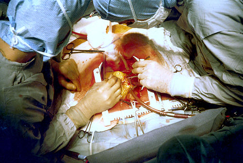
From the Greek [kheirurgia], a compound word meaning "a work done by hand". The Greek word [kheir/cheir] means "hand", and [ergon] means "work". The intent of the word is that of a medical treatment that is realized by the use of the hands and/or hand instrumentation.
Technology has advanced the evolution of surgery. Today minimally invasive surgical procedures, videoscopic procedures, and robotic-enhanced surgery are commonplace.
Images and links in the public domain, courtesy of:www.wikipedia.com
- Details
")
Click for a larger image
The arcuate line is the arch-shaped (hence the name) inferior border of the posterior sheath of the rectus abdominis muscle. This structure is seen in a laparoscopic (posterior) view (see image, label "B") and represents the transition from a superior area with well-formed aponeurotic posterior rectus sheath to an area devoid of the posterior rectus sheath.
At this point, the inferior (deep) epigastric vessels (see image, label "C") pass from deep to superficial, under the arcuate line and continue superiorly providing blood to the rectus abdominis muscle.
The arcuate line also represents a transition from a well-formed and stronger wall posterior to the rectus abdominis muscle to a weaker region, covered only by deep muscle fascia and transversalis fascia. This allows a surgeon to enter the preperitoneal region using a Totally Extraperitoneal (TEP) approach for a laparoscopic herniorrhaphy.
Label "A" shows the "corona mortis" anatomical variation.
Image property of: CAA.Inc. Artist: M. Zuptich
- Details

Click for a larger image
The inguinal (Poupart's) ligament has always been described as a separate, discrete, distinctive ligamentous structure. This is not so. The inguinal ligament is the thickened, incurved, lower free border of the external oblique aponeurosis. This structure extends between the anterior superior iliac spine (ASIS) superolaterally, and the pubic tubercle inferomedially. The inferomedial portion of the inguinal ligament send fibers towars the pectineal ligament (Cooper's ligament) and forms the lacunar (Gimbernat's) ligament.
Inferior to the inguinal ligament is an open region (subinguinal space) that allows passage of structures between the abdominopelvic region and the femoral region. Some of these structures are: Iliacus muscle, psoas major muscle.
Although described by Vesalius, Fallopius, and others it was the French anatomist and surgeon Francois Poupart (1661-1708) who described this structure in relation to hernia in his book "Chirurgie Complete" published in 1695.
Image property of: CAA.Inc. Artist: D.M. Klein
- Details
The suffix [-itis] originates from the Greek and means "inflammation". This suffix is also used to mean "infection", although inflammation is only one of the signs of infection. The symptoms and signs of infection are:
- Edema - localized swelling (tumor)
- Redness- Localized (rubor)
- Localized raise in temperature - Fever (calor)
- Pain - (dolor)
- Localized functional impairment (functio lesa)
The terms in parentheses are the Latin words used to describe these symptoms and sign.
Examples of uses of this suffix are:
- Hepatitis: Inflammation or infection of the liver
- Pancreatitis: Inflammation or infection of the pancreas
- Cholecystitis: Inflammation or infection of the gallbladder [chole-]="gall'; [cyst]="sac" or "bladder"
- Rhinitis: Inflammation or infection of the nose
- Pharyngotracheitis: Inflammation or infection of the pharynx and trachea
- Details
The suffix [-oid] originates from the Greek [oeides], meaning "similar to", "like", or "shaped like". This suffix can be found the the medical terms [sigmoid] meaning "similar or shaped like a sigma"; [sphenoid], meaning "shaped like a wedge"; [cricoid], meaning "shaped like a ring", and [arytenoid] also from the Greek [arytaina], meaning "similar to a ladle".
This suffix is also used in daily conversation, as the following examples illustrate:
- Android - "similar to a human", from the Greek [andros] human
- Anthropoid - similar to a man, from the Greek [anthropos], "man"
- Asteroid - "similar to a star", from the Greek [aster], "star"
- Arachnoid - "similar to a spider", from the latin [arachnid], spider. It refers to the spider-web look of this menynx