Medical Terminology Daily (MTD) is a blog sponsored by Clinical Anatomy Associates, Inc. as a service to the medical community. We post anatomical, medical or surgical terms, their meaning and usage, as well as biographical notes on anatomists, surgeons, and researchers through the ages. Be warned that some of the images used depict human anatomical specimens.
You are welcome to submit questions and suggestions using our "Contact Us" form. The information on this blog follows the terms on our "Privacy and Security Statement" and cannot be construed as medical guidance or instructions for treatment.
We have 779 guests and no members online

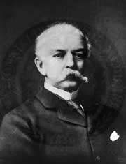
Jean George Bachmann
(1877 – 1959)
French physician–physiologist whose experimental work in the early twentieth century provided the first clear functional description of a preferential interatrial conduction pathway. This structure, eponymically named “Bachmann’s bundle”, plays a central role in normal atrial activation and in the pathophysiology of interatrial block and atrial arrhythmias.
As a young man, Bachmann served as a merchant sailor, crossing the Atlantic multiple times. He emigrated to the United States in 1902 and earned his medical degree at the top of his class from Jefferson Medical College in Philadelphia in 1907. He stayed at this Medical College as a demonstrator and physiologist. In 1910, he joined Emory University in Atlanta. Between 1917 -1918 he served as a medical officer in the US Army. He retired from Emory in 1947 and continued his private medical practice until his death in 1959.
On the personal side, Bachmann was a man of many talents: a polyglot, he was fluent in German, French, Spanish and English. He was a chef in his own right and occasionally worked as a chef in international hotels. In fact, he paid his tuition at Jefferson Medical College, working both as a chef and as a language tutor.
The intrinsic cardiac conduction system was a major focus of cardiovascular research in the late nineteenth and early twentieth centuries. The atrioventricular (AV) node was discovered and described by Sunao Tawara and Karl Albert Aschoff in 1906, and the sinoatrial node by Arthur Keith and Martin Flack in 1907.
While the connections that distribute the electrical impulse from the AV node to the ventricles were known through the works of Wilhelm His Jr, in 1893 and Jan Evangelista Purkinje in 1839, the mechanism by which electrical impulses spread between the atria remained uncertain.
In 1916 Bachmann published a paper titled “The Inter-Auricular Time Interval” in the American Journal of Physiology. Bachmann measured activation times between the right and left atria and demonstrated that interruption of a distinct anterior interatrial muscular band resulted in delayed left atrial activation. He concluded that this band constituted the principal route for rapid interatrial conduction.
Subsequent anatomical and electrophysiological studies confirmed the importance of the structure described by Bachmann, which came to bear his name. Bachmann’s bundle is now recognized as a key determinant of atrial activation patterns, and its dysfunction is associated with interatrial block, atrial fibrillation, and abnormal P-wave morphology. His work remains foundational in both basic cardiac anatomy and clinical electrophysiology.
Sources and references
1. Bachmann G. “The inter-auricular time interval”. Am J Physiol. 1916;41:309–320.
2. Hurst JW. “Profiles in Cardiology: Jean George Bachmann (1877–1959)”. Clin Cardiol. 1987;10:185–187.
3. Lemery R, Guiraudon G, Veinot JP. “Anatomic description of Bachmann’s bundle and its relation to the atrial septum”. Am J Cardiol. 2003;91:148–152.
4. "Remembering the canonical discoverers of the core components of the mammalian cardiac conduction system: Keith and Flack, Aschoff and Tawara, His, and Purkinje" Icilio Cavero and Henry Holzgrefe Advances in Physiology Education 2022 46:4, 549-579.
5. Knol WG, de Vos CB, Crijns HJGM, et al. “The Bachmann bundle and interatrial conduction” Heart Rhythm. 2019;16:127–133.
6. “Iatrogenic biatrial flutter. The role of the Bachmann’s bundle” Constán E.; García F., Linde, A.. Complejo Hospitalario de Jaén, Jaén. Spain
7. Keith A, Flack M. The form and nature of the muscular connections between the primary divisions of the vertebrate heart. J Anat Physiol 41: 172–189, 1907.
"Clinical Anatomy Associates, Inc., and the contributors of "Medical Terminology Daily" wish to thank all individuals who donate their bodies and tissues for the advancement of education and research”.
Click here for more information
- Details
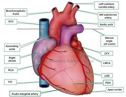
The aortic arch is a segment of the aorta that arches from the midline towards posterior and to the left. It presents with three branches. From proximal to distal they are the brachiocephalic trunk, the left common carotid artery, and the left subclavian artery. There are several anatomical variations of the branches of the aortic arch.
There is no clear anatomical landmark to denote the ending of the ascending aorta and the beginning of the aortic arch, as there is no clear anatomical landmark to denote the ending of the aortic arch and the beginning of the descending aorta. Anatomists use as a reference a horizontal plane that passes through the angle of Louis. Since this plane also separates the inferior from the superior mediastinum, the aortic arch is found in the superior mediastinum, while the ascending and descending aorta are found in the inferior mediastinum.
The aortic arch has anatomical relations with the bifurcation of the trachea, the pulmonary trunk and its bifurcation, and the left brachiocephalic vein. In its inferior surface, the aortic arch in the adult has the embryological remnant of the ductus arteriousus, called the ligamentum arteriosum.
The term "aortic arch" was coined and first used by Lorenz Heister (1683 1785)
Image property of: CAA.Inc.. Artist: Victoria G. Ratcliffe
- Details
This is a medical root term that originates from the Greek and means "vessel", as in a "container". The term is commonly misunderstood to mean "artery". The original meanings of the term in early Greek and Roman medicine where multiple. It was Lorenz Heister (1683-1758) who first used the term in its modern meaning. Applications of this root term include:
- Angiology: Study of vessels
- Angioma: Vessel tumor or mass, usually referred to a malformation of knotted vessels. The plural form is "angiomata"
- Angioplasty: Reshaping of a vessel
- Angiitis: Inflammation of a vessel. Note the double "i" in the word. This is the correct form of the term. "Angitis" is not correct!
- Angiogenesis: Creation or generation of vessels
- Neoangiogenesis: The prefix [neo-] means [new], therefore the term means "creation or generation of new vessels"
- Cholangiogram: The prefix [chol-] means "bile", while the suffix [-ogram] means "examination of". A [cholangiogram] is the "examination of a bile vessel"
- Cineangiogram: The prefix [cine-] means "movement", although we use it to mean "movie", while the suffix [-ogram] means "examination of". A [cineangiogram] is the "examination of a vessel in movement"
- Details
This is a medical root term that originates from the Greek "arthron" which means "joint". The term is used in many medical words. Applications of this root term include:
- Arthrotomy: Opening of a joint
- Arthritis: Inflammation (or infection) of a joint
- Arthrology: Study of a joint
- Arthrodesis: Fixation of a joint
- Arthropathy: A disease affecting a joint
- Arthroplasty: Reshaping of a joint
- Arthroscopy: Visualizing inside a joint with a scope
As a side note: What is the plural form for arthritis? Hover your cursor over the word to see the answer
- Details
This article is part of the series "A Moment in History" where we honor those who have contributed to the growth of medical knowledge in the areas of anatomy, medicine, surgery, and medical research.
Charles H. McBurney, MD (1845- 1913). British surgeon and anatomist, Dr. McBurney studied at Harvard University, and received his MD from the Colombia University in New York. At the forefront of the aseptic technique revolution, Dr. MacBurney, following Halsted's example, required the use of surgical gloves and strict aseptic technique in his operating room, considered the "first modern operating room in America"
His studies focused on appendicitis, and demonstrated a point of maximum tenderness at a point "exactly between an inch and a half and two inches from the anterior spinous process of the ileum on a straight line drawn between that process and the umbilicus". This point has become known as the eponymic "McBurney's point". There is a discrepancy between the original description of this point by McBurney and some medical publications. Continuing his research on the surgical approach to the inflamed vermiform appendix, in 1894 Dr. MacBurney presented an approach that used a small incision for appendectomy. This incision is know today as "McBurney's incision."
Sources:
1. "Charles Heber McBurney (1845 – 1913)" Yale,SH and Musana, KA Clin Med Res. 2005 August; 3(3): 187–189.
2. "Charles McBurney (1845–1913)—point, sign, and incision" JAMA 1966;197:1098–1099
3. "The first modern operating room in America" Clemons BJ AORN J. 2000 Jan;71(1):164-8, 170
Original image in the public domain, courtesy of the National Institutes of Health
- Details
The word [apex] is Latin and means "the top". It refers to the highest point in a mountain or in a pyramid; the point furthest from the base. The plural form is [apices]. There are several anatomical apices in the human body.
The cardiac apex (also known by the Latin term apex cordis) is a misunderstood term. It refers to the "top" of the heart, but this is clear only when you place the heart in such a way that the apex is actually pointing "up" (see image). In this position the heart is like a pyramid and the base will be the surface opposite the apex. The anatomical location of the apex of the heart is posterior to the left 5th intercostal space in adults, just medial to the left midclavicular line.
Image property of: CAA, Inc. Photographer: David M. Klein
- Details

Endoscope
The prefix [endo-] is of Greek origin and means "inner or within". There are many uses of the term as follows:
- Endocardium: the root term [card] means "heart" and the suffix [-ium] refers to a "layer or membrane" - Inner layer of the heart
- Endocrine: the suffix [-crine] means "secretion", the word meaning "inner secretion". Refers to a gland that deposits its secretions within the bloodstream. The products of endocrine glands are known generically as "hormones"
- Endometrium: the root term [-metr-] is Greek, meaning "uterus" . The word endometrium means "inner layer of the uterus"
- Endoscope: the term [-scope] refers to an instrument used for viewing. There is a consensus that a viewing instrument that enters through a natural body cavity will be called an "endoscope" (see image). All others will adopt the name of the cavity that is being viewed, as a laparoscope, a thoracoscope, an arthroscope, etc.


