![]()
Medical Terminology Daily (MTD) is a blog sponsored by Clinical Anatomy Associates, Inc. as a service to the medical community. We post anatomical, medical or surgical terms, their meaning and usage, as well as biographical notes on anatomists, surgeons, and researchers through the ages. Be warned that some of the images used depict human anatomical specimens.
You are welcome to submit questions and suggestions using our "Contact Us" form. The information on this blog follows the terms on our "Privacy and Security Statement" and cannot be construed as medical guidance or instructions for treatment.
We have 1279 guests online
")
Marcia Crocker Noyes
(1869 – 1946)
Further to my comment on old books and research that started with an interesting bookplate (Ex-Libris). I continued my research and found that the person in charge of the Osler library bookplate was a fascinating individual that today maybe a ghost in the MedChi library and building in Baltimore... This is certainly an article that can be called "A Moment in History"
Marcia Crocker Noyes was the librarian at The Maryland State Medical Society from 1896 to 1946 and was a founding member of the Medical Library Association.[1][2][3]
Sir William Osler, MD. a famous Johns Hopkins surgeon was a noted bibliophile and had a large personal collection of books on various topics. When he became the President of MedChi in 1896, he was dismayed at the condition of the library and knew that with the right person and some stewardship, it could become a significant collection. Sir William asked his friend, Dr. Bernard Steiner, a physician and President of the Enoch Pratt Free Library in Baltimore for suggestions of a librarian, and Dr. Steiner recommended Marcia Crocker Noyes. A native of New York, and a graduate of Hunter College, Marcia had moved to Baltimore for a lengthy visit with her sister, and took a “temporary” position at the Pratt Library, which turned into three years. Although she had no medical experience or background, she was enthusiastic, and most importantly, she was willing to move into the apartment provided for the librarian, who needed to be available 24 hours a day.
The image in this article is Ms. Noyes on her first year on the job. Marcia developed a book classification system for medical books, based on the Index Medicus, and called it the Classification for Medical Literature. The system uses the alphabet with capital letters for the major divisions of medicine and lower-case ones for the sub-sections. The system was used for many years, but it's now dated and the Faculty's original shelving scheme was never changed. The card catalogs still reflect her classification and many of the cards are written in Marcia's back-slanting handwriting.
Marcia knew enough to ask the Faculty's members about medical questions, terminology and literature. She gradually won over the predominantly male membership and they became her greatest allies; Sir William at the start, and then for nearly 40 years, Dr. John Ruhräh, a wealthy pediatrician with no immediate family of his own. She made a point of attending almost every Faculty function, and in 1904, under guidelines from the American Medical Association, Marcia was made the Faculty Secretary. For much of her first 10 years, she was the Faculty's only full-time employee, only being assisted by Mr. Caution, the Faculty's janitor. Later in life Marcia would say that she hired him because of his name!
Within ten years, the library had outgrown its space, and plans, spearheaded by Marcia and Sir William before his move to Oxford, were made to build a headquarters building, mainly to house the library's growing collection of medical books and journals.
Marcia was instrumental in the design and building of the new headquarters. She travelled to Philadelphia, New York and Boston to look at their medical society buildings, and eventually, the Philadelphia architectural firm, Ellicott & Emmart was selected to design and build the new Faculty building. Every detail of the building held her imprimatur, from the graceful staircase, to the light-filled reading room, and all of the myriad details of the millwork, marble tesserae, and most of all, the four-story cast iron stacks. She was on-site, climbing up unfinished staircases, checking out the progress of the building, which was built in less than one year at a cost of $90,000.
Among the features of the new building was a fourth-floor apartment for her. She referred to it as the "first penthouse in Baltimore" and it had a garden and rooftop terrace. The library collection eventually grew to more than 65,000 volumes from medical and specialty societies around the world. Journals were traded back and forth, and physicians eagerly anticipated the arrival of each new issue. At the same time, Marcia was involved in the Medical Library Association as one of eight founding members. The MLA promotes medical libraries and the exchange of information. One of the earliest mandates of the MLA was the Exchange, a distribution and trade service for those who had duplicates or little-used books in their collections. Initially, the Exchange was run out of the Philadelphia medical society, but in 1900 it was moved to Baltimore and Marcia oversaw it. Several hundred periodicals and journals were received and sent each month, a huge amount of work for a tiny staff. In 1904, the Faculty had run out of room to manage the Exchange, so it was moved to the Medical Society of the Kings County (Brooklyn). But without Marcia's excellent administrative skills, it floundered and in 1908, the MLA asked Marcia to take charge once again.
In 1909, when the new Faculty building opened, there was enough room to run the Exchange and with the help of MLA Treasurer, noted bibliophile and close friend, Dr. John Ruhräh, it once again became successful. Additionally, Marcia and Dr. Ruhräh combined forces to revive the MLA's bulletin, which had all but ceased publication in 1908, taking the Exchange with it. This duo maintained editorial control from 1911 until 1926. In 1934, around the time of Dr. Ruhräh's death, Marcia became the first “unmedicated” professional to head the MLA. During her tenure, the MLA incorporated, the first seal was adopted, and the annual meeting was held in Baltimore. Marcia wanted to write the history of the MLA once she retired from full-time work at the Faculty, but her health was beginning to fail. She had back problems and had suffered a serious burn on her shoulder as a young woman, possibly from her time running a summer camp, Camp Seyon, for young ladies in the Adirondack Mountains. In 1946, a celebration was planned to honor Marcia's 50 years at the Faculty. But she was adamant that the physicians wait until November, the actual date of her 50 years. However, they knew she was gravely ill, and might not make it until then, so a huge party was held in April. More than 250 physicians attended the celebration, but the ones she was closest to in the early years, were long gone. She was presented with a suitcase, a sum of money to use for travelling, and her favorite painting of Dr. John Philip Smith, a founder of the Medical College in Winchester, Virginia. It was painted by Edward Caledon Smith, a Virginia painter who had been a student of the painter Thomas Sully.[4] She adored this painting and vowed, jokingly, to take it with her wherever she went.
The painting was not to stay with her for very long, for she died in November 1946, and left it to the Faculty in her will. Her funeral was held in the Faculty's Osler Hall, named for her dear friend. More than 60 physicians served as her pallbearers, and she was buried at Baltimore's Green Mount Cemetery. In 1948, the MLA decided to establish an award in the name of Marcia Crocker Noyes. It was for outstanding achievement in medical library field and was to be awarded every two years, or when a truly worthy candidate was submitted. In 2014, the Faculty began giving a bouquet of flowers to the winner of the award in Marcia's name, and in honor of her work. Much evidence exists for this tradition, as we know that the physicians, especially Drs. Osler and Ruhräh, frequently gave her bouquets of flowers. Marcia also cultivated flower gardens at the Faculty and decorated the rooms with her work.
Today, the MedChi building is open for tours and if the rumors are to be believed Ms. Marcia Crocker Noyes is still at work in her beloved library as the "resident ghost" [1][5]

NOTE: This article has been modified from the original Wikipedia article on Marcia Crocker Noyes. The article itself is well-written with interesting images of the subject. I would encourage you to visit it. The second insert is from book 00736 in my personal library and shows in pencil, the incredibly small handwriting of Marsha C. Noyes.
Sources:
1. "Marcia, Marcia, Marcia" MedChi Archives blog.
2. "Marcia C. Noyes, Medical Librarian" (PDF). Bulletin of the Medical Library Association. 35 (1): 108–109. 1947. PMC 194645
3. Smith, Bernie Todd (1974). "Marcia Crocker Noyes, Medical Librarian: The Shaping of a Career" (PDF). Bulletin of the Medical Library Association. 62 (3): 314–324. PMC 198800Freely accessible. PMID 4619344.
4. Edward Caledon BRUCE (1825-1901)"
5. Behind the scenes tour MedChiBuilding
"Clinical Anatomy Associates, Inc., and the contributors of "Medical Terminology Daily" wish to thank all individuals who donate their bodies and tissues for the advancement of education and research”.
Click here for more information


- Details
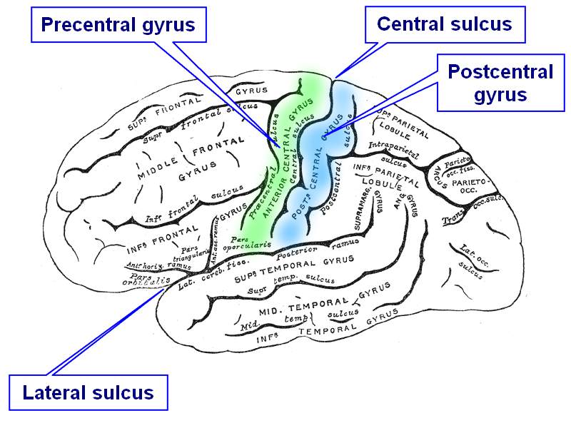
Sulcus/gyrus and brain, lateral view
These two different terms must be analyzed together. The Latin term [sulcus] means "groove or fissure". Its plural form is [sulci]. There are many anatomical sulci in the body, one of them being the costal sulcus in the ribs.
The second term [gyrus] is also Latin and means "circle or ring", as used in the words gyroscope or gyrations. In its adjective or descriptive form, [gyrus] is used to denote something "bent, curved, or broad-shouldered"1. The plural form is [gyri]. In the case of the brain a gyrus is formed as a mound or an elevation between the "valleys" of the sulci (see image). If you click on the image a secondary image depicting the lateral aspect of the brain will appear.
In the brain there are many sulci, the secondary image shows the lateral or Sylvian sulcus, and the central sulcus or sulcus of Rolando.
In relation to the central sulcus there are two gyri. The anteriorly situated precentral gyrus is considered the primary motor cortex and associated with voluntary motor activity (colored in green in the secondary image). The postcentral gyrus (colored in blue) is situated posterior to the central sulcus and is the primary sensory cortex, associated with somatic (bodily) conscious sensation.
Sources:
1. "The Origin of Medical Terms" Skinner, HA 1970 Hafner Publishing Co.
2. "Medical Meanings - A Glossary of Word Origins" Haubrich, WD. ACP Philadelphia
3 "Tratado de Anatomia Humana" Testut et Latarjet 8 Ed. 1931 Salvat Editores, Spain
4. "Anatomy of the Human Body" Henry Gray 1918. Philadelphia: Lea & Febiger
Initial image by:Albert Kok,courtesy of:Wikipedia.org. Second image modified from the original image by Henry Vandyke Carter, MD., courtesy of bartleby.com
Terms suggested by Sara Mueller.
- Details
This article is part of the series "A Moment in History" where we honor those who have contributed to the growth of medical knowledge in the areas of anatomy, medicine, surgery, and medical research.

Adam Christian Thebesius
Adam Christian Thebesius (1686- 1732). German physician and anatomist, Thebesius studied in the University of Leiden, Netherlands, where he received his doctorate in 1708 with the thesis "De circulo sanguinis in corde" (on the circulation of the blood in the heart). In 1713 he became a member of the Royal Academy of Natural Scientists (Kaiserliche Akademie der Naturforscher), where he adopted the Latin name "Eyryphon". Besides his natural sciences and medical research, Thebesius developed an interest in astrophysics.
Extremely interested in coronary circulation, Thebesius injected dyes and fluids in the coronary arteries, veins, and coronary sinus. Along with Raymond Vieussens (1635-1713) , Thebesius described all these structures. Today his name is attached to the eponymic Thebesian veins (venae cordi minima), and the Thebesian valve guarding the exit of the coronary sinus into the right atrium of the heart. Both these structures were mentioned in his 1708 doctoral thesis
Sources:
1. “The Role of the Thebesian Vessels in the Circulation of the Heart” Wearn, J.T. J Exp Med. 1928 January 31; 47(2): 293–315
2. The Story Behind the Word. Some Interesting Origins of Medical Terms. Wain,H. 1958.
3. The Origin of Medical Terms. Skinner, H.A. 1970
Original image in the public domain, courtesy of the National Library of Medicine
- Details
The ventricular system of the brain is an interconnected system of cavities and ducts within the brain through which cerebrospinal fluid (CSF) circulates. The CSF is produced in the choroid plexuses located within the ventricles.

Ventricular system of the brain

Brain dissection - 4th ventricle
There are two large curved lateral ventricles, each found within a cerebral hemisphere. They connect with the third ventricle via an opening called the "foramen of Monro". The third ventricle connects with the fourth ventricle by way of a slender canal called the "cerebral aqueduct" or the "aqueduct of Sylvius".
The third ventricle is found deep within the brain between the right and left diencephalic portion of the cerebrum.
The fourth ventricle is located between the pons of the brain stem anteriorly and the cerebellum posteriorly. This ventricle has a rhomboidal shape and it connects with the external aspect of the brain and the subarachnoid space. Failure of this CSF drainage from the ventricles to the subarachnoid space can lead to pathological accumulation of CSF within the ventricles and hydrocephalus.
The line drawn image shows a lateral view of the brain with a superimposed image of the ventricular system. If you click on this image, you will see a superior view of a cast of the ventricular system (by Retzius).
The dissection image shows a posterior view of the brain stem and the cerebellum which has been opened in the median plane to expose the 4th ventricle. Click on the image to see a larger version.
Line images in the public domain by Henry VanDyke Carter, MD. (Gray's Anatomy) Dissection images property of CAA, Inc. Photographer E. Klein
- Details
The cerebrospinal fluid (CSF) is a colorless, transparent fluid produced from the arterial flow of blood by the choroid plexuses found within the ventricular system of the brain. The CSF exits the ventricular system and enters the subarachnoid space and its cisterns. It is then absorbed at the level of the arachnoid granulations into the venous component of the cardiovascular system.
The CSF has many functions, some of them being protection, the creation of a fluid environment where the brain 'floats", cleansing, and others. For a more detailed description of the CSF, click here.
The CSF is produced at an average rate of 550-700ml/day. It is absorbed at the same rate. An imbalance between production and absorption of CSF (as well as a blockage within the ventricular system) can lead to an accumulation of CSF within the brain, causing hydrocephalus.
- Details
This article is part of the series "A Moment in History" where we honor those who have contributed to the growth of medical knowledge in the areas of anatomy, medicine, surgery, and medical research.
 Hippocrates
Hippocrates of Cos (460 BC - 370 BC). A Greek physician, Hippocrates was born on the Greek island of Cos (Kos) c. 460BC. Considered the "Father of Medicine" he removed Medicine from the realms of superstition and magic. He was the first to record medical writings and is considered the first one to use and maintain proper medical terminology. There are many writing attributed to Hippocrates, but there is no assurance that these were actually written by Hippocrates himself. Hippocrates changed the art of medical diagnosis by replacing supernatural precepts with observation-based methodology. Natural, rather than supernatural causes, would from here on explain all disease processes, what was known as Rational Medicine.
He is known for having set the oath that governs medical principles, the Hippocratic Oath, although there are many authors that contend that this oath was written long time after he died.
Sources:
1. "Hippocrates himself" JAMA. 1968;204(12):1138-1139
2. "Hippocrates: father of medicine" Tan, S Y (01/01/2002). Singapore medical journal(0037-5675), 43(1), p.5.
- Details

Anterior view of the thorax, showing surface
relations of bones, lungs (purple), pleura (blue),
and heart (red outline).
P. Pulmonary valve. A. Aortic valve.
B. Bicuspid valve. T. Tricuspid valve
This is a combined word arising from terms [atrium], [ventricle], and [sulcus]. For the etymology of each word, click on the corresponding link.
The atrioventricular sulcus, also know as the "coronary groove" or "coronary sulcus" is an evident incomplete groove between the atria and ventricles of the heart. It is complete posteriorly and is separated anterosuperiorly by the roots of the aorta and the pulmonary trunk. It contains the right coronary artery on the right side, and the circumflex artery on the left side, hence the name "coronary groove". These coronary arteries are not visible as they are usually covered by the epicardium and subepicardial fat.
The atrioventricular sulcus (and the corresponding coronaries) are also in relation to the deeper situated atrioventricular (AV) valves, the tricuspid valve on the right; and the mitral or bicuspid valve on the left side. The accompanying image depicts the location of the AV valves, and therefore the location of the AV sulcus. The image is an anterior view of the thorax, showing surface relations of bones, lungs (purple), pleura (blue), and heart (red outline). P. Pulmonary valve. A. Aortic valve. B. Bicuspid valve. T. Tricuspid valve
Sources:
1. "The Origin of Medical Terms" Skinner, HA 1970 Hafner Publishing Co.
2. "Medical Meanings - A Glossary of Word Origins" Haubrich, WD. ACP Philadelphia
3 "Tratado de Anatomia Humana" Testut et Latarjet 8 Ed. 1931 Salvat Editores, Spain
4. "Anatomy of the Human Body" Henry Gray 1918. Philadelphia: Lea & Febiger
Image modified by CAA, Inc. Original image by Henry Vandyke Carter, MD., courtesy of bartleby.com