Medical Terminology Daily (MTD) is a blog sponsored by Clinical Anatomy Associates, Inc. as a service to the medical community. We post anatomical, medical or surgical terms, their meaning and usage, as well as biographical notes on anatomists, surgeons, and researchers through the ages. Be warned that some of the images used depict human anatomical specimens.
You are welcome to submit questions and suggestions using our "Contact Us" form. The information on this blog follows the terms on our "Privacy and Security Statement" and cannot be construed as medical guidance or instructions for treatment.
We have 1169 guests and no members online

Jean George Bachmann
(1877 – 1959)
French physician–physiologist whose experimental work in the early twentieth century provided the first clear functional description of a preferential interatrial conduction pathway. This structure, eponymically named “Bachmann’s bundle”, plays a central role in normal atrial activation and in the pathophysiology of interatrial block and atrial arrhythmias.
As a young man, Bachmann served as a merchant sailor, crossing the Atlantic multiple times. He emigrated to the United States in 1902 and earned his medical degree at the top of his class from Jefferson Medical College in Philadelphia in 1907. He stayed at this Medical College as a demonstrator and physiologist. In 1910, he joined Emory University in Atlanta. Between 1917 -1918 he served as a medical officer in the US Army. He retired from Emory in 1947 and continued his private medical practice until his death in 1959.
On the personal side, Bachmann was a man of many talents: a polyglot, he was fluent in German, French, Spanish and English. He was a chef in his own right and occasionally worked as a chef in international hotels. In fact, he paid his tuition at Jefferson Medical College, working both as a chef and as a language tutor.
The intrinsic cardiac conduction system was a major focus of cardiovascular research in the late nineteenth and early twentieth centuries. The atrioventricular (AV) node was discovered and described by Sunao Tawara and Karl Albert Aschoff in 1906, and the sinoatrial node by Arthur Keith and Martin Flack in 1907.
While the connections that distribute the electrical impulse from the AV node to the ventricles were known through the works of Wilhelm His Jr, in 1893 and Jan Evangelista Purkinje in 1839, the mechanism by which electrical impulses spread between the atria remained uncertain.
In 1916 Bachmann published a paper titled “The Inter-Auricular Time Interval” in the American Journal of Physiology. Bachmann measured activation times between the right and left atria and demonstrated that interruption of a distinct anterior interatrial muscular band resulted in delayed left atrial activation. He concluded that this band constituted the principal route for rapid interatrial conduction.
Subsequent anatomical and electrophysiological studies confirmed the importance of the structure described by Bachmann, which came to bear his name. Bachmann’s bundle is now recognized as a key determinant of atrial activation patterns, and its dysfunction is associated with interatrial block, atrial fibrillation, and abnormal P-wave morphology. His work remains foundational in both basic cardiac anatomy and clinical electrophysiology.
Sources and references
1. Bachmann G. “The inter-auricular time interval”. Am J Physiol. 1916;41:309–320.
2. Hurst JW. “Profiles in Cardiology: Jean George Bachmann (1877–1959)”. Clin Cardiol. 1987;10:185–187.
3. Lemery R, Guiraudon G, Veinot JP. “Anatomic description of Bachmann’s bundle and its relation to the atrial septum”. Am J Cardiol. 2003;91:148–152.
4. "Remembering the canonical discoverers of the core components of the mammalian cardiac conduction system: Keith and Flack, Aschoff and Tawara, His, and Purkinje" Icilio Cavero and Henry Holzgrefe Advances in Physiology Education 2022 46:4, 549-579.
5. Knol WG, de Vos CB, Crijns HJGM, et al. “The Bachmann bundle and interatrial conduction” Heart Rhythm. 2019;16:127–133.
6. “Iatrogenic biatrial flutter. The role of the Bachmann’s bundle” Constán E.; García F., Linde, A.. Complejo Hospitalario de Jaén, Jaén. Spain
7. Keith A, Flack M. The form and nature of the muscular connections between the primary divisions of the vertebrate heart. J Anat Physiol 41: 172–189, 1907.
"Clinical Anatomy Associates, Inc., and the contributors of "Medical Terminology Daily" wish to thank all individuals who donate their bodies and tissues for the advancement of education and research”.
Click here for more information
- Details
The word [sesamoid] means "similar to a sesame". First used by Galen c.180AD, he describes small ovoid bones that are "similar to a sesame seed", referencing the seed of the plant sesamum indicum, the oil of which was used as a laxative at that time.
Sesamoid bones are found in the tendons of some muscles and are mostly inconstant. Largey and Bonnet proposed a classification for these bones as: accessory, capsuloligamentous, intratendineous, and mixed.
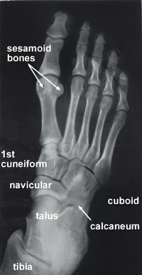
Of special interest to this article are the sesamoid bones found within the two tendons of the flexor hallucis brevis muscle in the base of the foot (see accompanying X-ray image). These bones, especially the medial sesamoid bone, was attributed religious, mystical, and magical powers since ancient times. This is due to the fact that this small bone is highly resistant to natural decomposition. A Hebrew medical text dated 210 BC, attributed to Ushaia presented a small bone he called "luz" as the "depository of the soul". Many other authors, including Vesalius (who called it Albadaran), believed that upon resurrection, the whole body could reform from this "seed" bone.
This belief was later reinforced by religious texts into the early Renaissance which stated that this bone was indestructible and its presence was enough to guarantee resurrection for believers.
Note: The original X-ray was published here courtesy of Wesley Norman, PhD in his website. The website is no longer available, so the image is now in the public domain via www.archive.org.
Sources:
1. "Les os sesamoides de l’hallux : du mythe a`la fonction" Largely,A; Bonnel, BE Med Chir Pied 2008 24: 28–38
2. "Tratado de Anatomia Humana" Testut et Latarjet 8 Ed. 1931 Salvat Editores, Spain
3. "De Humani Corporis Fabrica" Vesalius, Andrea. 1543 Oporinus
Thanks to the first year medical students at the University of Cincinnati who inspired this article. Dr. Miranda
- Details
This article is part of the series "A Moment in History" where we honor those who have contributed to the growth of medical knowledge in the areas of anatomy, medicine, surgery, and medical research.
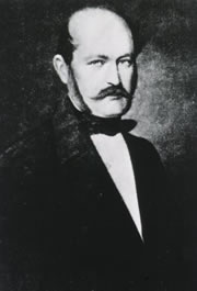
Ignaz Semmelweis
Ignaz Semmelweis, MD (1818- 1865). Born in Budapest as Ign?c F?l?p Semmelweis, he started his university studies as a lawyer, but changed to Medicine and in 1844, at the age of 26, attained his MD degree. in 1847 he was appointed as an assistant in Obstetrics, almost at the same time of the death of a friend (Kolletschka, a pathologist) who died of what appeared to be "puerperal fever", also known as "childbed fever" after being accidentally stabbed by a knife during the autopsy of a female who had died of that disease. Semmelweis reasoned that the disease somehow was transmitted via the wound and started a crusade to have surgeons and students clean their hands with a carbolized solution before examining a healthy pregnant woman.
Although the obstetric wards under his care reduced the rate of this disease to almost nothing, Semmelweis endured criticism from his teachers, colleagues, and peers, and he did not make any friends by calling "murderers" those who did not follow his ideas. murderers". An excerpt of a letter to one of this detractors reads: "I denounce you before God and the world as a murderer and the history of puerperal fever will not do you an injustice when for the service of having been the first to oppose my life-saving technique it perpetuates your name as a medical Nero". He did not publish his findings until later in life, and then received even more criticism.
In 1865 was committed to an mental asylum only to die a few days later. He was only 47 years old. The same year he died Joseph Lister performed the first operations using antiseptic technique.
Sources:
1. Newsom S." Pioneers in Infection Control - Semmelweis, Ignaz Philipp". The Journal of hospital infection. 1993-03-01;23:175-187.
2. Ellis, H. (2008). Ignaz Semmelweis: tragic pioneer in the prevention of puerperal sepsis. British Journal Of Hospital Medicine (London, England: 2005), 69(6), 358
3. " A Corner of History: Ignaz Philipp Semmelweis" Wynder, EL Prev Med 3" (4) Dec 1974, 574-580
4. "Ignaz Semmelweis; a hand-washing pioneer" P. Rangapa JAPI May 2010 58:328
Original image courtesy of Images from the History of Medicine
- Details
The term [muscle] arises from the Latin word [musculus] which derives from the Latin term [mus] meaning "mouse". We can only guess that, just as today, Roman fathers would show their biceps and forearm muscles to their children and tried to make them believe a mouse had gotten under their skin!. The root term for muscle is [-my-]. The corresponding combining form is [-myo-].
There are three types of muscle in the human body:
- Skeletal muscle: it is typical of muscles related to bones (skeletal) and they are voluntary.
- Smooth muscle: found in organs that act without volition (involuntary), such as the digestive system and glands.
- Cardiac muscle: found exclusively in the heart.
Skeletal and cardiac muscles have distinct striations visible under a microscope.
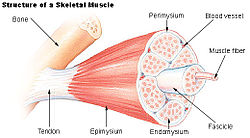
Muscles are formed by subunits, each one surrounded by a named membrane. One of the suffixes that means layer or membrane is [-sium]:
- Epimysium: Epi=outer; my=muscle; sium=membrane. The outer or external membrane (layer) of a muscle
- Perimysium: Peri=around; my=muscle; sium=membrane. A membrane around a muscle
- Endomysium: Endo= inner or internal; my=muscle; sium=membrane. The inner or internal membrane of a muscle-
Original image courtesy of Wikipedia. Click on the image for a larger version.
- Details
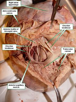
The trabeculae carnae is a meshwork of fleshy cords found in the inner aspect of the right and left ventricles of the heart.
The Latin term [trab] means "beam", and [trabeculum] refers to the group of beams that supports a roof, like an intertwined network. The plural for of [trabeculum] is [trabeculae].
The second term [carnae] is Latin for "meaty". The meaning of [trabeculae carnae] is the "meaty meshwork".
The trabeculae carnae are more evident and larger in the left ventricle than in the right ventricle, and larger and more complex towards the cardiac apex. The accompanying image shows the dissection of a human heart exposing the trabeculae carnae in the right ventricle.
Click on the image for a larger version
Image property of: CAA, Inc. Photographer: David M. Klein
- Details
Although not a medical root term, the root [-nym-] arises from the Greek [onoma] meaning "name". There are many terms that incorporate this root:
- Eponym: Use of a proper name to denote a structure
- Eunym: [Eu-] is a prefix that means "good", so it imeans a "good name". Also written as "euonym"
- Homonym: Same name
- Synomym: "A name with the same sense, or same meaning"
- Antomym: From the Greek [ant- and anti] meaning opposite. An opposite name
- Anonym: From the Greek [an- and ano-] meaning "without". Without a name (anonymous)
- Pseudonym: From the Greek [Pseudo-] meaning "false". A false name
- Toponym: From [topos], meaning place. The name of a place or location
- Details
This article is part of the series "A Moment in History" where we honor those who have contributed to the growth of medical knowledge in the areas of anatomy, medicine, surgery, and medical research.
Henry Koplik, MD (1858 -1927). American pediatrician and researcher, was born in 1858 in the city of New York. He received his MD from the College of Physicians and Surgeons at the Colombia University in New York. He spent several years studying in Berlin, Vienna, and Prague. Upon his return to the US he worked at the lower Manhattan Good Samaritan dispensary, where he later built a large pediatric outpatient clinic which became a model for the care of infants and children. In fact, under Dr Koplik's direction, this clinic became the world's first "milk depot" providing fresh milk and infant food for underprivileged mothers in the area. Dr. Koplik was one of the founders of the American Pediatric Society, and was one of its presidents.
Mostly remembered by the pathognomonic and eponymic "Koplik's spots", Dr Koplik had many other achievements. Some of them include the prophylaxis of a milk depot, the strict discipline in diagnosis and care of the pediatric patient, the discovery of the bacillus responsible for whooping cough, the prevention of cross-contamination at a pediatric ward, etc.
Dr. Koplik wrote a number of clinical and research papers on hygiene and public health, as well on a number of medical topics, plus a book on "Diseases of Infancy and Childhood".
Sources:
1. "Koplik's Spots for the Record: an Illustrated Historical Note" Brem, J; Clin Ped 1972 11:3 161-163
2. "Pediatric Profiles: Henry Koplik (1858-1927)" Bass, MH J Ped 1957 119-125
3. "The History of the First Milk Depot or Gouttes de Lait With Consultations in America" JAMA 50: 1574, 1914.
4. "Some Pediatric Eponyms: Koplik's Spots," W. R. Bett Brit. J. Child.Dis. 28: 127, 1931
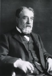
Original image in the public domain, courtesy of the National Institutes of Health.


