
Medical Terminology Daily (MTD) is a blog sponsored by Clinical Anatomy Associates, Inc. as a service to the medical community. We post anatomical, medical or surgical terms, their meaning and usage, as well as biographical notes on anatomists, surgeons, and researchers through the ages. Be warned that some of the images used depict human anatomical specimens.
You are welcome to submit questions and suggestions using our "Contact Us" form. The information on this blog follows the terms on our "Privacy and Security Statement" and cannot be construed as medical guidance or instructions for treatment.
We have 822 guests online
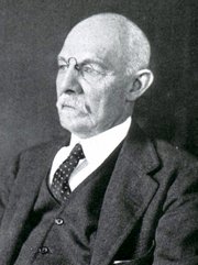
William S. Halsted, MD
(1852 – 1922)
American anatomist, teacher, and surgeon, William Stewart Halsted was born in New York City, USA to a wealthy family of English origin. His father was involved in charitable work and Governor and trustee to a city hospital. Not a brilliant student initially, Halsted took an undergraduate in Liberal Arts in Yale, CT., after which he entered the Medical College of Physicians at the Columbia College, where he excelled.
As a second-year medical student Halsted applied and obtained a position in surgery at a local hospital. In here he learned about Lister’s antiseptic technique and became an adamant proponent of it to reduce infection. In 1877 Halsted obtained his MD. After a short time as House Physician at the New York Hospital, Halsted traveled to Europe to further his education studying for two years at the Universities of Vienna, Leipzig, and W?rzburg.
Besides being at the forefront of surgical and antiseptic techniques (introducing the use of rubber gloves in surgery), Halsted was extremely concerned with the way medical students were taught in the US. He pioneered bedside clinical round discussions with the medical students after two years of basic sciences studies. Halsted developed the idea of a patient chart; he also developed the residency program for medical students in use today.
Halsted is probably the most influential researcher and surgeon at the turn of the century. He dedicated time to the study of intestinal anastomoses and the use of silk as a suture material. His experimental work in 1887 proved that the inclusion of the submucosa layer in an anastomosis was mandatory, as well that a single layered anastomosis was enough to attain closure. Perhaps Halsted’s most important contribution was the application and use of the scientific method to surgical questions. Halsted’s principles set the standards used today in surgical suturing and surgical stapling.
He also pioneered the development and surgical techniques for radical mastectomy as a treatment for breast cancer.
As a side effect of this studied in anesthesia and the use of cocaine for anesthesia, Halsted became addicted to this substance, a problem that followed him through the years. Without impairing his capacity as a researcher and a surgeon, Halsted eventually recovered. He died in Baltimore in 1922 as a complication to surgery.
Sources:
1. Dubay, A. D., & Franz, G. M. (2003). Acute Wound Healing: The Biology of Acute Wound Failure. Surg Clin NA, 83, 463-481.
2. Halsted, W. S. (1887). Circular Suture of the Intestine - An Experimental Study. Am J Med Sci, 436-461.
3. “William Stewart Halsted: his life and contributions to surgery” Osborne, P. Lancet Oncol 2007; 8: 256–65
4. “William Stewart Halsted: Surgical pioneer” Burress, P Endoc Today (2010), 8: (2) 22
5. “William Stewart Halsted (1852–1922) Neurological stamp” Haas, LF J Neurol Neurosurg Psych 2000;69:641
Original image courtesy of "Images from the History of Medicine" at www.nih.gov
"Clinical Anatomy Associates, Inc., and the contributors of "Medical Terminology Daily" wish to thank all individuals who donate their bodies and tissues for the advancement of education and research”.
Click here for more information


- Details
Click for a larger image
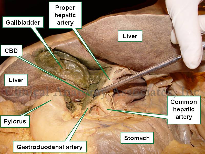
UPDATED: The [common bile duct] also known as the [ductus choledocus]. is part of the hepatobiliary tree, taking bile from the gallbladder and liver to the second portion of the duodenum. The common bile duct begins at the junction of the common hepatic duct with the cystic duct, it continues inferiorly, usually to the right of the proper hepatic artery and anterior to the portal vein. It then passes posterior to the first portion of the duodenum, is surrounded by pancreatic tissue and ends at the hepatopancreatic ampulla of (Vater).
The junction of the common bile duct with the hepatopancreatic ampulla of (Vater) is the narrowest portion of the hepatobiliary tree, The lodging of a gallstone at this junction can be the cause for choledocholitiasis and jaundice.
In the accompanying image the common bile duct is elevated with a probe. The lesser omentum has been removed to show the common bile duct and vascular structures that are found between the two peritoneal layers that form the lesser omentum.
- Details

Inferior surface of the liver
The [gallbladder] is a bile transient storage organ, part of the hepatobiliary tree, situated in the anteroinferior aspect of the liver. The gallbladder is found in a depression on the inferior aspect of the right lobe of the liver, the gallbladder fossa or fossa vesicae felleae.
In the gallbladder we describe its dome-shaped fundus, the body of the organ, and the neck which is the area that opens into the cystic duct. Close to the neck, the gallbladder has a small pouch (Hartmann's pouch) which is important for surgeons during a laparoscopic cholecystectomy, as this is where they will lock one of the instruments that allows them to manipulate the gallbladder for dissection of the organ from the gallbladder fossa (the gallbladder bed). The other surgical grasper is placed at the gallbladder fundus.
The gallbladder is composed by three layers. From deep to superficial they are:
• Mucosa: Characterized by a columnar epithelium. Towards the neck of the gallbladder the mucosa creates spiral ridges that continue in to the cystic duct.
• Fibromuscular layer: This layer is composed by connective tissue and smooth muscle, mostly longitudinal
• Serosa: This is an incomplete layer and is formed by visceral peritoneum covering the area of the gallbladder not in contact with the liver. In an unusual anatomical variation, the serosa layer can be almost complete, forming a pseudomesentery that may contains some veins.
The gallbladder receives its blood supply by way of the cystic artery, a branch of the right hepatic artery. The venous return is by way of multiple small veins that empty into the liver venous system. In some cases, these veins may form large sinuses between the liver and the gallbladder causing potential troublesome bleeding during a cholecystectomy. For those who like medical history, Dr. Eric Muhe performed the first laparoscopic cholecystectomy on September 12, 1985! We are but a few days from the 30th anniversary!
For more information on terminology on "gall-", "bile", "chol", and "chole", click here.
Sources:
1 "Tratado de Anatomia Humana" Testut et Latarjet 8 Ed. 1931 Salvat Editores, Spain
2. "Anatomy of the Human Body" Henry Gray 1918. Philadelphia: Lea & Febiger
Image modified by CAA, Inc. Original image courtesy of bartleby.com
- Details
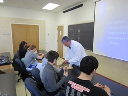

Last Friday April 17, I prepared and delivered a lecture on "Surgical Sutures, Needles, and Knots" which included a hands-on workshop on knots and wound closure on simulated tissue.
This was presented at the invitation of the Pre-Health club of the Mount Saint Joseph University in Cincinnati, OH. I am always glad to be invited to do these presentations as they allow me to maintain contact with the future generation of Health Care Professionals.
Of course this is a very short presentation compared to the longer course that Clinical Anatomy Associates, Inc. delivers for medical companies, but it shows these future professionals the complexity of the world of wound closure, healing, surgical sutures, needles, and knots.
We ended the lab with the challenge to do a two-layer closure of a simulated wound. Most of the attendees did a pretty good job. Congratulations!
My personal thanks to Dr. Eric Johnson who coordinated the meeting, and to the Pre-Health Club for their invitation. For more pictures of the meeting, see the Facebook album page of "Medical Terminology Daily"
- Details
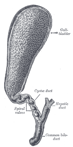
Gallbladder and cystic duct
The cystic duct is a tubular structure that connects the neck of the gallbladder to the extrahepatic ductal system. It is 2-4 cm. in length and its lumen is about 2.6 +/- 0.7 mmm. The shape of the cystic duct varies, as it can be straight, angled, or acutely curved.
The mucosa of the cystic duct presents with 2-10 crescent-shaped folds that create a spiral-shaped inner structure referred to as the "Valve of Heister", first described by Lorenz Heister in 1732. These folds become smaller and scarcer towards the distal portion of the duct.
The cystic duct can present with several anatomical variations, from total absence where the neck of the gallbladder empties directly in to the common bile duct, to duplication, and even rare occasions where the cystic duct empties separately into the duodenal lumen.
The cystic duct is an important surgical landmark as it is one of the boundaries of the cystohepatic triangle or "Triangle of Calot", described by Jean-Francois Calot (1861 - 1944), which determines the location of the cystic artery, a critical structure that needs to be ligated and transected during a cholecystectomy.
Sources:
1 "Cystic Duct and Heister’s “Valves” Dasgupta,C, Stringer, MD, Clin Anat (2005) 18:81–87
2. "Tratado de Anatomia Humana" Testut et Latarjet 8 Ed. 1931 Salvat Editores, Spain
3. "Anatomy of the Human Body" Henry Gray 1918. Philadelphia: Lea & Febiger
Image modified by CAA, Inc. Original image courtesy of bartleby.com
- Details
The root term [-phor-] arises from the Greek word [φέρω] meaning “to bear”, “well”, and “healthy”. The meaning of the word today in medical terminology is “well-being”. The addition of the adjectival suffix [-ia] meaning “pertaining to” add to form [-phoria] meaning “pertaining to well-being” or “feeling well”. It is used in terms such as:
• Euphoria: The prefix [eu-] means “good”. The sensation of feeling good, of good well-being
• Dysphoria: The prefix [dys-] means “abnormal”. Abnormal feelings of well-being. In psychiatry these dysphoric moments can be seen in bipolar patients.
- Details

Click for a larger image
The term [intervertebral] means "between vertebrae", and [foramen] means "opening". The intervertebral foramina are bilateral openings between adjacent vertebrae. Each intervertebral foramen is found between adjacent pedicles ("P" in the large image), bound by the inferior vertebral notch and the superior vertebral notch of adjacent pedicles.
Although the term [intervertebral foramen] has been used for a long time, the concept has evolved to a more modern "intervertebral canal" or as some clinicians call it, the "lateral canal". The reason for this is that the intervertebral foramen is actually a tunnel whose length is determined by the width of the pedicles. This intervertebral canal has marked differences between the lateral, middle, and medial structures contained in the intervertebral canal.
Some of these structure are nerve roots, the dorsal root ganglion, the initial portion of the spinal nerve, dural sac, arteries, veins, recurrent nerves, fat, and a complex system of transforaminal and intraforaminal ligaments1. The structures contained in the intervertebral foramen can be compressed if the height of the intervertebral discs is compromised, or by a herniation of the intervertebral disc. The diameter of the intervertebral canal can also be reduced by bone and joint pathology.
If you hover over the image, the intervertebral foramen will be highlighted. For a larger version click on the image.
Images property of: CAA.Inc. Photographer: D.M. Klein
1 Thoracic and lumbar intraforaminal ligaments Akdemir, G.; J Neurosurg Spine 13:351-355, 2010