![]()
Medical Terminology Daily (MTD) is a blog sponsored by Clinical Anatomy Associates, Inc. as a service to the medical community. We post anatomical, medical or surgical terms, their meaning and usage, as well as biographical notes on anatomists, surgeons, and researchers through the ages. Be warned that some of the images used depict human anatomical specimens.
You are welcome to submit questions and suggestions using our "Contact Us" form. The information on this blog follows the terms on our "Privacy and Security Statement" and cannot be construed as medical guidance or instructions for treatment.
We have 622 guests online
")
Marcia Crocker Noyes
(1869 – 1946)
Further to my comment on old books and research that started with an interesting bookplate (Ex-Libris). I continued my research and found that the person in charge of the Osler library bookplate was a fascinating individual that today maybe a ghost in the MedChi library and building in Baltimore... This is certainly an article that can be called "A Moment in History"
Marcia Crocker Noyes was the librarian at The Maryland State Medical Society from 1896 to 1946 and was a founding member of the Medical Library Association.[1][2][3]
Sir William Osler, MD. a famous Johns Hopkins surgeon was a noted bibliophile and had a large personal collection of books on various topics. When he became the President of MedChi in 1896, he was dismayed at the condition of the library and knew that with the right person and some stewardship, it could become a significant collection. Sir William asked his friend, Dr. Bernard Steiner, a physician and President of the Enoch Pratt Free Library in Baltimore for suggestions of a librarian, and Dr. Steiner recommended Marcia Crocker Noyes. A native of New York, and a graduate of Hunter College, Marcia had moved to Baltimore for a lengthy visit with her sister, and took a “temporary” position at the Pratt Library, which turned into three years. Although she had no medical experience or background, she was enthusiastic, and most importantly, she was willing to move into the apartment provided for the librarian, who needed to be available 24 hours a day.
The image in this article is Ms. Noyes on her first year on the job. Marcia developed a book classification system for medical books, based on the Index Medicus, and called it the Classification for Medical Literature. The system uses the alphabet with capital letters for the major divisions of medicine and lower-case ones for the sub-sections. The system was used for many years, but it's now dated and the Faculty's original shelving scheme was never changed. The card catalogs still reflect her classification and many of the cards are written in Marcia's back-slanting handwriting.
Marcia knew enough to ask the Faculty's members about medical questions, terminology and literature. She gradually won over the predominantly male membership and they became her greatest allies; Sir William at the start, and then for nearly 40 years, Dr. John Ruhräh, a wealthy pediatrician with no immediate family of his own. She made a point of attending almost every Faculty function, and in 1904, under guidelines from the American Medical Association, Marcia was made the Faculty Secretary. For much of her first 10 years, she was the Faculty's only full-time employee, only being assisted by Mr. Caution, the Faculty's janitor. Later in life Marcia would say that she hired him because of his name!
Within ten years, the library had outgrown its space, and plans, spearheaded by Marcia and Sir William before his move to Oxford, were made to build a headquarters building, mainly to house the library's growing collection of medical books and journals.
Marcia was instrumental in the design and building of the new headquarters. She travelled to Philadelphia, New York and Boston to look at their medical society buildings, and eventually, the Philadelphia architectural firm, Ellicott & Emmart was selected to design and build the new Faculty building. Every detail of the building held her imprimatur, from the graceful staircase, to the light-filled reading room, and all of the myriad details of the millwork, marble tesserae, and most of all, the four-story cast iron stacks. She was on-site, climbing up unfinished staircases, checking out the progress of the building, which was built in less than one year at a cost of $90,000.
Among the features of the new building was a fourth-floor apartment for her. She referred to it as the "first penthouse in Baltimore" and it had a garden and rooftop terrace. The library collection eventually grew to more than 65,000 volumes from medical and specialty societies around the world. Journals were traded back and forth, and physicians eagerly anticipated the arrival of each new issue. At the same time, Marcia was involved in the Medical Library Association as one of eight founding members. The MLA promotes medical libraries and the exchange of information. One of the earliest mandates of the MLA was the Exchange, a distribution and trade service for those who had duplicates or little-used books in their collections. Initially, the Exchange was run out of the Philadelphia medical society, but in 1900 it was moved to Baltimore and Marcia oversaw it. Several hundred periodicals and journals were received and sent each month, a huge amount of work for a tiny staff. In 1904, the Faculty had run out of room to manage the Exchange, so it was moved to the Medical Society of the Kings County (Brooklyn). But without Marcia's excellent administrative skills, it floundered and in 1908, the MLA asked Marcia to take charge once again.
In 1909, when the new Faculty building opened, there was enough room to run the Exchange and with the help of MLA Treasurer, noted bibliophile and close friend, Dr. John Ruhräh, it once again became successful. Additionally, Marcia and Dr. Ruhräh combined forces to revive the MLA's bulletin, which had all but ceased publication in 1908, taking the Exchange with it. This duo maintained editorial control from 1911 until 1926. In 1934, around the time of Dr. Ruhräh's death, Marcia became the first “unmedicated” professional to head the MLA. During her tenure, the MLA incorporated, the first seal was adopted, and the annual meeting was held in Baltimore. Marcia wanted to write the history of the MLA once she retired from full-time work at the Faculty, but her health was beginning to fail. She had back problems and had suffered a serious burn on her shoulder as a young woman, possibly from her time running a summer camp, Camp Seyon, for young ladies in the Adirondack Mountains. In 1946, a celebration was planned to honor Marcia's 50 years at the Faculty. But she was adamant that the physicians wait until November, the actual date of her 50 years. However, they knew she was gravely ill, and might not make it until then, so a huge party was held in April. More than 250 physicians attended the celebration, but the ones she was closest to in the early years, were long gone. She was presented with a suitcase, a sum of money to use for travelling, and her favorite painting of Dr. John Philip Smith, a founder of the Medical College in Winchester, Virginia. It was painted by Edward Caledon Smith, a Virginia painter who had been a student of the painter Thomas Sully.[4] She adored this painting and vowed, jokingly, to take it with her wherever she went.
The painting was not to stay with her for very long, for she died in November 1946, and left it to the Faculty in her will. Her funeral was held in the Faculty's Osler Hall, named for her dear friend. More than 60 physicians served as her pallbearers, and she was buried at Baltimore's Green Mount Cemetery. In 1948, the MLA decided to establish an award in the name of Marcia Crocker Noyes. It was for outstanding achievement in medical library field and was to be awarded every two years, or when a truly worthy candidate was submitted. In 2014, the Faculty began giving a bouquet of flowers to the winner of the award in Marcia's name, and in honor of her work. Much evidence exists for this tradition, as we know that the physicians, especially Drs. Osler and Ruhräh, frequently gave her bouquets of flowers. Marcia also cultivated flower gardens at the Faculty and decorated the rooms with her work.
Today, the MedChi building is open for tours and if the rumors are to be believed Ms. Marcia Crocker Noyes is still at work in her beloved library as the "resident ghost" [1][5]

NOTE: This article has been modified from the original Wikipedia article on Marcia Crocker Noyes. The article itself is well-written with interesting images of the subject. I would encourage you to visit it. The second insert is from book 00736 in my personal library and shows in pencil, the incredibly small handwriting of Marsha C. Noyes.
Sources:
1. "Marcia, Marcia, Marcia" MedChi Archives blog.
2. "Marcia C. Noyes, Medical Librarian" (PDF). Bulletin of the Medical Library Association. 35 (1): 108–109. 1947. PMC 194645
3. Smith, Bernie Todd (1974). "Marcia Crocker Noyes, Medical Librarian: The Shaping of a Career" (PDF). Bulletin of the Medical Library Association. 62 (3): 314–324. PMC 198800Freely accessible. PMID 4619344.
4. Edward Caledon BRUCE (1825-1901)"
5. Behind the scenes tour MedChiBuilding
"Clinical Anatomy Associates, Inc., and the contributors of "Medical Terminology Daily" wish to thank all individuals who donate their bodies and tissues for the advancement of education and research”.
Click here for more information


- Details
The root term [-men-] originates from the Latin word [mensis] meaning “month”. Earlier forms of this term probably arise from the Greek [μήνας] (minas), also meaning “month”, but with the connotation of “lunar month” or “moon”.
Since a woman’s menstrual cycle is on average 28 days (ranging from 31 to 35 days) and a lunar month is 29 days and 12 hours in length, the root term [-men-] has been associated with a woman’s menstrual cycle, and menstruation. The term can be found in many words such as:
• Menses: The period of flow in a menstrual cycle. The “period”
• Amenorrhea: The prefix [a-] means “without” or “absence of”. The suffix [-(o)rrhea] means "flow". Without menstrual flow.
• Dysmenorrhea: The prefix [dys-] means “abnormal”. Abnormal menstrual flow
• Catamenial: Being or feeling sick during menses. Read more here
- Details
Click for a larger image
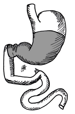
The Billroth II procedure is a variation to the Billroth I procedure pioneered by Dr. Theodor Billroth in 1881. The procedure is a a "subtotal gastrectomy" where gastrointestinal continuity after the resection is attained with an anastomosis between the stomach and the jejunum, a gastrojejunostomy.
The procedure was originally performed as a way to resect peptic ulcers caused by hyperacidity. Billroth removed up to 70% of the stomach. Modern variations of the procedure are less agressive, resecting only 50% of the distal stomach (a hemigastrectomy), or an antrectomy.
The reason for the Billroth II variation is the difficulty performing a gastroduodenostomy. This can be caused by a short abdominal esophagus, a short proximal gastric pouch or other reasons. The accompanying image shows the digestive tract before the resection. The area to the resected (specimen) is grayed out. If you hover your cursor over the image you will see the completed Billroth II procedure.
In the completed procedure you can see A: The stapled-closed duodenal stump. B: The gastrojejunostomy that allows passage of food from the stomach into the jejunum, and C: the staple-closed gastric stump that is not part of the gastrojejunostomy. Bear in mind that this sketch depicts only one of the many ways of performing this procedure
Images property of:CAA.Inc. Artist:Dr. E. Miranda
- Details
Hover your mouse on the image
The Billroth I procedure was pioneered by Dr. Theodor Billroth in 1881. The original procedure was described as a "subtotal gastrectomy" where gastrointestinal continuity after the resection was attained with an anastomosis between the stomach and the duodenum, a gastroduodenostomy.
The procedure was originally performed as a way to resect peptic ulcers caused by hyperacidity. Billroth removed up to 70% of the stomach. Modern variations of the procedure are less agressive, resecting only 50% of the distal stomach (a hemigastrectomy), or an antrectomy.
After Billroth's pioneering work, several variations on the procedure appeared (Polya, Hofmeister) as well as different techniques (open vs. laparoscopic), and the use of different materials, starting with carbolized silk to the modern endolaparoscopic surgical staplers.
The accompanying image shows the digestive tract before the resection. The area to the resected (specimen) is grayed out. If you hover your cursor over the image you will see the completed Billroth I procedure.
In the completed procedure you can see A: The stapled-closed proximal gastric pouch. B: The duodenum. The red arrow points to the gastroduodenostomy, that is, the anastomosis between the stomach and the duodenum which in this case was done in the posterior aspect of the proximal gastric pouch. Bear in mind that this sketch depicts only one of the many ways of performing this procedure
Images property of: CAA.Inc. Artist:Dr. E. Miranda
- Details
This article is part of the series "A Moment in History" where we honor those who have contributed to the growth of medical knowledge in the areas of anatomy, medicine, surgery, and medical research.

Original image courtesy of the
National Library of Medicine
Jean Louis Petit (1674 – 1750). French surgeon and anatomist, Jean Louis Petit was born in Paris in on March 13, 1674. His family rented an apartment at his house to Alexis Littre (1658 – 1726), a French anatomist. Petit became an apprentice of Littre at seven years of age, helping him in the dissections for his lectures and at an early age became the assistant in charge of the anatomic amphitheater.
Because of Petit’s dedication to anatomy and medicine, in 1690 at the age of sixteen, became a disciple of a famous Paris surgeon, Castel.
In 1692, Petit entered the French army and performed surgery in two military campaigns. By 1693 he started delivering lectures and was accepted as a great surgeon, being invited to the most difficult operations. In 1700 he was appointed Chief Surgeon of the Military School in Paris and in the same year he received the degree of Master of Surgery from the Faculty of Paris.
In 1715 he was made a member of the Royal Academy of Sciences and an honorary member of the Royal Society of London. He was appointed by the King as the first Director General of the Royal Academy of Surgery when it was founded in 1731.
Petit’s written works are of historical importance. “Traite des Maladies des Os” ( A Treatise on Bone Diseases); “Traite des Maladies Chirurgicales et des Operation” (A Treatise on Surgical Diseases and their Operations” This last book was published posthumously in 1774. He also published a monograph on hemorrhage, another on lachrymal fistula, and others.
He was one of the first to perform choIecystotomy and mastoidotomy. His original tourniquet design for amputations saved many in the battlefield and the design of the same surgical instrument today has not changed much since its invention by him.
His name is remembered in the lumbar triangle, also called the "triangle of Petit", and the abdominal hernia that can ensue through that area of weakness, the lumbar hernia or "Petit's hernia".
Sources:
1. “Jean Louis Petit – A Sketch of his Life, Character, and Writings” Hayne, AP San Fran Western Lancet 1875 4: 446-454
2. “Oeuvres compl?tes de Jean-Louis Petit” 1837 Imprimerie de F. Chapoulaud
3. Extraits de l'eloge de Jean-Louis Petit Ius dans Ia seance publique de I' Academie royale de chirurgie du 26 mai 1750” Louis A. Chirurgie 2001: 126 : 475- 81
- Details
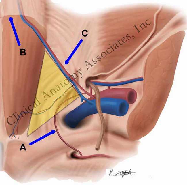
Corona mortis (A)
Important for inguinal hernia anatomy and surgery, this term is Latin from [corona] meaning "crown' and [mortis] meaning "death'; the "crown or circle of death". The corona mortis (blue arrow) refers to an anatomical variation1, a vascular anastomosis between the obturator and the external iliac vascular systems that passes over Cooper's pectineal ligament and posterior to the lacunar (Gimbernat's) ligament.
In some cases, the corona mortis is the actual obturator artery that arises from the inferior epigastric artery instead of the internal iliac artery. It can also arise from the external iliac artery. In both cases, it has been called an "aberrant obturator artery". This could be a misnomer, as this anatomical variation can be present in up to 25% of the cases. When present, the corona mortis can be injured when a surgeon looks to enlarge the femoral ring by opening the lacunar ligament from the anterior aspect. In this approach the "corona mortis" is not visible, as it is found immediately posterior to the lacunar ligament. This vascular structure could even be endangered in a laparoscopic procedure for inguinal of femoral hernia repair and a staple or tack is driven blindly into the pectineal (Cooper's) ligament.
Berberoglu states that "although these tiny anastomoses... have been described in classical anatomy textbooks, these texts neglect to mention that theses anastomoses can be life-threatening".
In some rare cases, the corona mortis (aberrant obturator artery) coexists with the normal obturator artery. Although called a [corona], this anatomical structure is an incomplete circle. In the image, the [corona mortis] is labeled "A".
Sources:
1. Rusu et al: "Anatomical considerations on the corona mortis" Surg Radiol Anat (2010) 32:17–24
2. Berberoglu et al: "An anatomic study in seven cadavers and an endoscopic study in 28 patients" Surg Endosc (2001) 15:72-75
Image property of:CAA.Inc.. Artist:M. Zuptich
- Details
UPDATED: The word [catamenial] is Greek. The prefix [cata-] arises from [, from [κάτω] (kato) meaning "down", "down because of", or "down to", the root term )[-men-] from [μήνας] (menas) meaning "month", referring to "lunar month" or to a female's menstrual cycle, which is usually just about a lunar month long, and the adjectival suffix [-ial] means "pertaining to".
[Catamenial] then means "to be down (sick) during a menstrual cycle" and refers to a condition that recurs in reference to menses.
Examples of the use of this word are:
• Catamenial depression or catamenial psychosis
• Catamenial pneumothorax - related to endometriosis
• Catamenial epilepsy or seizures
The links will open scholarly articles that use this word.