![]()
Medical Terminology Daily (MTD) is a blog sponsored by Clinical Anatomy Associates, Inc. as a service to the medical community. We post anatomical, medical or surgical terms, their meaning and usage, as well as biographical notes on anatomists, surgeons, and researchers through the ages. Be warned that some of the images used depict human anatomical specimens.
You are welcome to submit questions and suggestions using our "Contact Us" form. The information on this blog follows the terms on our "Privacy and Security Statement" and cannot be construed as medical guidance or instructions for treatment.
We have 578 guests and no members online
")
Marcia Crocker Noyes
(1869 – 1946)
Further to my comment on old books and research that started with an interesting bookplate (Ex-Libris). I continued my research and found that the person in charge of the Osler library bookplate was a fascinating individual that today maybe a ghost in the MedChi library and building in Baltimore... This is certainly an article that can be called "A Moment in History"
Marcia Crocker Noyes was the librarian at The Maryland State Medical Society from 1896 to 1946 and was a founding member of the Medical Library Association.[1][2][3]
Sir William Osler, MD. a famous Johns Hopkins surgeon was a noted bibliophile and had a large personal collection of books on various topics. When he became the President of MedChi in 1896, he was dismayed at the condition of the library and knew that with the right person and some stewardship, it could become a significant collection. Sir William asked his friend, Dr. Bernard Steiner, a physician and President of the Enoch Pratt Free Library in Baltimore for suggestions of a librarian, and Dr. Steiner recommended Marcia Crocker Noyes. A native of New York, and a graduate of Hunter College, Marcia had moved to Baltimore for a lengthy visit with her sister, and took a “temporary” position at the Pratt Library, which turned into three years. Although she had no medical experience or background, she was enthusiastic, and most importantly, she was willing to move into the apartment provided for the librarian, who needed to be available 24 hours a day.
The image in this article is Ms. Noyes on her first year on the job. Marcia developed a book classification system for medical books, based on the Index Medicus, and called it the Classification for Medical Literature. The system uses the alphabet with capital letters for the major divisions of medicine and lower-case ones for the sub-sections. The system was used for many years, but it's now dated and the Faculty's original shelving scheme was never changed. The card catalogs still reflect her classification and many of the cards are written in Marcia's back-slanting handwriting.
Marcia knew enough to ask the Faculty's members about medical questions, terminology and literature. She gradually won over the predominantly male membership and they became her greatest allies; Sir William at the start, and then for nearly 40 years, Dr. John Ruhräh, a wealthy pediatrician with no immediate family of his own. She made a point of attending almost every Faculty function, and in 1904, under guidelines from the American Medical Association, Marcia was made the Faculty Secretary. For much of her first 10 years, she was the Faculty's only full-time employee, only being assisted by Mr. Caution, the Faculty's janitor. Later in life Marcia would say that she hired him because of his name!
Within ten years, the library had outgrown its space, and plans, spearheaded by Marcia and Sir William before his move to Oxford, were made to build a headquarters building, mainly to house the library's growing collection of medical books and journals.
Marcia was instrumental in the design and building of the new headquarters. She travelled to Philadelphia, New York and Boston to look at their medical society buildings, and eventually, the Philadelphia architectural firm, Ellicott & Emmart was selected to design and build the new Faculty building. Every detail of the building held her imprimatur, from the graceful staircase, to the light-filled reading room, and all of the myriad details of the millwork, marble tesserae, and most of all, the four-story cast iron stacks. She was on-site, climbing up unfinished staircases, checking out the progress of the building, which was built in less than one year at a cost of $90,000.
Among the features of the new building was a fourth-floor apartment for her. She referred to it as the "first penthouse in Baltimore" and it had a garden and rooftop terrace. The library collection eventually grew to more than 65,000 volumes from medical and specialty societies around the world. Journals were traded back and forth, and physicians eagerly anticipated the arrival of each new issue. At the same time, Marcia was involved in the Medical Library Association as one of eight founding members. The MLA promotes medical libraries and the exchange of information. One of the earliest mandates of the MLA was the Exchange, a distribution and trade service for those who had duplicates or little-used books in their collections. Initially, the Exchange was run out of the Philadelphia medical society, but in 1900 it was moved to Baltimore and Marcia oversaw it. Several hundred periodicals and journals were received and sent each month, a huge amount of work for a tiny staff. In 1904, the Faculty had run out of room to manage the Exchange, so it was moved to the Medical Society of the Kings County (Brooklyn). But without Marcia's excellent administrative skills, it floundered and in 1908, the MLA asked Marcia to take charge once again.
In 1909, when the new Faculty building opened, there was enough room to run the Exchange and with the help of MLA Treasurer, noted bibliophile and close friend, Dr. John Ruhräh, it once again became successful. Additionally, Marcia and Dr. Ruhräh combined forces to revive the MLA's bulletin, which had all but ceased publication in 1908, taking the Exchange with it. This duo maintained editorial control from 1911 until 1926. In 1934, around the time of Dr. Ruhräh's death, Marcia became the first “unmedicated” professional to head the MLA. During her tenure, the MLA incorporated, the first seal was adopted, and the annual meeting was held in Baltimore. Marcia wanted to write the history of the MLA once she retired from full-time work at the Faculty, but her health was beginning to fail. She had back problems and had suffered a serious burn on her shoulder as a young woman, possibly from her time running a summer camp, Camp Seyon, for young ladies in the Adirondack Mountains. In 1946, a celebration was planned to honor Marcia's 50 years at the Faculty. But she was adamant that the physicians wait until November, the actual date of her 50 years. However, they knew she was gravely ill, and might not make it until then, so a huge party was held in April. More than 250 physicians attended the celebration, but the ones she was closest to in the early years, were long gone. She was presented with a suitcase, a sum of money to use for travelling, and her favorite painting of Dr. John Philip Smith, a founder of the Medical College in Winchester, Virginia. It was painted by Edward Caledon Smith, a Virginia painter who had been a student of the painter Thomas Sully.[4] She adored this painting and vowed, jokingly, to take it with her wherever she went.
The painting was not to stay with her for very long, for she died in November 1946, and left it to the Faculty in her will. Her funeral was held in the Faculty's Osler Hall, named for her dear friend. More than 60 physicians served as her pallbearers, and she was buried at Baltimore's Green Mount Cemetery. In 1948, the MLA decided to establish an award in the name of Marcia Crocker Noyes. It was for outstanding achievement in medical library field and was to be awarded every two years, or when a truly worthy candidate was submitted. In 2014, the Faculty began giving a bouquet of flowers to the winner of the award in Marcia's name, and in honor of her work. Much evidence exists for this tradition, as we know that the physicians, especially Drs. Osler and Ruhräh, frequently gave her bouquets of flowers. Marcia also cultivated flower gardens at the Faculty and decorated the rooms with her work.
Today, the MedChi building is open for tours and if the rumors are to be believed Ms. Marcia Crocker Noyes is still at work in her beloved library as the "resident ghost" [1][5]

NOTE: This article has been modified from the original Wikipedia article on Marcia Crocker Noyes. The article itself is well-written with interesting images of the subject. I would encourage you to visit it. The second insert is from book 00736 in my personal library and shows in pencil, the incredibly small handwriting of Marsha C. Noyes.
Sources:
1. "Marcia, Marcia, Marcia" MedChi Archives blog.
2. "Marcia C. Noyes, Medical Librarian" (PDF). Bulletin of the Medical Library Association. 35 (1): 108–109. 1947. PMC 194645
3. Smith, Bernie Todd (1974). "Marcia Crocker Noyes, Medical Librarian: The Shaping of a Career" (PDF). Bulletin of the Medical Library Association. 62 (3): 314–324. PMC 198800Freely accessible. PMID 4619344.
4. Edward Caledon BRUCE (1825-1901)"
5. Behind the scenes tour MedChiBuilding
"Clinical Anatomy Associates, Inc., and the contributors of "Medical Terminology Daily" wish to thank all individuals who donate their bodies and tissues for the advancement of education and research”.
Click here for more information


- Details
Click for a larger image
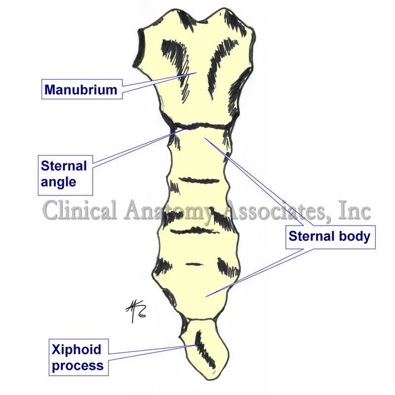
UPDATED: The word [manubrium] is Latin and mean "handle", referring to the area where a person holds an instrument or device. To exemplify this, in Spanish the vernacular use of the word [manubrio] refers to the handles of bicycle or even the steering wheel of a car.
In anatomy, the term is used with the same meaning. In the malleus, a hammer-like ossicle of the middle ear, the manubrium is the handle-like extension of the bone that attaches to the tympanic membrane.
In the case of the sternum, the [manubrium sterni] is the superior portion bound by the sternal angle (of Louis) inferiorly. The use of the word manubrium can be explained because in early anatomy, the sternum was known by the Latin term [gladius] referring to the similarity of the sternum to the short sword of the gladiators. The area where you hold the sword is the handle, ergo, manubrium.
The manubrium has a superior and median notch called the "suprasternal notch" or the "jugular notch". It is important because in the case of a mediastinoscopy, the incision is made just superior to this landmark. The manubrium articulates superolaterally with the clavicle and inferolaterally with the superior aspect of the cartilage of the second rib. The rest of the rib cartilage articulates with the body of the sternum.
Image property of:CAA.Inc.. Artist: Mark J. Zuptich
- Details
The word [induration] arises from the Latin words induratio, meaning "thick or hard" and indurare, meaning "hardening".
It refers to a pathological hardening of tissues caused by tumoration or edema, increase of fibrous or connective tissue, or other causes. It is a good, descriptive term when stating a patient's symptoms. The term has been in use in English since the 14th century.
Note: The links to Google Translate include an icon that will allow you to hear the pronunciation of the word.
- Details
At the beginning of 1998, Clinical Anatomy Associates, Inc. was formed as an Ohio Corporation. Our mission is to deliver industry relevant, cutting-edge Training, Marketing, and R&D services that will enable our clients to gain a competitive advantage. Over the past two decades, Clinical Anatomy Associates, Inc. has become the go-to R&D resource for feasibility studies that require cadaver studies and anatomical research. We are also a preferred training solution for Sales Representatives, Distributors, Engineers, Clinicians, and Marketing Managers in the areas of Medical Terminology, Clinical Anatomy, and Surgical Procedures. Our expertise allows us to deliver training in a variety of medical and anatomical topics.
In 2012 Dr. Efrain A. Miranda, CEO of Clinical Anatomy Associates started "Medical Terminology Daily" (MTD), a website/blog as a service to the medical community, medical students, and the medical industry. MTD posts medical or surgical terms, its meaning and usage, as well as biographical notes on anatomists, surgeons, and researchers through the ages. These posts are also shared on Facebook to a group of followers.
20 year anniversary for Clinical Anatomy Associates, Inc. and 6 years for Medical Terminology Daily! Help us congratulate our staff and specially the contributors and friends of Medical Terminology Daily.
Our thanks to all our customers, friends, and contributors for an amazing 20 years!!! Looking forward to more!!
- Details
- Written by: Prof. Claudio R. Molina, MSc
Click for a larger image
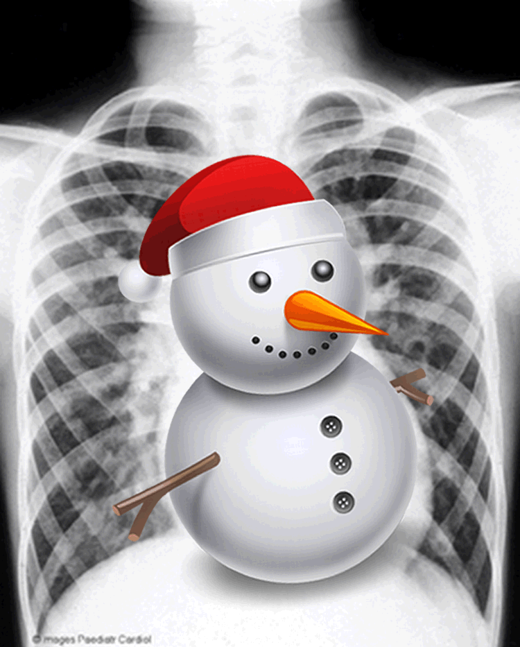
The “snowman sign” is a particular image on a chest X-Ray image, which is seen in anomalous pulmonary venous drainage and coarctation of the aorta which causes a Total Anomalous Pulmonary Venous Return (TAPVR).
This abnormality occurs when the pulmonary veins fail to drain into the left atrium and instead form an aberrant connection with some others cardiovascular structures. Such abnormalities account for approximately 2% of cardiac malformations.
There are four types of TAPVR; type 1 is the most common (and the one that creates the snowman sign). In this case the pulmonary veins terminate at the supracardiac level, emptying into the right atrium by way of an anomalous pulmonary venous drainage into the superior vena cava (SVC), and the left brachiocephalic vein (by way of a vertical vein). The confluence of these veins dilates the right brachiocephalic vein, which appears as a dilated vessel on the right of the upper mediastinal edge. When seen in an AP Chest X-Ray, the TAPVAR type 1, resembles a snowman; the dilated vertical vein on the left, the right brachiocephalic vein superiorly, and the SVC on the right form the head of the snowman, the body is formed by the enlarged right atrium.
Article written by: Prof. Claudio R. Molina, MsC.
Sources:
1. Emma C. Ferguson, Rajesh Krishnamurthy, and Sandra A. A. Oldham. (2007) Classic Imaging Signs of Congenital Cardiovascular Abnormalities. RadioGraphics 27:5, 1323-1334.
2. Somerville, J., & Grech, V. (2009). The chest x-ray in congenital heart disease 1. Total anomalous pulmonary venous drainage and coarctation of the aorta. Images in Paediatric Cardiology, 11(1), 7–9.
- Details

Click for a larger image
Kabourophobia is the fear of crabs and lobsters.
The etymology of the word [kabourophobia] comes from the Greek word [καβουρης] (pronounced “kavouris”), meaning [crab], and the suffix [-phobia], also from the Greek, arises from the word [φοβία] (pronounced “fovía”)
Kabourophobia is an extremely rare phobia, but it was brought to the public’s attention when a modern pop singer stated that she was afraid of crabs. Also, a prank (maybe acted) was shown on video on the internet with a man surrounded by lobsters.
Kabourophobia is very specific, and it can also be a part of a wider phobia called ostraconophobia, which is the fear of crustaceans, adding shrimp, oysters, clams, crabs, lobsters, etc.
An interesting point is that the word [crab] in Greek has another acception, that is the word [Καρκίνος] (pronounced “karkinos”), which is the root for the medical term [cancer].
We thank Jackie Miranda-Klein for her contribution suggesting this word.
- Details

Galen of Pergamon
(129AD - 200AD
The word sympathetic is the adjectival form of sympathy. This word arises from the Greek [συμπάθεια]and is composed of [syn/sym] meaning “together” and [pathos], a word which has been used to mean “disease”. In reality “pathos” has to do more with the “feeling of self”. Based on this, the word sympathy means “together in feeling”, which is what we use today.
How the term got to be used to denote a component of the so-called autonomic nervous system is part of the history of Medicine and Anatomy.
Galen of Pergamon (129AD-200AD), whose teachings on Medicine and Anatomy lasted as indisputable for almost 1,500 years, postulated that nerves were hollow and allowed for “animal spirits” to travel between organs and allowed the coordinated action of one with the other, in “sympathy” with one another. As the knowledge of the components of the nervous system grew, this concept of “sympathy” stayed, becoming a staple of early physiological theories on the action of the nervous system.
Jacobus Benignus Winslow (1669-1760) named three “sympathetic nerves” one of them was the facial nerve (the small sympathetic), the other the vagus nerve, which he called the “middle sympathetic”, and the last was what was known then as the “intercostalis nerve of Willis” or “large sympathetic", today’s sympathetic chain. Other nerves that worked coordinated with this “sympathetics” were considered to work in parallel with it. It is from this concept that the term “parasympathetic” arises.
Interestingly, the ganglia on the sympathetic chain were for years known as “small brains” and it was postulated that there was a separate multi-brain system coordinating the action of the thoracic and abdominopelvic viscera. The coordination between this “autonomous nervous system” and the rest of the body was made by way of the white and gray rami communicantes.
Today we know that there is only one brain and only one nervous system with an autonomic component which has a “sympathetic” component that is mostly in charge of the “fight or flight” reaction and a “parasympathetic” component that has a “slow down” or “depressor” function. Both work coordinated, so I guess Galen was not "off the mark" after all.
So, we still use the terms “sympathetic” and “parasympathetic”, but the origin of these terms has been blurred by history.
Sources:
1. "Claudius Galenus of Pergamum: Surgeon of Gladiators. Father of Experimental Physiology" Toledo-Pereyra, LH; Journal of Investigative Surgery, 15:299-301, 2002
2. "The Origin of Medical Terms" Skinner, HA 1970 Hafner Publishing Co.
3. "Medical Meanings:A Glossary of Word Origins" Haubrish, WS American College of Physicians Philadelphia, 1997
4. "The History of the Discovery of the Vegetative (Autonomic) Nervous System" Ackerknecht, EH Medical History, 1974 Vol 18.
Original image courtesy of Images from the History of Medicine at nih.gov
Note: The links to Google Translate include an icon that will allow you to hear the pronunciation of the word.