
Medical Terminology Daily (MTD) is a blog sponsored by Clinical Anatomy Associates, Inc. as a service to the medical community. We post anatomical, medical or surgical terms, their meaning and usage, as well as biographical notes on anatomists, surgeons, and researchers through the ages. Be warned that some of the images used depict human anatomical specimens.
You are welcome to submit questions and suggestions using our "Contact Us" form. The information on this blog follows the terms on our "Privacy and Security Statement" and cannot be construed as medical guidance or instructions for treatment.
We have 2791 guests online
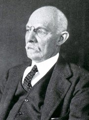
William S. Halsted, MD
(1852 – 1922)
American anatomist, teacher, and surgeon, William Stewart Halsted was born in New York City, USA to a wealthy family of English origin. His father was involved in charitable work and Governor and trustee to a city hospital. Not a brilliant student initially, Halsted took an undergraduate in Liberal Arts in Yale, CT., after which he entered the Medical College of Physicians at the Columbia College, where he excelled.
As a second-year medical student Halsted applied and obtained a position in surgery at a local hospital. In here he learned about Lister’s antiseptic technique and became an adamant proponent of it to reduce infection. In 1877 Halsted obtained his MD. After a short time as House Physician at the New York Hospital, Halsted traveled to Europe to further his education studying for two years at the Universities of Vienna, Leipzig, and W?rzburg.
Besides being at the forefront of surgical and antiseptic techniques (introducing the use of rubber gloves in surgery), Halsted was extremely concerned with the way medical students were taught in the US. He pioneered bedside clinical round discussions with the medical students after two years of basic sciences studies. Halsted developed the idea of a patient chart; he also developed the residency program for medical students in use today.
Halsted is probably the most influential researcher and surgeon at the turn of the century. He dedicated time to the study of intestinal anastomoses and the use of silk as a suture material. His experimental work in 1887 proved that the inclusion of the submucosa layer in an anastomosis was mandatory, as well that a single layered anastomosis was enough to attain closure. Perhaps Halsted’s most important contribution was the application and use of the scientific method to surgical questions. Halsted’s principles set the standards used today in surgical suturing and surgical stapling.
He also pioneered the development and surgical techniques for radical mastectomy as a treatment for breast cancer.
As a side effect of this studied in anesthesia and the use of cocaine for anesthesia, Halsted became addicted to this substance, a problem that followed him through the years. Without impairing his capacity as a researcher and a surgeon, Halsted eventually recovered. He died in Baltimore in 1922 as a complication to surgery.
Sources:
1. Dubay, A. D., & Franz, G. M. (2003). Acute Wound Healing: The Biology of Acute Wound Failure. Surg Clin NA, 83, 463-481.
2. Halsted, W. S. (1887). Circular Suture of the Intestine - An Experimental Study. Am J Med Sci, 436-461.
3. “William Stewart Halsted: his life and contributions to surgery” Osborne, P. Lancet Oncol 2007; 8: 256–65
4. “William Stewart Halsted: Surgical pioneer” Burress, P Endoc Today (2010), 8: (2) 22
5. “William Stewart Halsted (1852–1922) Neurological stamp” Haas, LF J Neurol Neurosurg Psych 2000;69:641
Original image courtesy of "Images from the History of Medicine" at www.nih.gov

"Clinical Anatomy Associates, Inc., and the contributors of "Medical Terminology Daily" wish to thank all individuals who donate their bodies and tissues for the advancement of education and research”.
Click here for more information


- Details
This medical term is Greek and is composed of [γιατρός] (iatros) meaning "doctor", "physician", or "healer" and the suffix [-(o)genic], meaning "creation", "born of", or "beggining". An iatrogenic condition is that which is caused or created by the doctor or the hospital.
This is an expensive word, as iatrogenic conditions may lead to a lawsuit!
- Details
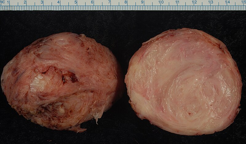
Click for a larger image
The word [leiomyoma] is of Greek origin with combined root terms. The term [-lei(o)-] arises from the Greek [λείος] meaning "smooth", the other root is [μυς] (mys) meaning "muscle". The suffix [-oma] means "tumor" or "mass". A [leiomyoma] is a "smooth muscle tumor". The medical plural form is [leiomyomata], or it can be [leiomyomas].
Since smooth muscle is involuntary muscle, leiomyomata are usually found in viscera. The most common leiomyomata are found in the uterus (see image), in the muscular or submucosal layer of the digestive system, mostly jejunum and ileum, and gallbladder, or in smooth muscle of the skin. The term itself does not imply that leiomyomata are cancerous, and most leiomyomata are not.
- Details
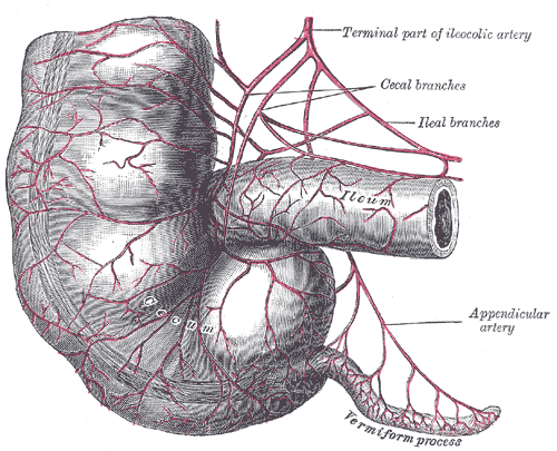
Terminal ileum, cecum,
and vermiform appendix
The [mesoappendix] is a triangular-shaped double-layered peritoneal membrane related to the vermiform appendix. One of the sides attaches to the vermiform appendix, the other is free, and the third one attaches to the ileum and the cecum. This last attachment varies in extension, giving the cecum varying degrees of mobility.
The mesoappendix contains the appendicular artery. This artery arises either from the ileocolic artery or the from the posterior ileocecal artery. The mesoappendix also contains the appendicular veins, lymphatics, lymphatic nodes, and fat.
In the female, there can be an extension of the mesoappendix that communicates with the broad ligament of the uterus. It is called the appendiculoovarian ligament, or Clado's ligament. This ligament may contain the appendiculoovarian artery, an anastomosis between the appendicular artery and the ovarian artery. The lymphatics contained in this appendiculoovarian ligament can also establish a lymphatic communication between the ovary and the vermiform appendix.
Sources:
1 "Tratado de Anatomia Humana" Testut et Latarjet 8 Ed. 1931 Salvat Editores, Spain
2. "Anatomy of the Human Body" Henry Gray 1918. Philadelphia: Lea & Febiger Image modified by CAA, Inc. Original image by Henry Vandyke Carter, MD., courtesy of bartleby.com
- Details
This article is part of the series "A Moment in History" where we honor those who have contributed to the growth of medical knowledge in the areas of anatomy, medicine, surgery, and medical research.

Click for a larger image
Alessandra Giliani (1307 – 1326). Italian prosector and anatomist. Alessandra Giliani is the first woman to be on record as being an anatomist and prossector. She was born on 1307 in the town of Persiceto in northern Italy.
She was admitted to the University of Bologna circa 1323. Most probably she studied philosophy and the foundations of anatomy and medicine. She studied under Mondino de Luzzi (c.1270 – 1326), one of the most famous teachers at Bologna.
Giliani was the prosector for the dissections performed at the Bolognese “studium” in the Bologna School of Anatomy. She developed a technique (now lost to history) to highlight the vascular tree in a cadaver using fluid dyes which would harden without destroying them. Giliani would later paint these structures using a small brush. This technique allowed the students to see even small veins.
Giliani died at the age of 19 on March 26, 1326, the same year that her teacher Mondino de Luzzi died. It is said that she was buried in front of the Madonna delle Lettere in the church of San Pietro e Marcellino at the Hospital of Santa Maria del Mareto in Florence by Otto Agenius Lustrulanus, another assistant to Modino de Luzzi.
Some ascribe to Agenius a love interest in Giliani because of the wording of the plaque that is translated as follows:
"In this urn enclosed are the ashes of the body of
Alessandra Giliani, a maiden of Persiceto.
Skillful with her brush in anatomical demonstrations
And a disciple equaled by few,
Of the most noted physician, Mondino de Luzzi,
She awaits the resurrection.
She lived 19 years: She died consumed by her labors
March 26, in the year of grace 1326.
Otto Agenius Lustrulanus, by her taking away
Deprived of his better part, inconsolable for his companion,
Choice and deservinging of the best from himself,
Has erected this plaque"
Sir William Osler says of Alessandra Giliani “She died, consumed by her labors, at the early age of nineteen, and her monument is still to be seen”
The teaching of anatomy in the times of Mondino de Luzzi and Alessandra Giliani required the professor to be seated on a high chair or “cathedra” from whence he would read an anatomy book by Galen or another respected author while a prosector or “ostensor” would demonstrate the structures to the student. The professor would not consider coming down from the cathedra to discuss the anatomy shown. This was changed by Andreas Vesalius.
The image in this article is a close up of the title page of Mondino’s “Anothomia Corporis Humani” written in 1316, but published in 1478. Click on the image for a complete depiction of this title page. I would like to think that the individual doing the dissection looking up to the cathedra and Mondino de Luzzi is Alessandra Giliani… we will never know.
The life and death of Alessandra Giliani has been novelized in the fiction book “A Golden Web” by Barbara Quick.
Sources
1. “Books of the Body: Anatomical Ritual and Renaissance Learning” Carlino, A. U Chicago Press, 1999
2. “Encyclopedia of World Scientists” Oakes, EH. Infobase Publishing, 2002
3. “The Biographical Dictionary of Women in Science”Harvey, J; Ogilvie, M. Vol1. Routledge 2000
4. “The Evolution of Modern Medicine” Osler, W. Yale U Press 1921
5. “The Mondino Myth” Pilcher, LS. 1906
Original image courtesy of NLM

- Details

Click for a larger image
The word [peritoneum] has a Greek origin [περίτόνοςαιον]. Loosely translated it has the prefix [peri-] meaning "around", the root [-ton-] from the Greek [tonos], meaning "to stretch", and the suffix [-eum] meaning "a membrane". It is "a membrane that is stretched around".
The peritoneum is a thin serosal membranous sac found in the abdominopelvic cavity. Histologically it is composed of a layer of mesothelium supported by a layer of connective tissue. Being a serosal sac, it contains in its interior a small amount of peritoneal fluid. The pathological accumulation of peritoneal fluid is called ascitis.
Although the peritoneum is one continuous membrane, and because of its relation to the organs and the abdominal wall, the peritoneum is described as formed by two components:
• Parietal peritoneum: The parietal peritoneum is that portion of the peritoneal sac related to or in contact with the walls of the abdomen and the pelvis.
• Visceral peritoneum: The visceral peritoneum is that portion of the peritoneal sac related to or in contact with the abdominopelvic viscera. I this case the peritoneum encases the viscera almost completely and is referred to as their serosa layer i,e: serosa layer of the ileum.
The double-layered portions of the peritoneal sac that stretch between organs or between organs and the abdominal wall are known by different names. They can be called an abdominopelvic [ligament], a [mesentery], a [meso..(something)], or an [omentum]. These structures are covered in separate articles. Some of these structures are:
- Falciform ligament: A sickle-shaped double fold of peritoneum related to the liver
- Ligament of Treitz: Also known as the "suspensory ligament of the duodenum"
- Infundibulopelvic ligament: A fold of peritoneum containing the ovarian arteries and veins
- Lesser omentum: The lesser omentum is one of the two double-folds of peritoneum related to the stomach
- Mesosigmoid: A double peritoneal membrane related to the sigmoid colon
- Transverse mesocolon: A double peritoneal membrane related to the transverse colon
- Mesoappendix: A double peritoneal membrane related to the vermiform appendix, etc.
Sources:
1. "Clinically Oriented Anatomy" Moore, KL. 3r Ed. Williams & Wilkins 1992
2. "The origin of Medical Terms" Skinner, AH, 1970
3. "Tratado de Anatomia Humana" Testut et Latarjet 8 Ed. 1931 Salvat Editores, Spain
Image modified from the original from Testut and Latajet, 1931. Public domain.
Thanks to Dr. Randall Wolf for suggesting this article
- Details
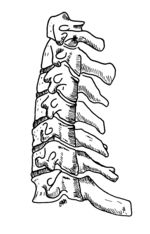
A derivate of the Latin root [cervix] or [cervicis] meaning "neck". The word [cervical] means "pertaining to the neck".
The term is used in many areas and structures of the human body:
• Cervical spine: refers to the spinal column region formed by the seven cervical vertebrae. See image
• Uterine cervix: The inferior region of the uterus which projects partially into the vagina.
• Cervical rib: An anatomic variation where one or more supernumerary ribs are found related to the lower cervical vertebrae. This anomaly can cause clinical symptoms.
Images property of: CAA.Inc. Artist: Dr. E. Miranda