![]()
Medical Terminology Daily (MTD) is a blog sponsored by Clinical Anatomy Associates, Inc. as a service to the medical community. We post anatomical, medical or surgical terms, their meaning and usage, as well as biographical notes on anatomists, surgeons, and researchers through the ages. Be warned that some of the images used depict human anatomical specimens.
You are welcome to submit questions and suggestions using our "Contact Us" form. The information on this blog follows the terms on our "Privacy and Security Statement" and cannot be construed as medical guidance or instructions for treatment.
We have 1512 guests online
")
Marcia Crocker Noyes
(1869 – 1946)
Further to my comment on old books and research that started with an interesting bookplate (Ex-Libris). I continued my research and found that the person in charge of the Osler library bookplate was a fascinating individual that today maybe a ghost in the MedChi library and building in Baltimore... This is certainly an article that can be called "A Moment in History"
Marcia Crocker Noyes was the librarian at The Maryland State Medical Society from 1896 to 1946 and was a founding member of the Medical Library Association.[1][2][3]
Sir William Osler, MD. a famous Johns Hopkins surgeon was a noted bibliophile and had a large personal collection of books on various topics. When he became the President of MedChi in 1896, he was dismayed at the condition of the library and knew that with the right person and some stewardship, it could become a significant collection. Sir William asked his friend, Dr. Bernard Steiner, a physician and President of the Enoch Pratt Free Library in Baltimore for suggestions of a librarian, and Dr. Steiner recommended Marcia Crocker Noyes. A native of New York, and a graduate of Hunter College, Marcia had moved to Baltimore for a lengthy visit with her sister, and took a “temporary” position at the Pratt Library, which turned into three years. Although she had no medical experience or background, she was enthusiastic, and most importantly, she was willing to move into the apartment provided for the librarian, who needed to be available 24 hours a day.
The image in this article is Ms. Noyes on her first year on the job. Marcia developed a book classification system for medical books, based on the Index Medicus, and called it the Classification for Medical Literature. The system uses the alphabet with capital letters for the major divisions of medicine and lower-case ones for the sub-sections. The system was used for many years, but it's now dated and the Faculty's original shelving scheme was never changed. The card catalogs still reflect her classification and many of the cards are written in Marcia's back-slanting handwriting.
Marcia knew enough to ask the Faculty's members about medical questions, terminology and literature. She gradually won over the predominantly male membership and they became her greatest allies; Sir William at the start, and then for nearly 40 years, Dr. John Ruhräh, a wealthy pediatrician with no immediate family of his own. She made a point of attending almost every Faculty function, and in 1904, under guidelines from the American Medical Association, Marcia was made the Faculty Secretary. For much of her first 10 years, she was the Faculty's only full-time employee, only being assisted by Mr. Caution, the Faculty's janitor. Later in life Marcia would say that she hired him because of his name!
Within ten years, the library had outgrown its space, and plans, spearheaded by Marcia and Sir William before his move to Oxford, were made to build a headquarters building, mainly to house the library's growing collection of medical books and journals.
Marcia was instrumental in the design and building of the new headquarters. She travelled to Philadelphia, New York and Boston to look at their medical society buildings, and eventually, the Philadelphia architectural firm, Ellicott & Emmart was selected to design and build the new Faculty building. Every detail of the building held her imprimatur, from the graceful staircase, to the light-filled reading room, and all of the myriad details of the millwork, marble tesserae, and most of all, the four-story cast iron stacks. She was on-site, climbing up unfinished staircases, checking out the progress of the building, which was built in less than one year at a cost of $90,000.
Among the features of the new building was a fourth-floor apartment for her. She referred to it as the "first penthouse in Baltimore" and it had a garden and rooftop terrace. The library collection eventually grew to more than 65,000 volumes from medical and specialty societies around the world. Journals were traded back and forth, and physicians eagerly anticipated the arrival of each new issue. At the same time, Marcia was involved in the Medical Library Association as one of eight founding members. The MLA promotes medical libraries and the exchange of information. One of the earliest mandates of the MLA was the Exchange, a distribution and trade service for those who had duplicates or little-used books in their collections. Initially, the Exchange was run out of the Philadelphia medical society, but in 1900 it was moved to Baltimore and Marcia oversaw it. Several hundred periodicals and journals were received and sent each month, a huge amount of work for a tiny staff. In 1904, the Faculty had run out of room to manage the Exchange, so it was moved to the Medical Society of the Kings County (Brooklyn). But without Marcia's excellent administrative skills, it floundered and in 1908, the MLA asked Marcia to take charge once again.
In 1909, when the new Faculty building opened, there was enough room to run the Exchange and with the help of MLA Treasurer, noted bibliophile and close friend, Dr. John Ruhräh, it once again became successful. Additionally, Marcia and Dr. Ruhräh combined forces to revive the MLA's bulletin, which had all but ceased publication in 1908, taking the Exchange with it. This duo maintained editorial control from 1911 until 1926. In 1934, around the time of Dr. Ruhräh's death, Marcia became the first “unmedicated” professional to head the MLA. During her tenure, the MLA incorporated, the first seal was adopted, and the annual meeting was held in Baltimore. Marcia wanted to write the history of the MLA once she retired from full-time work at the Faculty, but her health was beginning to fail. She had back problems and had suffered a serious burn on her shoulder as a young woman, possibly from her time running a summer camp, Camp Seyon, for young ladies in the Adirondack Mountains. In 1946, a celebration was planned to honor Marcia's 50 years at the Faculty. But she was adamant that the physicians wait until November, the actual date of her 50 years. However, they knew she was gravely ill, and might not make it until then, so a huge party was held in April. More than 250 physicians attended the celebration, but the ones she was closest to in the early years, were long gone. She was presented with a suitcase, a sum of money to use for travelling, and her favorite painting of Dr. John Philip Smith, a founder of the Medical College in Winchester, Virginia. It was painted by Edward Caledon Smith, a Virginia painter who had been a student of the painter Thomas Sully.[4] She adored this painting and vowed, jokingly, to take it with her wherever she went.
The painting was not to stay with her for very long, for she died in November 1946, and left it to the Faculty in her will. Her funeral was held in the Faculty's Osler Hall, named for her dear friend. More than 60 physicians served as her pallbearers, and she was buried at Baltimore's Green Mount Cemetery. In 1948, the MLA decided to establish an award in the name of Marcia Crocker Noyes. It was for outstanding achievement in medical library field and was to be awarded every two years, or when a truly worthy candidate was submitted. In 2014, the Faculty began giving a bouquet of flowers to the winner of the award in Marcia's name, and in honor of her work. Much evidence exists for this tradition, as we know that the physicians, especially Drs. Osler and Ruhräh, frequently gave her bouquets of flowers. Marcia also cultivated flower gardens at the Faculty and decorated the rooms with her work.
Today, the MedChi building is open for tours and if the rumors are to be believed Ms. Marcia Crocker Noyes is still at work in her beloved library as the "resident ghost" [1][5]

NOTE: This article has been modified from the original Wikipedia article on Marcia Crocker Noyes. The article itself is well-written with interesting images of the subject. I would encourage you to visit it. The second insert is from book 00736 in my personal library and shows in pencil, the incredibly small handwriting of Marsha C. Noyes.
Sources:
1. "Marcia, Marcia, Marcia" MedChi Archives blog.
2. "Marcia C. Noyes, Medical Librarian" (PDF). Bulletin of the Medical Library Association. 35 (1): 108–109. 1947. PMC 194645
3. Smith, Bernie Todd (1974). "Marcia Crocker Noyes, Medical Librarian: The Shaping of a Career" (PDF). Bulletin of the Medical Library Association. 62 (3): 314–324. PMC 198800Freely accessible. PMID 4619344.
4. Edward Caledon BRUCE (1825-1901)"
5. Behind the scenes tour MedChiBuilding
"Clinical Anatomy Associates, Inc., and the contributors of "Medical Terminology Daily" wish to thank all individuals who donate their bodies and tissues for the advancement of education and research”.
Click here for more information


- Details
Click for a larger image
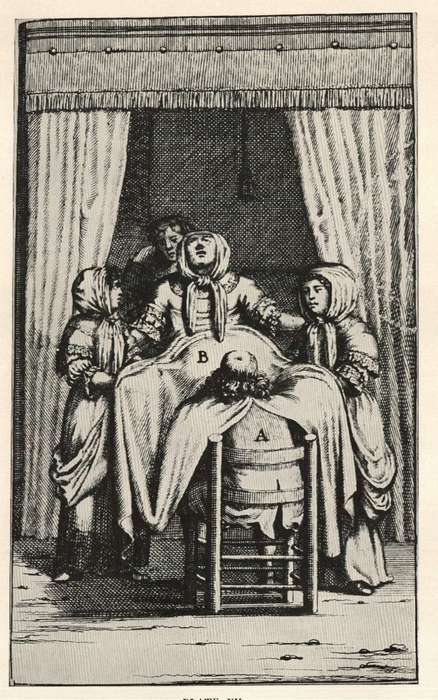
The term [obstetrics] arises from the Latin word [obstare] which means "to stand close to" or "to stand in front of". Since a midwife "stands" in front of the patient for child delivery, the word was used in its feminine singular form [obstetrix]. The Latin plural is [obstetrices]; The evolution of the term into modern "obstetrics" is quite clear.
Through the ages the term used for someone who helps deliver a child has changed. Andreas Vesalius used the term [obstetrices nutrices] meaning "nurse midwife". The term "midwife" was used for a long time, being replaced today for the word "obstetrics" and "obstetrician".
Although all female reproductive system care was originally the domain of midwives, through time two distinct specialties have evolved. Gynecology refers to the medical specialty that studies and treats the female reproductive system. Obstetrics, deals with the care of the pregnant patient and delivery of the fetus. Male-widwives in Europe were allowed access to the patient only with the use of a "modesty blanket". This is plate XV from the 1681 book "Korte en Bondige Van Der Voortteeling en Kinderbaren" by Samuel Janson.
As an interesting side note in history, the first male physician to attempt to work as a man-midwife was Dr. Wertt from Hamburg. Dr Wertt decided to disguise himself as a woman to attend patients. When he was discovered, the punishment was "swift and salutary": He was burned at the stake.
Word suggested and edited by: Dr. Sanford S. Osher , MTD Contributor- Details
This article is part of the series "A Moment in History" where we honor those who have contributed to the growth of medical knowledge in the areas of anatomy, medicine, surgery, and medical research.

Avicenna
Avicenna (980 AD – 1037 AD) Persian physician, philosopher, mathematician, naturalist, geologist, musical theorist, astronomer and poet. Ab? Al? al-Hysayn ibn-‘Abd-All?h ibn-S?na, also known as “ibn-S?na“ and as “Avicenna” was born in Afshaneh, near the city of Bokhara (in old Persia, what today is Iran) in 980AD.
Intellectually gifted, Avicenna studied philosophy and the Islam religion, with early studies in medicine. By age 18 he was already a famous physician. With access to the royal library Avicenna continued his studies and traveled through what today is Iran. Avicenna had government positions, becoming prime minister. Jailed for political reasons Avicenna wrote a large number of his medical, philosophical, and astronomical publications while in jail.
Of over 450 total medical books attributed to Avicenna, his most famous publication was the “al-Qanun-fi-al-Tibb” or the “Canon of Medicine”, consisting of five books on principles of medicine, diseases, drugs, and compound medicines. The Canon was translated into Latin and later into other languages, remaining an important book for at least until the 16th century.
He used the term “vermis” and spoke of the “tailed nucleus”, known to us as the “caudate nucleus”. Avicenna died in 1037AD at 57 years of age. He was buried in the city of Hamadh?n, where his tomb still exists. Avicenna has been called the “prince of physicians”.
Sources
1. “Avicenna” Koontz AR JAMA. 1962;179(1):99
2. "Honoring Avicenna, the Great Persian Physician on the World's Postage Stamps". Afshar, A. Arch Iranian Med (1029-2977), 13 (5), 447
3. "Avicenna and the Canon of Medicine: A millennial tribute" West J Med 133:367-370, Oct 1980
4. “Avicenna (980–1037 AD) Zargaran, A, et al. J Neurol (2012) 259:389–390 5. “Avicenna” JAMA 177:704 (1961)
Original image courtesy of NLM
- Details

Click for a larger image
The root term [-salping-] arises from the Greek [σάλπιγγα] or [salpinx], meaning "trumpet" or "bugle". Because of their trumpet shape, there are two anatomical structures where this term applies, one is the uterine or Fallopian tube, and the other is the auditory or Eustachian tube.
This root term is used in medical terms such as:
- Salpingitis: An inflammation or infection of the Fallopian (uterine) tube
- Salpingectomy: Removal of one or both Fallopian tubes
- Salpingopharyngeus: A muscle that extends between the Eustachian (auditory) tube and the pharynx
- Salpingooophorectomy: This combined root term adds the root [-oophor-] meaning ovary. The suffix [-ectomy] means "removal". The removal of the uterine tubes and the ovaries. This operation can be unilateral or bilateral.
Because of the rules of combination for root terms, the commonly used hyphenated [salpingo-oophorectomy] form of this word is not correct and should not be used.
Image modified from the original from Testut and Latajet, 1931. Public domain.
- Details
")
Click for a larger image
The term [scalpel] arises from the Latin word [scalpellum], meaning “a small knife”. Celsus (c.25BC - c.50BC) in De Re Medicina, uses the term [scalpellus] to refer to a surgeon’s knike.
The use of knives in Medicine is very old. 10,000 year old Mesolithic skulls have been found with craniotomies made with flint stone knives. The first one to describe surgical knives in recorded history was Hippocrates of Cos (460 BC - 370 BC).
Surgical knives were developed in a multitude of shape and forms, some of them veritable works of art. These scalpels were made by cutlery makers on demand by surgeons and had a fixed blade that had to be sharpened constantly, as surgery requires a very sharp blade.
The invention of the disposable razor by King Gillette in 1904 changed this. In 1910, Dr. John Benjamin Murphy (1857 – 1916) invented a handle that could hold a single-sided or a double-sided safety razor blade.
The design was improved by the invention of a disposable, sterilized surgical blade that could be easily installed on a reusable, sterilizable metal handle. Today sterile, disposable handle-blade combination scalpels of varying shapes and sizes are available, ensuring the surgeon the sharpest scalpel every time.
Sources
1. “The surgical knife” Ochsner J. Tex Heart Inst J. 2009; 36(5): 441–443
2. "History of the Surgical Blade" Shuja, A. Indep Rev Oct-Dec 2012;14(10-12)
3. "Masters of the Scalpel: The History of Surgery"" Riedman SR, R MacNally 1962
Images in the public domain, courtesy of Wikipedia
- Details

Click for a larger image
The [ureters] are two long, thin, bilateral muscular tubes that extend from the pelvis of the ipsilateral kidney to the posteroinferior aspect of the urinary bladder. The ureters are retroperitoneal structures and their function is the transport of urine between the kidney and the urinary bladder.
The word arises from the Greek [ουρητήρ], meaning "urinary duct". The term was originally used to denote both the urethra (in its singular form) and the ureters (in its plural form). Because of the problems using the term, the word [urethra] was created.
The ureter is composed of three layers: a mucosa, a thick muscular layer, and an adventitia. The muscular layer is itself composed of an deep longitudinal layer and a superficial circular layer formed by spiral smooth muscle fibers. In the distal portion of the ureter there is a third layer added to the ones mentioned, a thick longitudinal layer that extends into the walls of the urinary bladder. Since the ureter is a retroperitoneal organ, in the areas where the ureter is in contact with the parietal peritoneum, it can be said that the ureter presents with a fourth layer. The muscular construction of the ureters in layers allows for peristalsis that helps the flow of urine.
In the female the pelvic ureter passes just inferior to the uterine artery, as situation that surgeons refer to as "water under the bridge". Proper identification of the ureter is critical to avoid damage to this structure during a total hysterectomy. Surgeons will use the peristaltic movement of the ureter to identify it.
In its trajectory the ureters descend anterior to the psoas major muscle, pass over the pelvic brim, just anterior to the origin of the internal iliac artery, then hug the lateral pelvic wall and enter the urinary bladder from a posteroinferior aspect. As it descends the ureters receive blood supply from different arteries: renal arteries, gonadal (testicular or ovarian) arteries, aorta, common iliac arteries, and the inferior vesical arteries.
The ureters receive sympathetic and parasympathetic innervation from the autonomic nervous system renal plexus. Pieretti (2018) described the presence of intramural neuronal bodies (ganglionated plexi) within the ureter.
The image shows the longitudinal section of a kidney. 1-Renal pyramid, 3-Renal artery, 4-Renal vein, 5-Renal hilum, 6-Renal pelvis, 7- Ureter, 8-Minor calyx, 9-Renal capsule, 14-Minor calyx, 15- Major calyx, 16-Renal papilla, 17-Renal column .
Sources
1. "Gray's Anatomy"38th British Ed. Churchill Livingstone 1995
2. "Tratado de Anatomia Humana" Testut et Latarjet 8 Ed. 1931 Salvat Editores, Spain
3. "The origin of Medical Terms" Skinner, AH, 1970
4. "Histology; a Text and Atlas" Ross MH 3rd Ed. Williams and Wilkins 1995
Images in the public domain, courtesy of Wikipedia
- Details

Click for a larger image
The root term [-mamm-] arises from the Latin [mamma] (Pl. mammae), meaning "breast". The synonymous term [-mast-] arises from the Greek. This prefix is used in medical terms such as:
- Mammal: An animal with breasts or that feeds trough breast milk
- Mammary: Pertaining to the breast
- Mammoplasty: The suffix [-(o)plasty] is used to mean "surgical reshaping". Surgical reshaping of the breast. Can also be referred to as a mastoplasty
- Mammogram: The suffix [-(o)gram] means "to record", or "to write". The results of a breast examination, usually a film obtained with imaging technology
- Mammography: The suffix [-(o)graphy] means "to record", or "to examine". A breast examination, usually with imaging technology
- Mammillary bodies: Two small, round structures found in the anterior aspect of the mesencephalon (midbrain) between the crura cerebri. See accompanying image. Click on the image for a larger depiction.
- Mammillary processes: Also known as the mammilary tubercles. These are small bony processes found in the transverse processes of the lumbar vertebrae
Image property of: CAA.Inc.Photography: E. Klein