![]()
Medical Terminology Daily (MTD) is a blog sponsored by Clinical Anatomy Associates, Inc. as a service to the medical community. We post anatomical, medical or surgical terms, their meaning and usage, as well as biographical notes on anatomists, surgeons, and researchers through the ages. Be warned that some of the images used depict human anatomical specimens.
You are welcome to submit questions and suggestions using our "Contact Us" form. The information on this blog follows the terms on our "Privacy and Security Statement" and cannot be construed as medical guidance or instructions for treatment.
We have 932 guests online
")
Marcia Crocker Noyes
(1869 – 1946)
Further to my comment on old books and research that started with an interesting bookplate (Ex-Libris). I continued my research and found that the person in charge of the Osler library bookplate was a fascinating individual that today maybe a ghost in the MedChi library and building in Baltimore... This is certainly an article that can be called "A Moment in History"
Marcia Crocker Noyes was the librarian at The Maryland State Medical Society from 1896 to 1946 and was a founding member of the Medical Library Association.[1][2][3]
Sir William Osler, MD. a famous Johns Hopkins surgeon was a noted bibliophile and had a large personal collection of books on various topics. When he became the President of MedChi in 1896, he was dismayed at the condition of the library and knew that with the right person and some stewardship, it could become a significant collection. Sir William asked his friend, Dr. Bernard Steiner, a physician and President of the Enoch Pratt Free Library in Baltimore for suggestions of a librarian, and Dr. Steiner recommended Marcia Crocker Noyes. A native of New York, and a graduate of Hunter College, Marcia had moved to Baltimore for a lengthy visit with her sister, and took a “temporary” position at the Pratt Library, which turned into three years. Although she had no medical experience or background, she was enthusiastic, and most importantly, she was willing to move into the apartment provided for the librarian, who needed to be available 24 hours a day.
The image in this article is Ms. Noyes on her first year on the job. Marcia developed a book classification system for medical books, based on the Index Medicus, and called it the Classification for Medical Literature. The system uses the alphabet with capital letters for the major divisions of medicine and lower-case ones for the sub-sections. The system was used for many years, but it's now dated and the Faculty's original shelving scheme was never changed. The card catalogs still reflect her classification and many of the cards are written in Marcia's back-slanting handwriting.
Marcia knew enough to ask the Faculty's members about medical questions, terminology and literature. She gradually won over the predominantly male membership and they became her greatest allies; Sir William at the start, and then for nearly 40 years, Dr. John Ruhräh, a wealthy pediatrician with no immediate family of his own. She made a point of attending almost every Faculty function, and in 1904, under guidelines from the American Medical Association, Marcia was made the Faculty Secretary. For much of her first 10 years, she was the Faculty's only full-time employee, only being assisted by Mr. Caution, the Faculty's janitor. Later in life Marcia would say that she hired him because of his name!
Within ten years, the library had outgrown its space, and plans, spearheaded by Marcia and Sir William before his move to Oxford, were made to build a headquarters building, mainly to house the library's growing collection of medical books and journals.
Marcia was instrumental in the design and building of the new headquarters. She travelled to Philadelphia, New York and Boston to look at their medical society buildings, and eventually, the Philadelphia architectural firm, Ellicott & Emmart was selected to design and build the new Faculty building. Every detail of the building held her imprimatur, from the graceful staircase, to the light-filled reading room, and all of the myriad details of the millwork, marble tesserae, and most of all, the four-story cast iron stacks. She was on-site, climbing up unfinished staircases, checking out the progress of the building, which was built in less than one year at a cost of $90,000.
Among the features of the new building was a fourth-floor apartment for her. She referred to it as the "first penthouse in Baltimore" and it had a garden and rooftop terrace. The library collection eventually grew to more than 65,000 volumes from medical and specialty societies around the world. Journals were traded back and forth, and physicians eagerly anticipated the arrival of each new issue. At the same time, Marcia was involved in the Medical Library Association as one of eight founding members. The MLA promotes medical libraries and the exchange of information. One of the earliest mandates of the MLA was the Exchange, a distribution and trade service for those who had duplicates or little-used books in their collections. Initially, the Exchange was run out of the Philadelphia medical society, but in 1900 it was moved to Baltimore and Marcia oversaw it. Several hundred periodicals and journals were received and sent each month, a huge amount of work for a tiny staff. In 1904, the Faculty had run out of room to manage the Exchange, so it was moved to the Medical Society of the Kings County (Brooklyn). But without Marcia's excellent administrative skills, it floundered and in 1908, the MLA asked Marcia to take charge once again.
In 1909, when the new Faculty building opened, there was enough room to run the Exchange and with the help of MLA Treasurer, noted bibliophile and close friend, Dr. John Ruhräh, it once again became successful. Additionally, Marcia and Dr. Ruhräh combined forces to revive the MLA's bulletin, which had all but ceased publication in 1908, taking the Exchange with it. This duo maintained editorial control from 1911 until 1926. In 1934, around the time of Dr. Ruhräh's death, Marcia became the first “unmedicated” professional to head the MLA. During her tenure, the MLA incorporated, the first seal was adopted, and the annual meeting was held in Baltimore. Marcia wanted to write the history of the MLA once she retired from full-time work at the Faculty, but her health was beginning to fail. She had back problems and had suffered a serious burn on her shoulder as a young woman, possibly from her time running a summer camp, Camp Seyon, for young ladies in the Adirondack Mountains. In 1946, a celebration was planned to honor Marcia's 50 years at the Faculty. But she was adamant that the physicians wait until November, the actual date of her 50 years. However, they knew she was gravely ill, and might not make it until then, so a huge party was held in April. More than 250 physicians attended the celebration, but the ones she was closest to in the early years, were long gone. She was presented with a suitcase, a sum of money to use for travelling, and her favorite painting of Dr. John Philip Smith, a founder of the Medical College in Winchester, Virginia. It was painted by Edward Caledon Smith, a Virginia painter who had been a student of the painter Thomas Sully.[4] She adored this painting and vowed, jokingly, to take it with her wherever she went.
The painting was not to stay with her for very long, for she died in November 1946, and left it to the Faculty in her will. Her funeral was held in the Faculty's Osler Hall, named for her dear friend. More than 60 physicians served as her pallbearers, and she was buried at Baltimore's Green Mount Cemetery. In 1948, the MLA decided to establish an award in the name of Marcia Crocker Noyes. It was for outstanding achievement in medical library field and was to be awarded every two years, or when a truly worthy candidate was submitted. In 2014, the Faculty began giving a bouquet of flowers to the winner of the award in Marcia's name, and in honor of her work. Much evidence exists for this tradition, as we know that the physicians, especially Drs. Osler and Ruhräh, frequently gave her bouquets of flowers. Marcia also cultivated flower gardens at the Faculty and decorated the rooms with her work.
Today, the MedChi building is open for tours and if the rumors are to be believed Ms. Marcia Crocker Noyes is still at work in her beloved library as the "resident ghost" [1][5]

NOTE: This article has been modified from the original Wikipedia article on Marcia Crocker Noyes. The article itself is well-written with interesting images of the subject. I would encourage you to visit it. The second insert is from book 00736 in my personal library and shows in pencil, the incredibly small handwriting of Marsha C. Noyes.
Sources:
1. "Marcia, Marcia, Marcia" MedChi Archives blog.
2. "Marcia C. Noyes, Medical Librarian" (PDF). Bulletin of the Medical Library Association. 35 (1): 108–109. 1947. PMC 194645
3. Smith, Bernie Todd (1974). "Marcia Crocker Noyes, Medical Librarian: The Shaping of a Career" (PDF). Bulletin of the Medical Library Association. 62 (3): 314–324. PMC 198800Freely accessible. PMID 4619344.
4. Edward Caledon BRUCE (1825-1901)"
5. Behind the scenes tour MedChiBuilding
"Clinical Anatomy Associates, Inc., and the contributors of "Medical Terminology Daily" wish to thank all individuals who donate their bodies and tissues for the advancement of education and research”.
Click here for more information


- Details
This term has two roots terms and a suffix. [-sacr-] means "sacred" , but in this case refers to the sacral bone or sacrum. [-colp-] means "vagina", and the suffix [-opexy] means "fixation", "surgical fixation", or "suspension". As always, the [-o-] between the initial two root terms means "and". Following the rules to combine root terms, the word [sacrocolpopexy] means "fixation of the sacrum and vagina". The term "sacrocolposuspension" is synonymous with "sacrocolpopexy"
The fixation or suspension can be attained by the use of sutures, surgical staples, bone tacks, mesh, etc. or a combination of these devices. A sacrocolpopexy can be needed in the case of a weakened or damaged pelvic diaphragm that can lead to urinary incontinency and recurrent urinary infections, among other problems.
• For a procedural video of sacrocolpopexy click here. WARNING: The video is age-restricted
- Details
This article is part of the series "A Moment in History" where we honor those who have contributed to the growth of medical knowledge in the areas of anatomy, medicine, surgery, and medical research.

Click for a larger image
Mondino de Luzzi (ca.1270 – 1326). Italian anatomist, born Raimondo de Luzzi in the city of Bologna circa 1270. He was also known as Mondino, Remondino, or Mundinus de Leutiis, de Lentiis, de Lucci, and other variations of his name. His father Nerino Franzoli was an apothecary, and Mondino also started working as such.
In 1290 he enrolled in the Medical School at the University of Bologna obtaining his medical degree circa 1290. Mondino stayed at the university, where he continued to teach until his death in 1326.
His major publication is “Anothomia Corporis Humani”, written circa 1316 and found only in manuscript form. It was finally printed in movable type in 1478, making it easily available to the public. While some authors like Singer, 1925 contend that this is his only publication, others discuss the possibility that Mondino de’ Luzzi wrote other books that have been adjudicated to other authors as at the time the name “Mondino” was very common.
“Anothomia Corporis Humani” is the first anatomical book based on actual dissections, and the book was organized almost as a dissection manual, explaining dissection techniques to visualize specific structures. Initially this book had no illustrations, but some were added in later publications.
With over 40 editions, the last one in 1668, this book was used for almost 250 years. Mondino restarted human dissections in medical schools almost 1,500 years the medical school of Alexandria, leading many to call Mondino the “restorer of anatomy”.
It is said that Leonardo da Vinci (1452 – 1519) used one of Mondino’s books as a dissection manual to guide his own. Because Mondino followed Galen’s dictums and teachings, he was harshly criticized for his errors by Andreas Vesalius (1514 – 1564).
Although it is not clear if Mondino himself performed the actual dissections (he says he did), it is clear that he directed them. We know of two of his assistants: Otto Agenio Lustrolanus and Alessandra Giliani, the first woman prosector and anatomist. When Mondino died the same year as Alessandra Giliani, the expectation was that his assistant would continue the work of the master. Sadly Otto Agenio Lustrolanus died before he was 30 years old.
In the introduction to “Anothomia” Mondino says: "A work upon any science or art-as saith Galen-is issued for three reasons: First, that one may satisfy his friends. Second, that he may exercise his best mental powers. Third, that he may be saved from the oblivion incident to old age. Therefore, moved by these three causes, I have proposed to my pupils to compose a certain work on Medicine.”
"And because a knowledge of the parts to be subjected to medicine (which is the human body, and the names of its various divisions) is a part of medical science, as saith Averrhoes in his first chapter, in the section on the definition of medicine, for this reason among others I have set out to lay before you the knowledge of the parts of the human body which is derived from anatomy, not attempting to use a lofty style, but the rather that which is suitable to a manual procedure."
Sources:
1. “Mondino de' Luzzi's commentary on the Canones Generales of Mesue the Younger” Welborn, MC. Isis , 22: 1 (1934) , 8-11
2. “Medieval neuroanatomy: the text of Mondino dei Luzzi and the plates of Guido da Vigevano” Orly R. J Hist Neurosci. 1997 6 (2):113-123
3. “Mondino de Luzzi (1270-1326) Restaurador de la Disecci?n Anat?mica” Rever?n, RR. Informe Medico 2007; 9 (12):589-592
4. “The history and illustration of anatomy in the Middle Ages” Gurunluoglu, R, et al. J Med Biogr 2013 21: 219 – 229
5. “The Mondino Myth” Pilcher, LS. 1906
Original image courtesy of NLM
- Details
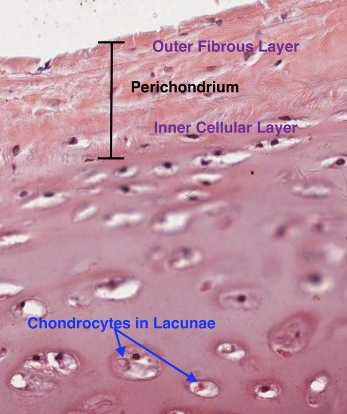
Hyaline cartilage
Cartilage is a type of avascular tissue with a highly specialized extracellular matrix that contains chondrocytes. The condrocytes produce and maintain the extracellular matrix. The root term meaning [cartilage] is [-chondr-], which originatesfrom the Greek [χόνδρος] or [chondros] meaning "cartilage" or "gristle". The Latin equivalent is [cartilago] giving us the synonymous root term [-cartilag-], from which [cartilage] arises.
The matrix contains large amounts of glicosaminoglycans, which allows for easy diffusion of substances from surrounding structures and blood vessels. Being avascular, cartilage does not have its own blood supply. The extracellular matrix also has large quantities of hyaluronic acid, which allows cartilage a weight-bearing capacity. This is why cartilage is particularly useful in bony joints and covering articular surfaces.
There are three types of cartilage present in the human body:
• Elastic cartilage: This type of cartilage is characterized by elastic fibers, usually in layers or lamellae
• Fibrocartilage: This type of cartilage is characterized by large bundles of collagen, making it look and feel fibrous
• Hyaline cartilage: This type of cartilage is characterized by an homogenous amorphous matrix
Thanks to Dr. Stephen Gallik for the mage and links. For more information on mammalian hystology, you can visit Dr. Gallik's website here.
- Details

The root term [-cheil-] derivates from the Greek word [χείλος (keilos]] meaning "lip". There are other medical root terms that also mean lip, but they arise from the Latin words [labellum, labrum, and labra]. There are many medical terms that include the root [-cheil-]:
• Cheilitis: The suffix [-itis] means "inflammation". Inflammation of the lips
• Cheilitis simplex: A very medical way of saying "chapped lips". See accompanying image.
• Cheiloplasty: The suffix [-(o)plasty] means "surgical reshaping". A surgical reshaping or plastic surgery of the lips
• Angular cheilitis: Inflammation of the angle of the mouth, sometimes causing a fissure
• Cheilognathopalatoschisis: This wors combines several roots: [-cheil-], meaning "lip", [-gnath-] meaning "jaw", [-palat-], meaning "palate", while the suffix [-schisis] means "to split". A split or separation of the lip, jaw, and the hard and soft palate.
- Details
The root term [-lapar-] arises from the Greek word [λαπάρα] which means "flank or "loin"". It refers to the lateral region of the abdomen between the costal margin superiorly and the iliac crest inferiorly. In its pure etymological meaning the root term [lapar], as in "laparotomy" or "laparoscopy" should be used to denote a surgical action in only two of the abdominal regions, the right and left lumbar abdominal regions (or flank regions). The suffix [-otomy] originates from the Greek [τέμνω] (tomos) which means "to cut" or "to open".
The first modern use of the term [laparotomy] referring to "an abdominal incision" was in January 1878 by Thomas Bryant, FRCS in his book "A Manual for the Practice of Surgery". This of course caused an upheaval with language purists, as a true laparotomy is a flank incision only. Nonetheless the meaning of the term as suggested by Bryant has been in use since. Today any abdominal incision is a laparotomy.