![]()
Medical Terminology Daily (MTD) is a blog sponsored by Clinical Anatomy Associates, Inc. as a service to the medical community. We post anatomical, medical or surgical terms, their meaning and usage, as well as biographical notes on anatomists, surgeons, and researchers through the ages. Be warned that some of the images used depict human anatomical specimens.
You are welcome to submit questions and suggestions using our "Contact Us" form. The information on this blog follows the terms on our "Privacy and Security Statement" and cannot be construed as medical guidance or instructions for treatment.
We have 1058 guests online
")
Marcia Crocker Noyes
(1869 – 1946)
Further to my comment on old books and research that started with an interesting bookplate (Ex-Libris). I continued my research and found that the person in charge of the Osler library bookplate was a fascinating individual that today maybe a ghost in the MedChi library and building in Baltimore... This is certainly an article that can be called "A Moment in History"
Marcia Crocker Noyes was the librarian at The Maryland State Medical Society from 1896 to 1946 and was a founding member of the Medical Library Association.[1][2][3]
Sir William Osler, MD. a famous Johns Hopkins surgeon was a noted bibliophile and had a large personal collection of books on various topics. When he became the President of MedChi in 1896, he was dismayed at the condition of the library and knew that with the right person and some stewardship, it could become a significant collection. Sir William asked his friend, Dr. Bernard Steiner, a physician and President of the Enoch Pratt Free Library in Baltimore for suggestions of a librarian, and Dr. Steiner recommended Marcia Crocker Noyes. A native of New York, and a graduate of Hunter College, Marcia had moved to Baltimore for a lengthy visit with her sister, and took a “temporary” position at the Pratt Library, which turned into three years. Although she had no medical experience or background, she was enthusiastic, and most importantly, she was willing to move into the apartment provided for the librarian, who needed to be available 24 hours a day.
The image in this article is Ms. Noyes on her first year on the job. Marcia developed a book classification system for medical books, based on the Index Medicus, and called it the Classification for Medical Literature. The system uses the alphabet with capital letters for the major divisions of medicine and lower-case ones for the sub-sections. The system was used for many years, but it's now dated and the Faculty's original shelving scheme was never changed. The card catalogs still reflect her classification and many of the cards are written in Marcia's back-slanting handwriting.
Marcia knew enough to ask the Faculty's members about medical questions, terminology and literature. She gradually won over the predominantly male membership and they became her greatest allies; Sir William at the start, and then for nearly 40 years, Dr. John Ruhräh, a wealthy pediatrician with no immediate family of his own. She made a point of attending almost every Faculty function, and in 1904, under guidelines from the American Medical Association, Marcia was made the Faculty Secretary. For much of her first 10 years, she was the Faculty's only full-time employee, only being assisted by Mr. Caution, the Faculty's janitor. Later in life Marcia would say that she hired him because of his name!
Within ten years, the library had outgrown its space, and plans, spearheaded by Marcia and Sir William before his move to Oxford, were made to build a headquarters building, mainly to house the library's growing collection of medical books and journals.
Marcia was instrumental in the design and building of the new headquarters. She travelled to Philadelphia, New York and Boston to look at their medical society buildings, and eventually, the Philadelphia architectural firm, Ellicott & Emmart was selected to design and build the new Faculty building. Every detail of the building held her imprimatur, from the graceful staircase, to the light-filled reading room, and all of the myriad details of the millwork, marble tesserae, and most of all, the four-story cast iron stacks. She was on-site, climbing up unfinished staircases, checking out the progress of the building, which was built in less than one year at a cost of $90,000.
Among the features of the new building was a fourth-floor apartment for her. She referred to it as the "first penthouse in Baltimore" and it had a garden and rooftop terrace. The library collection eventually grew to more than 65,000 volumes from medical and specialty societies around the world. Journals were traded back and forth, and physicians eagerly anticipated the arrival of each new issue. At the same time, Marcia was involved in the Medical Library Association as one of eight founding members. The MLA promotes medical libraries and the exchange of information. One of the earliest mandates of the MLA was the Exchange, a distribution and trade service for those who had duplicates or little-used books in their collections. Initially, the Exchange was run out of the Philadelphia medical society, but in 1900 it was moved to Baltimore and Marcia oversaw it. Several hundred periodicals and journals were received and sent each month, a huge amount of work for a tiny staff. In 1904, the Faculty had run out of room to manage the Exchange, so it was moved to the Medical Society of the Kings County (Brooklyn). But without Marcia's excellent administrative skills, it floundered and in 1908, the MLA asked Marcia to take charge once again.
In 1909, when the new Faculty building opened, there was enough room to run the Exchange and with the help of MLA Treasurer, noted bibliophile and close friend, Dr. John Ruhräh, it once again became successful. Additionally, Marcia and Dr. Ruhräh combined forces to revive the MLA's bulletin, which had all but ceased publication in 1908, taking the Exchange with it. This duo maintained editorial control from 1911 until 1926. In 1934, around the time of Dr. Ruhräh's death, Marcia became the first “unmedicated” professional to head the MLA. During her tenure, the MLA incorporated, the first seal was adopted, and the annual meeting was held in Baltimore. Marcia wanted to write the history of the MLA once she retired from full-time work at the Faculty, but her health was beginning to fail. She had back problems and had suffered a serious burn on her shoulder as a young woman, possibly from her time running a summer camp, Camp Seyon, for young ladies in the Adirondack Mountains. In 1946, a celebration was planned to honor Marcia's 50 years at the Faculty. But she was adamant that the physicians wait until November, the actual date of her 50 years. However, they knew she was gravely ill, and might not make it until then, so a huge party was held in April. More than 250 physicians attended the celebration, but the ones she was closest to in the early years, were long gone. She was presented with a suitcase, a sum of money to use for travelling, and her favorite painting of Dr. John Philip Smith, a founder of the Medical College in Winchester, Virginia. It was painted by Edward Caledon Smith, a Virginia painter who had been a student of the painter Thomas Sully.[4] She adored this painting and vowed, jokingly, to take it with her wherever she went.
The painting was not to stay with her for very long, for she died in November 1946, and left it to the Faculty in her will. Her funeral was held in the Faculty's Osler Hall, named for her dear friend. More than 60 physicians served as her pallbearers, and she was buried at Baltimore's Green Mount Cemetery. In 1948, the MLA decided to establish an award in the name of Marcia Crocker Noyes. It was for outstanding achievement in medical library field and was to be awarded every two years, or when a truly worthy candidate was submitted. In 2014, the Faculty began giving a bouquet of flowers to the winner of the award in Marcia's name, and in honor of her work. Much evidence exists for this tradition, as we know that the physicians, especially Drs. Osler and Ruhräh, frequently gave her bouquets of flowers. Marcia also cultivated flower gardens at the Faculty and decorated the rooms with her work.
Today, the MedChi building is open for tours and if the rumors are to be believed Ms. Marcia Crocker Noyes is still at work in her beloved library as the "resident ghost" [1][5]

NOTE: This article has been modified from the original Wikipedia article on Marcia Crocker Noyes. The article itself is well-written with interesting images of the subject. I would encourage you to visit it. The second insert is from book 00736 in my personal library and shows in pencil, the incredibly small handwriting of Marsha C. Noyes.
Sources:
1. "Marcia, Marcia, Marcia" MedChi Archives blog.
2. "Marcia C. Noyes, Medical Librarian" (PDF). Bulletin of the Medical Library Association. 35 (1): 108–109. 1947. PMC 194645
3. Smith, Bernie Todd (1974). "Marcia Crocker Noyes, Medical Librarian: The Shaping of a Career" (PDF). Bulletin of the Medical Library Association. 62 (3): 314–324. PMC 198800Freely accessible. PMID 4619344.
4. Edward Caledon BRUCE (1825-1901)"
5. Behind the scenes tour MedChiBuilding
"Clinical Anatomy Associates, Inc., and the contributors of "Medical Terminology Daily" wish to thank all individuals who donate their bodies and tissues for the advancement of education and research”.
Click here for more information


- Details
This article is part of the series "A Moment in History" where we honor those who have contributed to the growth of medical knowledge in the areas of anatomy, medicine, surgery, and medical research.

Prof Dr. Eric Muhe
Prof. Dr. Eric Mühe (1938 - 2005) Much has been written about who gets the glory of having performed the first laparoscopic cholecystectomy. Many famous names are in the annals of Surgery listed as pioneers in this field: Perissat, Berci, Mouret, Dubois, Sepulveda, Reddick, McKernan, Saye, Lizana, and others. Prof. Dr. Eric Mühe was usually not mentioned, but there is no doubt that on September 12, 1985, Dr. Mühe performed a laparoscopic cholecystectomy using a device of his own design (Galloscope) which lifted the anterior abdominal wall, maintained pneumoperitoneum, and doubled as a laparoscope.
Dr. Mühe was an avid cyclist and built and repaired his own bicycles. He once told me that he was looking at bicycle metal tube, and when looking through it, he thought that he could obtain access to the abdomen and the gallbladder with minimal changes. I am privileged to have known him and I was sad to learn that he passed in 2005. Dr. Miranda
For more information in downloadable PDF format: CLICK HERE, or HERE
- Details

Click for a larger image
The [transverse processes] are two lateral bony processes found in most vertebrae, with the exception of the coccygeal vertebrae.
Transverse processes have regional variations. The transverse processes in the cervical vertebrae are heavily modified with anterior and posterior roots forming the boundaries of the foramina transversaria. They are "gutter-shaped" to accommodate the spinal nerves, and angled at 60 degrees anteriorly with a 15 degree inferior tilt.
In the thoracic region the transverse processes point posterolaterally and present with an articular facet (costal facet) for the costal tubercle of a rib, forming the costotransverse joint. The transverse processes of T11 and T12 do not have a costal facet.
The transverse processes in the lumbar region point slightly posterolateral, and they are larger. The longest transverse processes in the lumbar region are those of L3. The transverse processes of L5 are atypical in that they also point superolaterally at almost 45 degrees. This is because these transverse processes are attached to the posterior superior iliac spine and iliac crest by the iliolumbar ligament, contributing to the prevention of forward displacement of L5 over S1.
The sacral transverse processes contribute to the larger mass of the sacrum fusing to form the intermediate sacral crest on its posterior surface.
Thanks to Dr. Mary M Tuchscherer for her comments and improvement of this article.
Image property of: CAA.Inc. Photographer: David M. Klein
- Details
This article is part of the series "A Moment in History" where we honor those who have contributed to the growth of medical knowledge in the areas of anatomy, medicine, surgery, and medical research.

Michael Severtus
Michael Servetus (1511 -1553) was a Spanish theologian, physician, and anatomist. He is also known as Miguel Servet, Miguel Serveto, and Michel de Villeneuve. Servetus had studies in a multitude of fields, including catography, mathematics, pharmacology, astronomy, etc. He was born in 1511 in Aragon, Spain. Servetus started his studies in law in 1531and Medicine in 1536, where he excelled as an anatomist. Just as Andreas Vesalius and William Harvey, he clashed with the Galenic vision of anatomy and physiology. He correctly stated the theory of pulmonary circulation, but with no logical proof as Harvey.
Servetus was openly critical of the catholic church, publishing three books that openly questioned the Holy Trinity dogma. Servetus published his findings on pulmonary circulation in a controversial book "Cristianismi Restitutio", where pulmonary circulation was only one of the points he made which were mostly his position on the Holy Trinity and questioning the idea that everyone was predestined, as the catholic church professed at that time. His anatomical views were the least of his problems; because of this open criticism of Galen and the church. Servetus was burnt at the stake in Geneva on October 27, 1553.
Original image courtesy of National Library of Medicine.
- Details
Click for a larger image
The [vertebral endplate] is the term used to denote a structure formed by the superior (and inferior) aspect of the vertebral body and a layer of hyaline cartilage related to it. The vertebral endplate is formed by the cortical bone of the anular epiphysis, the central depression and the hyaline cartilage that fills the depresssion.
The vertebral endplate is important for the health of the intervertebral disk (IVD). Since the IVD loses its blood supply early in life all the nutrition to the IVD has to pass through the cortical bone and the hyaline cartilage. Note that hyaline cartilage is avascular. Separation of the components of the vertebral endplate due to trauma or other pathology can cause the IVD to become pathological.
The vertebral endplate also plays an important role in the biomechanics of spine movement. Being slightly incurved, the endplate acts as a shock absorber, bending up to 1/2 mm. Damage to the endplate can reduce this function and set the stage for failure of the endplate and a potential vertebral fracture.
Sometimes the IVD can herniate through the endplate causing what is known as "Schmorl's bodies".
Image property of: CAA.Inc.Photographer: David M. Klein
- Details
The medical word [avascular] means "without vessels" and refers to structures that do not have vessels providing it with blood supply. Avascular structures, like hyaline cartliage, receive their oxygen and nutrients by diffusion from nearby structures.
The preffix [-a-] means "without", and the root term [-vascul-] means "vessels".
- Details
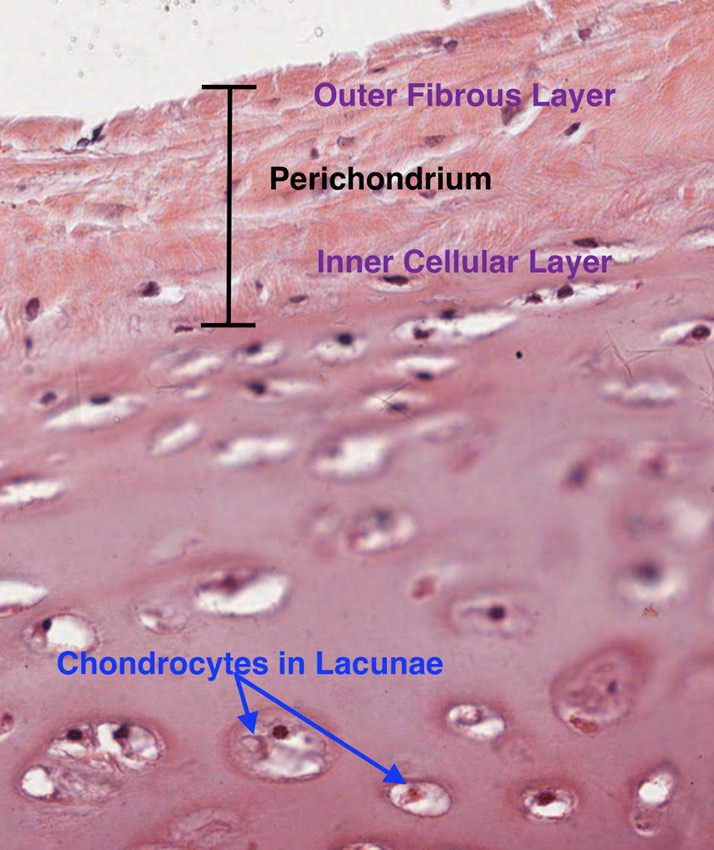
Hyaline cartilage
Hyaline cartilage is a type of cartilage characterized by a very homogenous avascular matrix. It is the most common type of cartilage. Hyaline cartilage has a bluish glassy look to it, hence the name.
Within the matrix of hyaline cartilage there are spaces called "lacunae" wich contain chondrocytes. These produce and maintain the extracellular matrix. Hyaline cartilage is found covering articular surfaces allowing for effortless sliding of the articular surfaces one against the other. Hyaline cartilage is avascular.
The accompanying image is a histology slide of hyaline cartilage. For more information on hyaline cartilage, read the article "The Importance of Hyaline Cartilage"by Dr. Stephen Gallik.
The word "hyaline" is a derivate of the Greek [υαλώδης] (yalódis) meaning "glassy".
Thanks to Dr. Stephen Gallik for the mage and links. For more information on mammalian hystology, you can visit Dr. Gallik's website here.
Note: Google Translate includes the symbol (?). Clicking on it will allow you to hear the pronunciation of the word.