
Medical Terminology Daily (MTD) is a blog sponsored by Clinical Anatomy Associates, Inc. as a service to the medical community. We post anatomical, medical or surgical terms, their meaning and usage, as well as biographical notes on anatomists, surgeons, and researchers through the ages. Be warned that some of the images used depict human anatomical specimens.
You are welcome to submit questions and suggestions using our "Contact Us" form. The information on this blog follows the terms on our "Privacy and Security Statement" and cannot be construed as medical guidance or instructions for treatment.
We have 2334 guests online
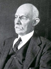
William S. Halsted, MD
(1852 – 1922)
American anatomist, teacher, and surgeon, William Stewart Halsted was born in New York City, USA to a wealthy family of English origin. His father was involved in charitable work and Governor and trustee to a city hospital. Not a brilliant student initially, Halsted took an undergraduate in Liberal Arts in Yale, CT., after which he entered the Medical College of Physicians at the Columbia College, where he excelled.
As a second-year medical student Halsted applied and obtained a position in surgery at a local hospital. In here he learned about Lister’s antiseptic technique and became an adamant proponent of it to reduce infection. In 1877 Halsted obtained his MD. After a short time as House Physician at the New York Hospital, Halsted traveled to Europe to further his education studying for two years at the Universities of Vienna, Leipzig, and W?rzburg.
Besides being at the forefront of surgical and antiseptic techniques (introducing the use of rubber gloves in surgery), Halsted was extremely concerned with the way medical students were taught in the US. He pioneered bedside clinical round discussions with the medical students after two years of basic sciences studies. Halsted developed the idea of a patient chart; he also developed the residency program for medical students in use today.
Halsted is probably the most influential researcher and surgeon at the turn of the century. He dedicated time to the study of intestinal anastomoses and the use of silk as a suture material. His experimental work in 1887 proved that the inclusion of the submucosa layer in an anastomosis was mandatory, as well that a single layered anastomosis was enough to attain closure. Perhaps Halsted’s most important contribution was the application and use of the scientific method to surgical questions. Halsted’s principles set the standards used today in surgical suturing and surgical stapling.
He also pioneered the development and surgical techniques for radical mastectomy as a treatment for breast cancer.
As a side effect of this studied in anesthesia and the use of cocaine for anesthesia, Halsted became addicted to this substance, a problem that followed him through the years. Without impairing his capacity as a researcher and a surgeon, Halsted eventually recovered. He died in Baltimore in 1922 as a complication to surgery.
Sources:
1. Dubay, A. D., & Franz, G. M. (2003). Acute Wound Healing: The Biology of Acute Wound Failure. Surg Clin NA, 83, 463-481.
2. Halsted, W. S. (1887). Circular Suture of the Intestine - An Experimental Study. Am J Med Sci, 436-461.
3. “William Stewart Halsted: his life and contributions to surgery” Osborne, P. Lancet Oncol 2007; 8: 256–65
4. “William Stewart Halsted: Surgical pioneer” Burress, P Endoc Today (2010), 8: (2) 22
5. “William Stewart Halsted (1852–1922) Neurological stamp” Haas, LF J Neurol Neurosurg Psych 2000;69:641
Original image courtesy of "Images from the History of Medicine" at www.nih.gov

"Clinical Anatomy Associates, Inc., and the contributors of "Medical Terminology Daily" wish to thank all individuals who donate their bodies and tissues for the advancement of education and research”.
Click here for more information


- Details

Click for a larger image
The root term [-auricul-] arises from the Latin word [Auricula], which is a diminutive of [auris] meaning "ear". Auricula means "a little ear".
The adjectival form [auricular] is used in several places in human anatomy. One of them is the [auricular surface of the iliac bone] (see image) referring to a roughened area in the medial aspect of the iliac bone where it articulates with the sacrum, forming the sacroiliac joint. The Latin term for this articular surface is facies auricularis ossis ilii. A corresponding auricular surface is found in the sacral bone, the facies auricularis ossis sacri.
[Auricular] is also used in the term [auricular cartilage], referring to the fibrocartilage found in the ear or [pinna].
A shortened version is the root term [-aur(i)-], meaning the same. It is used in the word [auricle] referring to the atrial appendages of the heart. These appendages when seen from the outside of the heart really look like little "boxer's ears". This image (click here) shows the left atrial appendage (LAA) or left auricle.
By the way, did you know that one of the digits was called "digitus auricularis"? Read the story here.
Image property of: CAA.Inc.Photographer: David M. Klein
- Details
This article is part of the series "A Moment in History" where we honor those who have contributed to the growth of medical knowledge in the areas of anatomy, medicine, surgery, and medical research.

Click for a another image
Enrique Finochietto, MD (1881 – 1948) Argentinian surgeon and researcher, Enrique Finochietto was born in 1881 in the city of Buenos Aires. He studied in both public and private schools, entering medical school at age 16. He received his medical degree from the University of Buenos Aires in 1904.
Shortly after graduation, he became an intern at the Rawson hospital of Buenos Aires. Dr. Finochietto would become part of the staff of this hospital and be part of its staff all his life. He traveled to Europe to study with surgeons such as Calot, Kocher, Roux, and others, visiting France, Switzerland, Austria, and Italy.
Upon his return he dedicated himself to surgery and endoscopy, and with the help of his brother Ricardo started research and invented many medical and surgical devices, including a motorized surgical table, endoscopic devices and surgical instruments, some of which are in use today throughout the world.
During World War I Dr. Finochietto worked for no fee (ad honorem) as surgeon-in-chief at the Argentine Hospital in Paris. For his dedication and work, France awarded him the Legion d’Honneur and the Red Cross. He came back to Argentina and then traveled to the USA where he visited with the Mayo Brothers and Harvey Cushing. One of Dr. Finochietto’s mottos was “Only the surgeon who goes beyond his obligations serves his duty”.
Although mostly remembered for the Finochietto rib spreader, Dr. Finochietto tackled most types of surgery during his career, including neurosurgery, gastrointestinal, thyroid, thoracic, and orthopedic.
Dr. Finochietto died on February 17, 1948. His legacy in Argentinian surgery lives on through his school of thought, research and disciples.
If you click on Dr. Finochietto’s image (courtesy of Wikipedia) you will see an image of his namesake rib spreader (courtesy of Surgiway, France ).
Sources:
1. “Memoir: Enrique Finochietto, MD (1881 – 1948)” DeBakey. ME. Ann Surg. 1948 Aug; 128(2): 319–320
2. “Enrique Finochietto” The Legacy of Surgery in Argentina” De La Fuente, SG. J Surg Ed (2007)64:2; 120-123
3. “El separador intercostal de Enrique Finochietto, el hospital Rawson, la escuela quir?rgica, y una an?cdota que los relaciona” Saadia, A. Rev Arg Cir Cardiovasc (2009) 3:3 149-150
4. “Rese?as hist?ricas: Enrique Finochietto” Parquet RA. Acta Gastroenterol Latinoam (2009); 39:18
5. “Surgery in Argentina” Beveraggi, EM. Arch Surg (1999) 134”438-444
6. “The Legends Behind Cardiothoracic Surgical Instruments” Ailawadi, G et al. Ann Thorac Surg (2010) 89:5; 1693-1700
- Details
The term [facet] is English and derives from the French word [facette] meaning "a small face". The French term is itself derived from the Latin [facies], meaning "face".
The term facet in anatomy is used to refer to a small smooth surface, usually an articular surface. Another use of the word, still meaning "a small face" can be found in the jeweler's term [facet] used to describe one of the many small surfaces in a diamond.
In spinal anatomy, the term “a facet joint” is most commonly used, but the term should be pronounced with the accent on the first syllable as in “fácet”! And just to be a bit more correct, the proper term for a so-called “facet joint” is “zygapophyseal joint”. This term is one of several medical terms that are used and/or pronounced incorrectly.
- Details
Click for a larger image
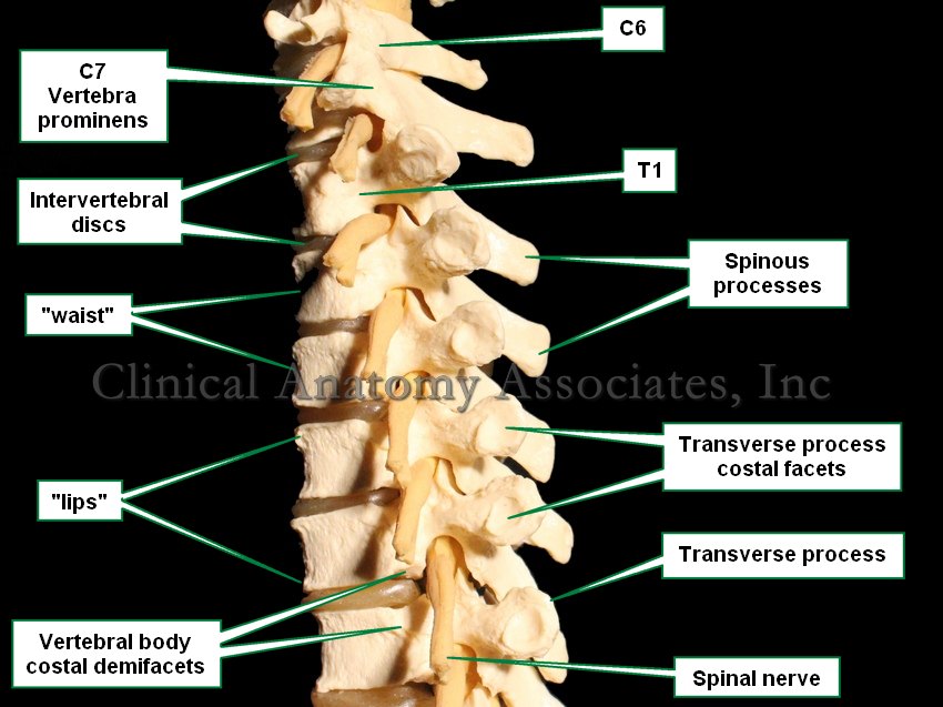
The [spinous process] is a single, median posterior bony process found in most vertebrae, with the exception of C1, and coccygeal vertebrae.
The spinous processes have regional variations according to the spinal region. Cervical vertebrae usually have a bifid spinous process, while C7 (vertebra prominens) has the longest spinous process in the cervical region
In the thoracic region the spinous processes are thin, slender and they overlap each other, while in the lumbar region these processes are thick and square. The spinous processes in the sacral region become progressively smaller.
Image property of: CAA.Inc.Photographer: David M. Klein
- Details
The Latin phrase [in situ] means "in its place". Refers to a structure that is in its place and undisturbed.
- Details
This article is part of the series "A Moment in History" where we honor those who have contributed to the growth of medical knowledge in the areas of anatomy, medicine, surgery, and medical research.
Click for a larger image
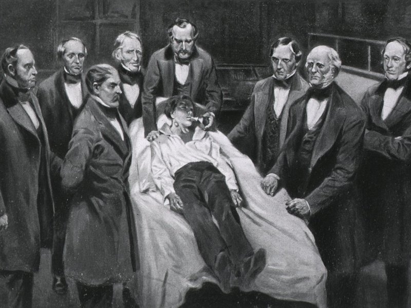
When I started the sidebar segment entitled "A Moment in History", I thought that it would be only biographical articles. On January 28, 2014, traveling from Austin TX to Cincinnati I was reading a wonderful book: "Masters of the Scalpel" (1962) by Sarah R. Riedman Ph.D. In one of the chapters Dr. Riedman writes about the first use of anesthesia in Surgery. Because of the author's style, as I read the following excerpt, I found myself wondering how it would have felt to be there while history was being made:
"On the morning of October 16, 1846, all but one of the principals were ready in the amphitheatre: Dr. Warren, the senior surgeon, his assistants and strong-armed men who were to hold the patient down, the students, and other spectators were waiting; the patient was brought in, pale with fright.
There was no turning back: everything was set for the removal of the tumor on his jaw. Only Morton (Dr. William T. G. Morton) had not arrived. Dr. Warren was ready to proceed without him, announcing to the onlookers: "As Dr. Morton has not arrived, I presume he is otherwise engaged." And he was - putting the finishing touches on his inhaler in the instrument maker's shop.
Just as the skeptical audience burst into laughter at Dr. Warren's remark, Morton appeared. The operation was held up for a few minutes while Morton prepared a sponge soaked in ether which he placed in the inhaling globe, temporarily corked.
As he came forward to the operating table on which the patient was strapped as always before an operation, Dr. Warren turned to Morton, saying: "Well sir! Your patient is ready."
But Morton wished to gain the patient's confidence. Pointing to Eben Frost ( A patient from whom Dr. Morton had removed a tooth under ether) who in gratitude had come along to the hospital, he said to the pale man: "There is a man who has been operated on under this chemical, and can tell you that it worked." Frost gladly complied.
"Are you afraid?" Morton asked the patient. Whether from courage or confidence, the patient replied, "No, I will do as you tell me."
Click for a larger image
Morton then put the neck of the flask to the patient's mouth, instructing him to breathe. Slowly the patient went under, his arms and legs jerking in a way probably familiar to frequenters at "ether frolics." As yet no one suspected what the chemical was. After several minutes, the patient was asleep and relaxed. It was now Morton's cue in the drama, as he turned to Dr. Warren. "Sir, your patient is ready."
Warren made the incision. He, like the witnesses, was ready for the bloodcurdling screams so familiar in the operating room. But the patient uttered not a sound.
The operation over, the patient slowly regained consciousness. When questioned by Morton, he readily admitted having felt no pain.
Dr. Warren then broke the silence with the famous words: "Gentlemen, this is no humbug!" And Dr. Henry J. Bigelow: "I have seen something today that will be heard round the world."
The first page of a new chapter in the story of surgery was turned that day."
While looking out the airplane window to the passing cities below, I thought about the millions of people that had been affected by this "Moment in History" and that it needed to be shared and retold to the generations to follow.
The operation took place in the first operating room built at the Massachusetts General Hospital. This room has been preserved and is today known as the "Ether Dome". You can read an article on a visit I made to this historic place. Dr. Miranda
Biographical note: Sarah Regal Riedman was born on April 20, 1902 in Kishiniev, Rumania and became a U.S. citizen in 1918. In 1926, she received a bachelor's degree from Hunter College, followed by a Masters of Science degree from New York University in 1928. In 1935, Ms. Riedman received her Ph.D. from Columbia University. She taught at Hunter College from 1926 to 1930, and at Brooklyn College from 1930 to 1952. At Brooklyn College, she was an instructor, and later an assistant professor of biology.
Ms. Riedman began writing science books for children in 1947, with the publication of "How Man Discovered His Body". Between 1947 and 1983, she wrote or co- wrote approximately forty books. We have not been able to find further information on her. Any contribution to her biography will be most welcome.
Original image (public domain) courtesy of NLM
