
Medical Terminology Daily (MTD) is a blog sponsored by Clinical Anatomy Associates, Inc. as a service to the medical community. We post anatomical, medical or surgical terms, their meaning and usage, as well as biographical notes on anatomists, surgeons, and researchers through the ages. Be warned that some of the images used depict human anatomical specimens.
You are welcome to submit questions and suggestions using our "Contact Us" form. The information on this blog follows the terms on our "Privacy and Security Statement" and cannot be construed as medical guidance or instructions for treatment.
We have 429 guests online
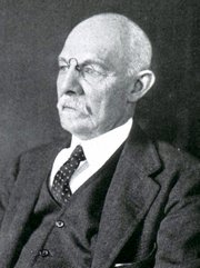
William S. Halsted, MD
(1852 – 1922)
American anatomist, teacher, and surgeon, William Stewart Halsted was born in New York City, USA to a wealthy family of English origin. His father was involved in charitable work and Governor and trustee to a city hospital. Not a brilliant student initially, Halsted took an undergraduate in Liberal Arts in Yale, CT., after which he entered the Medical College of Physicians at the Columbia College, where he excelled.
As a second-year medical student Halsted applied and obtained a position in surgery at a local hospital. In here he learned about Lister’s antiseptic technique and became an adamant proponent of it to reduce infection. In 1877 Halsted obtained his MD. After a short time as House Physician at the New York Hospital, Halsted traveled to Europe to further his education studying for two years at the Universities of Vienna, Leipzig, and W?rzburg.
Besides being at the forefront of surgical and antiseptic techniques (introducing the use of rubber gloves in surgery), Halsted was extremely concerned with the way medical students were taught in the US. He pioneered bedside clinical round discussions with the medical students after two years of basic sciences studies. Halsted developed the idea of a patient chart; he also developed the residency program for medical students in use today.
Halsted is probably the most influential researcher and surgeon at the turn of the century. He dedicated time to the study of intestinal anastomoses and the use of silk as a suture material. His experimental work in 1887 proved that the inclusion of the submucosa layer in an anastomosis was mandatory, as well that a single layered anastomosis was enough to attain closure. Perhaps Halsted’s most important contribution was the application and use of the scientific method to surgical questions. Halsted’s principles, also known as "Halsted's Rules of Surgery", set the standards used today in surgical suturing and surgical stapling.
He also pioneered the development and surgical techniques for radical mastectomy as a treatment for breast cancer.
As a side effect of this studied in anesthesia and the use of cocaine for anesthesia, Halsted became addicted to this substance, a problem that followed him through the years. Without impairing his capacity as a researcher and a surgeon, Halsted eventually recovered. He died in Baltimore in 1922 as a complication to surgery.
Sources:
1. Dubay, A. D., & Franz, G. M. (2003). Acute Wound Healing: The Biology of Acute Wound Failure. Surg Clin NA, 83, 463-481.
2. Halsted, W. S. (1887). Circular Suture of the Intestine - An Experimental Study. Am J Med Sci, 436-461.
3. “William Stewart Halsted: his life and contributions to surgery” Osborne, P. Lancet Oncol 2007; 8: 256–65
4. “William Stewart Halsted: Surgical pioneer” Burress, P Endoc Today (2010), 8: (2) 22
5. “William Stewart Halsted (1852–1922) Neurological stamp” Haas, LF J Neurol Neurosurg Psych 2000;69:641
Original image courtesy of "Images from the History of Medicine" at www.nih.gov

"Clinical Anatomy Associates, Inc., and the contributors of "Medical Terminology Daily" wish to thank all individuals who donate their bodies and tissues for the advancement of education and research”.
Click here for more information


- Details
The word [sinus] is Latin and refers to a "pocket" or a "curved surface or structure". In medical terms the word [sinus] is used more in the meaning of "pocket" and refers to a cavity that has only one entrance (or exit) both in anatomy and surgery.
The term is used properly when referring to pocket-like structures such as the "sinuses of Valsalva" or the "prostatic sinus", as well as the sinuses found in the cranium such as the frontal sinus, the ethmoidal sinus, the sphenoid sinus, etc.
The word is incorrectly used to denote structures that have more than one entrance (or exit) such as the coronary sinus or the venous sinuses found in the cranial dura mater.
The corresponding root term is [-sin-] which then can be used in words such as [sinotubular junction]
- Details
![Coronary Arteries. The [*] indicates the left coronary artery](/images/MTD/LargeImages/coronaryarterieslabels_lg.jpg "Coronary Arteries. The [*] indicates the left coronary artery")
Click for a larger image
UPDATED: The [anterior interventricular artery] is one of the two branches of the left coronary artery, the other one being the circumflex artery (CFX). Although the name "anterior interventricular artery" is the anatomical name, this artery is most commonly known by its clinical name, the [left anterior descending artery], or LAD.
The LAD lies deep to the epicardium in the subepicardial fatty layer. It gives off several important branches, including one or two (sometimes three) large diagonal arteries that provide blood supply to the anterior aspect of the left ventricle. Also of importance, the LAD provides several septal perforator arteries that provide blood supply to the anterior 2/3rds of the interventricular septum.
Because of the extensive arterial territory of the LAD, and its involvement with the left ventricle and the interventricular septum, the LAD is sometimes called by a non-clinical term: "the widow-maker".
There can be interesting anatomical variations in the coronary arteries of the heart. For a detail on these anatomical variations, click here. Heart and coronary artery anatomy is one of the many lecture topics presented by CAA, Inc
Image property of:CAA.Inc.. Artist:Victoria G. Ratcliffe
- Details
Click for a larger image
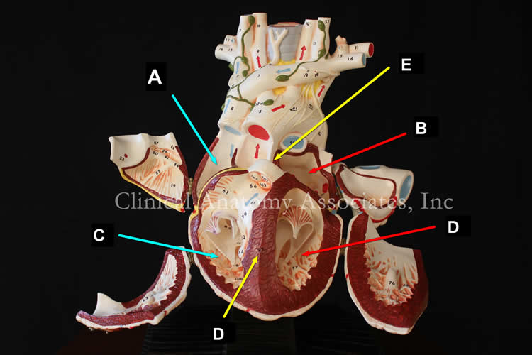
UPDATED: The prefix [inter-] means "between". The root term [ventricul-] refers to a ventricle, from the Latin [ventriculus], meaning "little sac" or "little belly". The word [septum] is Latin, and means "wall", "division", or "partition". The plural form for [septum] is [septa].
There are two septa in the heart. The interatrial septum (see superior image, item "E") is found between the atria, and the interventricular septum (see superior image, item "D"). The interventricular septum has two components, the muscular interventricular septum, and a small, superiorly situated membranous septum. The interventricular septum is quite muscular as seen in both images. Click on each image for a larger picture.
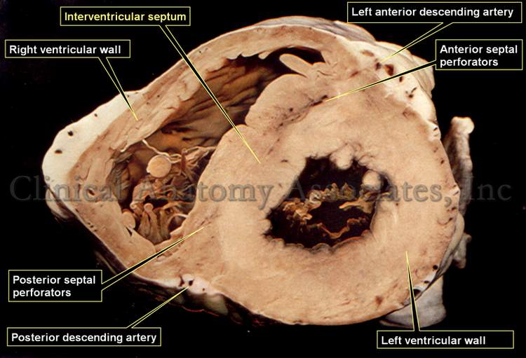
The cardiac interventricular septum receives blood supply from branches of both the right and left coronary arteries. The anterior 2/3rds of the interventricular septum receive blood supply through anterior septal perforators that arise from the left anterior descending artery (LAD), a branch of the left coronary artery.
Click for a larger image
The posterior 1/3rd of the interventricular septum receives blood supply by way of posterior septal perforators that arise from the posterior descending artery (PDA), a branch of the right coronary artery.
For a detail of the blood supply to the interventricular septum, click on the inferior image.
Superior image property of: CAA.Inc. Photographer: D.M. Klein.
Inferior image modified from "Slide Atlas of Cardiac Anatomy" (out of print): Library catalog.
- Details
From Latin, meaning “opposite to”. This term has been used since the late 1700’s in English literature to denote “the lowest point”.
This term will be of interest as we study the anatomy of the aortic valve and aortic root.
- Details

Click for a larger image
UPDATED: This word has a Greek origin from the term [ανεύρυσμα], composed of [ana] meaning "complete or throughout", and [eurus] meaning "wide", a "complete widening or dilation". It is used to refer to the dilation of an artery. Aneurysms can be formed in any artery, although they have some preferred sites. The most common aneurysms are found in the aorta, arterial circle of Willis, the root of the cerebral arteries, and internal carotid arteries.
Biomechanical studies suggest that once an aneurysm forms it will generally progress in its dilation until aneurysmal rupture. Because of turbulent flow within the aneurysm large clots are usually formed, which in turn can cause emboli.
The image shows an excised infrarenal abdominal aortic aneurysm (AAA). The two common iliac arteries can be seen. If you click on the image you will be able to see the same aneurysm opened through its posterior wall and the clot that was contained inside.
Since the definition of [aneurysm] is "a dilation of an artery", it is wrong to refer to the dilation of a vein as a "venous aneurysm. The proper term to refer to the dilation of a vein is [varix].
Image property of: CAA.Inc. Photographer: David M. Klein
- Details
Click for a larger image
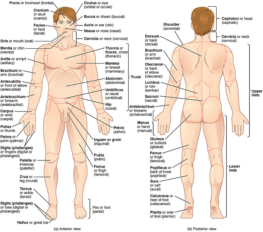
The [leg] is the anatomical region found between the knee joint superiorly and the ankle inferiorly. It contains two bones, the tibia and the fibula (os peroneus).
Since the Latin word [crus] means “pillar” or “leg”, the term [crural] is sometimes used to denote the leg region.
The wrong usage of the term [leg] to refer or denote all of the lower extremity is one of my pet peeves. There are many health care professionals who use this term wrongly and are therefore forced to use the terms “upper leg” to refer to the thigh, and “lower leg” to refer to the leg proper.
The posterior compartment of the leg contains the soleus and gastrocnemius muscles which cause a superficial elevation known as “the calf”. The Latin term [suram] means “calf”, so the term [sural] can be used to denote the posterior aspect of the leg.
The anterior aspect of the leg is marked by the bony anterior tibial crest or shin, a word that is sometimes wrongly used to mean “leg”.
Original image courtesy of Connexions (http://cnx.org) [CC-BY-3.0 (http://creativecommons.org/licenses/by/3.0)], via Wikimedia Commons
