![]()
Medical Terminology Daily (MTD) is a blog sponsored by Clinical Anatomy Associates, Inc. as a service to the medical community. We post anatomical, medical or surgical terms, their meaning and usage, as well as biographical notes on anatomists, surgeons, and researchers through the ages. Be warned that some of the images used depict human anatomical specimens.
You are welcome to submit questions and suggestions using our "Contact Us" form. The information on this blog follows the terms on our "Privacy and Security Statement" and cannot be construed as medical guidance or instructions for treatment.
We have 960 guests online
")
Marcia Crocker Noyes
(1869 – 1946)
Further to my comment on old books and research that started with an interesting bookplate (Ex-Libris). I continued my research and found that the person in charge of the Osler library bookplate was a fascinating individual that today maybe a ghost in the MedChi library and building in Baltimore... This is certainly an article that can be called "A Moment in History"
Marcia Crocker Noyes was the librarian at The Maryland State Medical Society from 1896 to 1946 and was a founding member of the Medical Library Association.[1][2][3]
Sir William Osler, MD. a famous Johns Hopkins surgeon was a noted bibliophile and had a large personal collection of books on various topics. When he became the President of MedChi in 1896, he was dismayed at the condition of the library and knew that with the right person and some stewardship, it could become a significant collection. Sir William asked his friend, Dr. Bernard Steiner, a physician and President of the Enoch Pratt Free Library in Baltimore for suggestions of a librarian, and Dr. Steiner recommended Marcia Crocker Noyes. A native of New York, and a graduate of Hunter College, Marcia had moved to Baltimore for a lengthy visit with her sister, and took a “temporary” position at the Pratt Library, which turned into three years. Although she had no medical experience or background, she was enthusiastic, and most importantly, she was willing to move into the apartment provided for the librarian, who needed to be available 24 hours a day.
The image in this article is Ms. Noyes on her first year on the job. Marcia developed a book classification system for medical books, based on the Index Medicus, and called it the Classification for Medical Literature. The system uses the alphabet with capital letters for the major divisions of medicine and lower-case ones for the sub-sections. The system was used for many years, but it's now dated and the Faculty's original shelving scheme was never changed. The card catalogs still reflect her classification and many of the cards are written in Marcia's back-slanting handwriting.
Marcia knew enough to ask the Faculty's members about medical questions, terminology and literature. She gradually won over the predominantly male membership and they became her greatest allies; Sir William at the start, and then for nearly 40 years, Dr. John Ruhräh, a wealthy pediatrician with no immediate family of his own. She made a point of attending almost every Faculty function, and in 1904, under guidelines from the American Medical Association, Marcia was made the Faculty Secretary. For much of her first 10 years, she was the Faculty's only full-time employee, only being assisted by Mr. Caution, the Faculty's janitor. Later in life Marcia would say that she hired him because of his name!
Within ten years, the library had outgrown its space, and plans, spearheaded by Marcia and Sir William before his move to Oxford, were made to build a headquarters building, mainly to house the library's growing collection of medical books and journals.
Marcia was instrumental in the design and building of the new headquarters. She travelled to Philadelphia, New York and Boston to look at their medical society buildings, and eventually, the Philadelphia architectural firm, Ellicott & Emmart was selected to design and build the new Faculty building. Every detail of the building held her imprimatur, from the graceful staircase, to the light-filled reading room, and all of the myriad details of the millwork, marble tesserae, and most of all, the four-story cast iron stacks. She was on-site, climbing up unfinished staircases, checking out the progress of the building, which was built in less than one year at a cost of $90,000.
Among the features of the new building was a fourth-floor apartment for her. She referred to it as the "first penthouse in Baltimore" and it had a garden and rooftop terrace. The library collection eventually grew to more than 65,000 volumes from medical and specialty societies around the world. Journals were traded back and forth, and physicians eagerly anticipated the arrival of each new issue. At the same time, Marcia was involved in the Medical Library Association as one of eight founding members. The MLA promotes medical libraries and the exchange of information. One of the earliest mandates of the MLA was the Exchange, a distribution and trade service for those who had duplicates or little-used books in their collections. Initially, the Exchange was run out of the Philadelphia medical society, but in 1900 it was moved to Baltimore and Marcia oversaw it. Several hundred periodicals and journals were received and sent each month, a huge amount of work for a tiny staff. In 1904, the Faculty had run out of room to manage the Exchange, so it was moved to the Medical Society of the Kings County (Brooklyn). But without Marcia's excellent administrative skills, it floundered and in 1908, the MLA asked Marcia to take charge once again.
In 1909, when the new Faculty building opened, there was enough room to run the Exchange and with the help of MLA Treasurer, noted bibliophile and close friend, Dr. John Ruhräh, it once again became successful. Additionally, Marcia and Dr. Ruhräh combined forces to revive the MLA's bulletin, which had all but ceased publication in 1908, taking the Exchange with it. This duo maintained editorial control from 1911 until 1926. In 1934, around the time of Dr. Ruhräh's death, Marcia became the first “unmedicated” professional to head the MLA. During her tenure, the MLA incorporated, the first seal was adopted, and the annual meeting was held in Baltimore. Marcia wanted to write the history of the MLA once she retired from full-time work at the Faculty, but her health was beginning to fail. She had back problems and had suffered a serious burn on her shoulder as a young woman, possibly from her time running a summer camp, Camp Seyon, for young ladies in the Adirondack Mountains. In 1946, a celebration was planned to honor Marcia's 50 years at the Faculty. But she was adamant that the physicians wait until November, the actual date of her 50 years. However, they knew she was gravely ill, and might not make it until then, so a huge party was held in April. More than 250 physicians attended the celebration, but the ones she was closest to in the early years, were long gone. She was presented with a suitcase, a sum of money to use for travelling, and her favorite painting of Dr. John Philip Smith, a founder of the Medical College in Winchester, Virginia. It was painted by Edward Caledon Smith, a Virginia painter who had been a student of the painter Thomas Sully.[4] She adored this painting and vowed, jokingly, to take it with her wherever she went.
The painting was not to stay with her for very long, for she died in November 1946, and left it to the Faculty in her will. Her funeral was held in the Faculty's Osler Hall, named for her dear friend. More than 60 physicians served as her pallbearers, and she was buried at Baltimore's Green Mount Cemetery. In 1948, the MLA decided to establish an award in the name of Marcia Crocker Noyes. It was for outstanding achievement in medical library field and was to be awarded every two years, or when a truly worthy candidate was submitted. In 2014, the Faculty began giving a bouquet of flowers to the winner of the award in Marcia's name, and in honor of her work. Much evidence exists for this tradition, as we know that the physicians, especially Drs. Osler and Ruhräh, frequently gave her bouquets of flowers. Marcia also cultivated flower gardens at the Faculty and decorated the rooms with her work.
Today, the MedChi building is open for tours and if the rumors are to be believed Ms. Marcia Crocker Noyes is still at work in her beloved library as the "resident ghost" [1][5]

NOTE: This article has been modified from the original Wikipedia article on Marcia Crocker Noyes. The article itself is well-written with interesting images of the subject. I would encourage you to visit it. The second insert is from book 00736 in my personal library and shows in pencil, the incredibly small handwriting of Marsha C. Noyes.
Sources:
1. "Marcia, Marcia, Marcia" MedChi Archives blog.
2. "Marcia C. Noyes, Medical Librarian" (PDF). Bulletin of the Medical Library Association. 35 (1): 108–109. 1947. PMC 194645
3. Smith, Bernie Todd (1974). "Marcia Crocker Noyes, Medical Librarian: The Shaping of a Career" (PDF). Bulletin of the Medical Library Association. 62 (3): 314–324. PMC 198800Freely accessible. PMID 4619344.
4. Edward Caledon BRUCE (1825-1901)"
5. Behind the scenes tour MedChiBuilding
"Clinical Anatomy Associates, Inc., and the contributors of "Medical Terminology Daily" wish to thank all individuals who donate their bodies and tissues for the advancement of education and research”.
Click here for more information


- Details
This article is part of the series "A Moment in History" where we honor those who have contributed to the growth of medical knowledge in the areas of anatomy, medicine, surgery, and medical research.

Don Antonio de Gimbernat i Arbos
UPDATED: Don Antonio de Gimbernat y Arbós (1734-1816). Spanish anatomist and surgeon. His complete name was Don Manuel Luis Antonio de Gimbernat y Arbós. He was born to a farmer’s family in 1734 in Cambrils (Tarragona), in what today is Cataluña. Gimbernat studied Latin and Philosophy at the University of Cervera, continuing his studies at the School of Surgery in Cádiz, where he graduated in 1762.
Gimbernat joined the Spanish Navy, but because of this capabilities, in 1765 he was offered the position of Anatomy Professor at the Royal School of Surgery in Barcelona. In 1768 he made an anatomical discovery that would render him immortal: he demonstrated the presence of the lacunar ligament. Furthermore he applied his knowledge of this ligament to improve on the surgical technique to reduce a strangulated femoral hernia. Gimbernat also discovered the lymph node found deep in the femoral ring (later to be known as Cloquet’s or Rosenmueller’s node)
In 1774 Gimbernat traveled through Europe to learn the latest surgical techniques. This trip was sponsored by King Carlos III. During his stay in London Gimbernat studied with John Hunter (1728 – 1793). In an attitude not common for a student at the time, at the end of one of Hunter's anatomical lectures on hernia, Gimbernat asked to go to the cadaver and demonstrate his findings. With approval of the teacher, he demonstrated for Hunter the lacunar ligament as well as his strangulated femoral hernia technique. Hunter watched the demonstration and at the end of it he just said "You are correct, sir".
Hunter was so impressed that from that day on he referred to the lacunar ligament as “Gimbernat’s ligament" and adopted his surgical technique. Gimbernat also showed Hunter his studies and technique to repair diaphragmatic hernias.
Manuel Gimbernat participated in the creation of the Spanish Royal School of Surgery, became a professor of surgery and orthopedics, and in 1789 he was named First Royal Surgeon and president of all the surgical schools in Spain.
In 1793, Gimbernat published his “ Nuevo Método de Operar en la Hernia Crural” dedicated to King Charles IV, which was translated as “A New Method of Operating for the Femoral Hernia”, into English in 1795.
In 1803 the Spanish king Carlos IV commissioned Don Francisco Javier de Balmis i Berenguer (1753 – 1819), a Spanish physician, to find a solution to the smallpox problem in the Spanish colonies in South America. While planning what was later to be known as the “Royal Philanthropic Vaccine Expedition” Balmis received critical contributions from Don Manuel Gimbernat.
All of his titles and positions were removed by King Fernando VII because Gimbernat was a supporter of Napoleon during his invasion of Spain in 1808. Sick, poor, blind, and with ailing mental faculties, Don Manuel Gimbernat died in Madrid on November 17, 1816.
Gimbernat was also a pioneer in ophthalmology, vascular surgery and urology. As for his incredible anatomical dissection capabilities, Gimbernat often said “mi autor más favorito es el cadaver humano" (my favorite author is the human body”
Personal note: My thanks to Dr. José Luis Bueno-López for his correction of the name of Gimbernat: "Although don Antonio de Gimbernat y Arbós was born in a town in Catalonia, Spain, he never wrote his name nor his contemporaries did, with the particle 'i' between his two family names (in the manner of the Catalan language) but with particle 'y' in the way of the Spanish language". There are many articles where Gimbernat's last name is written "Gimbernat i Arbos" (see link #3 on the Source section) which according to Dr. Bueno-López is incorrect. To read the article co-authored by Dr. Bueno-López on Gimbernat (#6 in our Sources section) click here.
Sources:
1. “Manuel Antonio de Gimbernat y Arbós. 1734-1816” Trauma (2012) 23: (1)
2. ” Gimbernat y Arbós, Antonio de (1734-1816) Loukas M et al World J Surg 2007; 31: 855-7
3. “Epónimos médicos: Ligamento de Gimbernat” Febrer JLF 1999 (Link)
4. “Antonio de Gimbernat (1734- 1816). Anatomist and surgeon” Puig-LaCalle J, Mart?-Pujol R. Arch Surg 1995; 130: 1017- 20
5. “Antonio de Gimbernat, 1734-1816” Matheson NM. Proc R Soc Med 1949; 42: 407-10.
6. "Antonio Gimbernat y Arbós: An Anatomist-surgeon of the Enlightenment (In the 220th Anniversary of his ‘‘A New Method of Operating the Crural Hernia’" Arraez-Aybar LA, Bueno-Lopez JL. Clin Anat (2013) 26:800–809
- Details
")
Click for a larger image
The [subarachnoid space] is the region or interval found between the deeply situated pia mater and the more superficial arachnoid mater. Extending between these two layers there is a fine meshwork or trabeculae of connective tissue strand. Also, between these two layers we find the CSF or cerebrospinal fluid. The subarachnoid space extends all around the brain and the spinal cord.
Where these two layers are in almost direct contact or apposition, the subarachnoid space is small, but there are areas deep in the sulci of the brain, where there can be slightly larger spaces. In fact, there are specific areas of the brain where the subarachnoid space is large enough (with consequent larger accumulations of CSF) that these areas are know as subarachnoid cisterns.
At the level of the posterior and lateral aspects of the medulla oblongata the subarachnoid space has communications with the internal cavities of the brain known as the ventricular system. The cerebrospinal fluid is produced deep within the ventricular system in a series of structures known as the choroid plexuses.
- Details
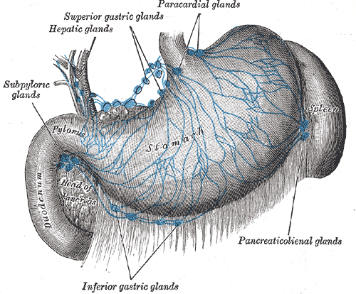
Lymphatics of the stomach
UPDATED: The Latin word [viscus] refers to an "internal organ". A better definition is that a [viscus] is a "single internal organ contained in a body cavity". Since there are three body cavities: craniospinal, thoracic, and abdominopelvic, each one of the organs in these cavities can be called a [viscus].
The plural form of [viscus] is [viscera]. It is a common mistake to use the plural form instead of the singular form when referring to a viscus. The sentence "the stomach is an abdominal viscera" is wrong. The correct sentence would be "the stomach is an abdominal viscus".
The adjective form [visceral] means "related to", or "pertaining to" a viscus or viscera.
The term [visceral] is also used to denote membranes that are related to a viscus. The [visceral] peritoneum is the portion of the peritoneal membrane that is found away from the abdominal wall and in relation to a viscus or viscera.
Original image courtesy of bartleby.com
- Details
![Four chamber section of the heart. By BrownCow. [Public domain], from Wikimedia Commons](/images/MTD/LargeImages/ductus_arteriosus_lg.jpg "Four chamber section of the heart")
Click for a larger image
The term [ductus arteriosus] is Latin and means “arterial duct”. The ductus arteriosus a component of fetal circulation and is a communication between the left pulmonary artery, close to its origin from the pulmonary trunk, and the inferior aspect of the aortic arch. This structure is also called the “duct of Botallus”.
The fetus oxygenates its blood using the placenta; therefore it does not need to use its lungs. The fetal blood is diverted from the right atrium to the left atrium through the foramen ovale. Still, some blood will pass from the right atrium to the right ventricle and then through the pulmonary valve into the pulmonary trunk. The ductus arteriosus will divert most of this blood, so that instead of going into the lungs, the blood will go into the aortic arch and then to the systemic circulation.
Once the baby is born, this right to left shunt is not needed and the foramen ovale will close immediately after birth, while the ductus arteriosus will slowly reduce its diameter to be completely closed in a few days. If this closure does not happen, the condition will be referred to as a “patent ductus arteriosus” or PDA.
Once the ductus arteriosus is close it is referred to as the “ligamentum arteriosus” or the “ligament of Botallus”, named after Leonardo Botallus (c.1530-????)
Sources:
1. "Gray's Anatomy"38th British Ed. Churchill Livingstone 1995
2. "Tratado de Anatomia Humana" Testut et Latarjet 8 Ed. 1931 Salvat Editores, Spain
Image by BrownCow. [Public domain], from Wikimedia Commons
- Details

Click for a larger image
The [prostatic sinuses] (sinus prostaticus) are bilateral depressions found lateral to the inferior portion of the median urethral crest (also known as the verumontanum). There are two prostatic sinuses within the prostatic urethra in the male
On the floor of these sinuses there are several perforations, representing the exit of the prostatic ducts (or ductules). These ducts bring prostatic fluid from the lateral lobes of the prostate into the urethra.
The image also shows the [prostatic utricle], also known as "utriculus prostaticus" or "utriculus", a small 6mm small dead-end channel found in the male prostatic urethra. The blue dotted line shows the cut edge of the urethra.
The word [utriculus] is Latin and means "little sac" or "little uterus".
Sources:
1. "The prostatic utricle is not a M?llerian duct remnant: immunohistochemical evidence for a distinct urogenital sinus origin" Shapiro E, Huang H, McFadden DE, et al. (2004) J Urol 172; 1753–1756
2. "Gray's Anatomy"38th British Ed. Churchill Livingstone 1995
3. "Tratado de Anatomia Humana" Testut et Latarjet 8 Ed. 1931 Salvat Editores, Spain
- Details
The suffix[-ceps] has a Latin origin from the word [caput], meaning "head" or "leader". This word evolved into [-capit-] as in [decapitation], [-capt-] as in [captain], and of course, [-ceps], meaning "head".
This suffix is used in many anatomical and medical terms such as:
• Biceps: Two heads. Both the biceps brachii muscle and the biceps femoris muscle have two muscular heads or components
• Triceps: Three heads. The triceps brachii muscle has three muscular heads or components. There is no triceps femoris in the human.
• Quadriceps: Four heads. The quadriceps femoris muscle has four muscular heads or components
An interesting side note is that the Latin word [caput] evolved through the ages as it was incorporated into other languages. In French it evolved into [chef] as the "head" or "leader" of a group of cooks, and then back to English as [chief].