Medical Terminology Daily (MTD) is a blog sponsored by Clinical Anatomy Associates, Inc. as a service to the medical community. We post anatomical, medical or surgical terms, their meaning and usage, as well as biographical notes on anatomists, surgeons, and researchers through the ages. Be warned that some of the images used depict human anatomical specimens.
You are welcome to submit questions and suggestions using our "Contact Us" form. The information on this blog follows the terms on our "Privacy and Security Statement" and cannot be construed as medical guidance or instructions for treatment.
We have 375 guests and no members online

Jean George Bachmann
(1877 – 1959)
French physician–physiologist whose experimental work in the early twentieth century provided the first clear functional description of a preferential interatrial conduction pathway. This structure, eponymically named “Bachmann’s bundle”, plays a central role in normal atrial activation and in the pathophysiology of interatrial block and atrial arrhythmias.
As a young man, Bachmann served as a merchant sailor, crossing the Atlantic multiple times. He emigrated to the United States in 1902 and earned his medical degree at the top of his class from Jefferson Medical College in Philadelphia in 1907. He stayed at this Medical College as a demonstrator and physiologist. In 1910, he joined Emory University in Atlanta. Between 1917 -1918 he served as a medical officer in the US Army. He retired from Emory in 1947 and continued his private medical practice until his death in 1959.
On the personal side, Bachmann was a man of many talents: a polyglot, he was fluent in German, French, Spanish and English. He was a chef in his own right and occasionally worked as a chef in international hotels. In fact, he paid his tuition at Jefferson Medical College, working both as a chef and as a language tutor.
The intrinsic cardiac conduction system was a major focus of cardiovascular research in the late nineteenth and early twentieth centuries. The atrioventricular (AV) node was discovered and described by Sunao Tawara and Karl Albert Aschoff in 1906, and the sinoatrial node by Arthur Keith and Martin Flack in 1907.
While the connections that distribute the electrical impulse from the AV node to the ventricles were known through the works of Wilhelm His Jr, in 1893 and Jan Evangelista Purkinje in 1839, the mechanism by which electrical impulses spread between the atria remained uncertain.
In 1916 Bachmann published a paper titled “The Inter-Auricular Time Interval” in the American Journal of Physiology. Bachmann measured activation times between the right and left atria and demonstrated that interruption of a distinct anterior interatrial muscular band resulted in delayed left atrial activation. He concluded that this band constituted the principal route for rapid interatrial conduction.
Subsequent anatomical and electrophysiological studies confirmed the importance of the structure described by Bachmann, which came to bear his name. Bachmann’s bundle is now recognized as a key determinant of atrial activation patterns, and its dysfunction is associated with interatrial block, atrial fibrillation, and abnormal P-wave morphology. His work remains foundational in both basic cardiac anatomy and clinical electrophysiology.
Sources and references
1. Bachmann G. “The inter-auricular time interval”. Am J Physiol. 1916;41:309–320.
2. Hurst JW. “Profiles in Cardiology: Jean George Bachmann (1877–1959)”. Clin Cardiol. 1987;10:185–187.
3. Lemery R, Guiraudon G, Veinot JP. “Anatomic description of Bachmann’s bundle and its relation to the atrial septum”. Am J Cardiol. 2003;91:148–152.
4. "Remembering the canonical discoverers of the core components of the mammalian cardiac conduction system: Keith and Flack, Aschoff and Tawara, His, and Purkinje" Icilio Cavero and Henry Holzgrefe Advances in Physiology Education 2022 46:4, 549-579.
5. Knol WG, de Vos CB, Crijns HJGM, et al. “The Bachmann bundle and interatrial conduction” Heart Rhythm. 2019;16:127–133.
6. “Iatrogenic biatrial flutter. The role of the Bachmann’s bundle” Constán E.; García F., Linde, A.. Complejo Hospitalario de Jaén, Jaén. Spain
7. Keith A, Flack M. The form and nature of the muscular connections between the primary divisions of the vertebrate heart. J Anat Physiol 41: 172–189, 1907.
"Clinical Anatomy Associates, Inc., and the contributors of "Medical Terminology Daily" wish to thank all individuals who donate their bodies and tissues for the advancement of education and research”.
Click here for more information
- Details
The word [infarction] arises from the Latin [infarcire] meaning "to fill" or "to stuff". The word reflects the fact that the "stuffing" of an artery with a clot can lead to cell death or necrosis. especially if the area supplied by the clogged vessel does not have collateral circulation.
It is a common misconception that the term "infarction" means by default a "myocardial infarction". This is not true, as an infarction can occur anywhere in the body, most commonly in areas without collateral circulation, as the heart and brain (distal to the arterial circle of Willis).
The term infarction is a synonym for [stroke] as both refer to the denial of blood supply to an area of the body.
- Details
This article is part of the series "A Moment in History" where we honor those who have contributed to the growth of medical knowledge in the areas of anatomy, medicine, surgery, and medical research.

Franz Anton Mesmer
Franz Anton Mesmer (1734 - 1815). A German physician, he was also known as Friedrich Anton Mesmer. He studied medicine at the University of Vienna; for his thesis, he developed the theory of “animal magnetism,” based on the works of Newton and gravity and his studies of astrology and the influence of magnetic fields on objects. His 1776 dissertation was titled “De Planetary Influxu” (On the Influence of the Planets)
After 10 years of a normal medical practice (for the times) Mesmer grew ever so impatient with the “classic” potions, salves, and bloodletting. He treated a woman of what today would be called “hysteria” or “somatization disorder” with every known medical treatment unsuccessfully; she improved after a treatment with magnets! leading Mesmer to believe that “animal magnetism” was the way to continue his career.
Mesmer developed a pseudotechnique to “magnetize” almost every element except steel, and his patient base grew. These patients all had some type of mental disorder that was susceptible to treatment by suggestion. Mesmer had discovered what today we know as the “placebo effect” and the basics of therapeutic hypnosis. Mesmer had so many patients that he had to treat them in “batches”, several at a time. Patients who were treated by Mesmer were said to have been “Mesmerized”.
Mesmer came under attack by the scientific establishment and when he could not prove his theories he was discredited. The fact that Mesmer used theatrics to further influence his suggestive patients did not help and he was labeled a “quack”. Mesmer retired a rich man to Switzerland where he died in 1815.
Sources:
1. "Franz Anton Mesmer and the Rise and Fallof Animal Magnetism: Dramatic Cures, Controversy, and Ultimately a Triumph for the Scientific Method" Lanska DJ, Lanska JT Brain, Mind and Medicine: Essays in Eighteenth-Century Neuroscience, 2007
2. "Early American mesmeric societies: a historical study" Gravitz, MA Am J Clin Hypn (1994) 37, 41–48
3. "Franz Anton Mesmer: The first psychotherapist of the modern age?" Traetta, L (2008) Int J Psychol 43 (3-4) 121
- Details
The root term [-styl-] is Greek and means "a pillar". The Latin term [stilos] means "a pointy structure". The suffix [oid] means "similar to". The word then means "similar to a pillar". In some cases the argument can be made that the Latin word "stylus" meaning a pen, is like a small pillar.
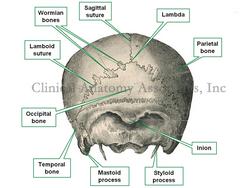
In anatomy, the styloid process is a pointy, slender pillar of bone that is found in the inferior aspect of the temporal bone. See accompanying image. Vesalius thought that a slender bony process of the ulna looked similar, so he called it also the "styloid process"
The root term [-styl-] can be found in muscles that are related to the styloid process of the temporal bone, such as: styloglossus, stylohyoid, and stylopharyngeus.
Article image in public domain, modified from Toldt's "Atlas of Human Anatomy", 1903.
- Details
The word [necrosis] arises from the Greek [nekros] meaning "a dead body" or "death". The suffix [-osis] means "condition", but with the connotation of "many". Literally, the term necrosis means "many deaths", but it used to refer to "cell death".
When necrosis is due to an arterial obstruction, the term becomes synonymous with "infarction".
- Details
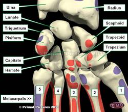
The trapezium bone is one of the four bones that comprise the distal row of the carpus or carpal bones that form the wrist. It is distinguished by a large groove on its anterior (volar) surface. It is found on the lateral (radial) aspect of the wrist between the scaphoid bone proximally and the first metacarpal distally (see image). It is also known as the "multangular bone" or the "os multangulum majus"
The trapezium bone articulates with four bones, including the scaphoid, trapezoid, and the first and second metacarpals.
The accompanying image shows the anterior (volar) surface of the wrist. Click on the image for a larger picture.
Image modified from the original: "3D Human Anatomy: Regional Edition DVD-ROM." Courtesy of Primal Pictures
- Details
The medical term [morphology] arises from the Greek root term [morphos] meaning "shape" or "form". The suffix [-ology] means "study of". In biological sciences [morphology] is the study of form.
Skinner1 defines morphology as the study of "the external configuration and structure of any part and the factors that influence its development and final form".
The term was first used in 1817
1. "The origin of Medical Terms" Skinner, AH, 1970


