
Medical Terminology Daily (MTD) is a blog sponsored by Clinical Anatomy Associates, Inc. as a service to the medical community. We post anatomical, medical or surgical terms, their meaning and usage, as well as biographical notes on anatomists, surgeons, and researchers through the ages. Be warned that some of the images used depict human anatomical specimens.
You are welcome to submit questions and suggestions using our "Contact Us" form. The information on this blog follows the terms on our "Privacy and Security Statement" and cannot be construed as medical guidance or instructions for treatment.
We have 3685 guests online
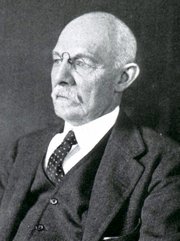
William S. Halsted, MD
(1852 – 1922)
American anatomist, teacher, and surgeon, William Stewart Halsted was born in New York City, USA to a wealthy family of English origin. His father was involved in charitable work and Governor and trustee to a city hospital. Not a brilliant student initially, Halsted took an undergraduate in Liberal Arts in Yale, CT., after which he entered the Medical College of Physicians at the Columbia College, where he excelled.
As a second-year medical student Halsted applied and obtained a position in surgery at a local hospital. In here he learned about Lister’s antiseptic technique and became an adamant proponent of it to reduce infection. In 1877 Halsted obtained his MD. After a short time as House Physician at the New York Hospital, Halsted traveled to Europe to further his education studying for two years at the Universities of Vienna, Leipzig, and W?rzburg.
Besides being at the forefront of surgical and antiseptic techniques (introducing the use of rubber gloves in surgery), Halsted was extremely concerned with the way medical students were taught in the US. He pioneered bedside clinical round discussions with the medical students after two years of basic sciences studies. Halsted developed the idea of a patient chart; he also developed the residency program for medical students in use today.
Halsted is probably the most influential researcher and surgeon at the turn of the century. He dedicated time to the study of intestinal anastomoses and the use of silk as a suture material. His experimental work in 1887 proved that the inclusion of the submucosa layer in an anastomosis was mandatory, as well that a single layered anastomosis was enough to attain closure. Perhaps Halsted’s most important contribution was the application and use of the scientific method to surgical questions. Halsted’s principles set the standards used today in surgical suturing and surgical stapling.
He also pioneered the development and surgical techniques for radical mastectomy as a treatment for breast cancer.
As a side effect of this studied in anesthesia and the use of cocaine for anesthesia, Halsted became addicted to this substance, a problem that followed him through the years. Without impairing his capacity as a researcher and a surgeon, Halsted eventually recovered. He died in Baltimore in 1922 as a complication to surgery.
Sources:
1. Dubay, A. D., & Franz, G. M. (2003). Acute Wound Healing: The Biology of Acute Wound Failure. Surg Clin NA, 83, 463-481.
2. Halsted, W. S. (1887). Circular Suture of the Intestine - An Experimental Study. Am J Med Sci, 436-461.
3. “William Stewart Halsted: his life and contributions to surgery” Osborne, P. Lancet Oncol 2007; 8: 256–65
4. “William Stewart Halsted: Surgical pioneer” Burress, P Endoc Today (2010), 8: (2) 22
5. “William Stewart Halsted (1852–1922) Neurological stamp” Haas, LF J Neurol Neurosurg Psych 2000;69:641
Original image courtesy of "Images from the History of Medicine" at www.nih.gov

"Clinical Anatomy Associates, Inc., and the contributors of "Medical Terminology Daily" wish to thank all individuals who donate their bodies and tissues for the advancement of education and research”.
Click here for more information


- Details

Click for a larger image
The anterior cerebral arteries are one of the two paired terminal branches of the internal carotid artery. Each anterior cerebral artery supplies arterial blood to the brain beyond the arterial circle of Willis. The vascular territory of the anterior cerebral artery supplies the medial surfaces of the frontal and parietal cerebral lobes as well as the medial border of the same lobes.
The anterior cerebral artery gives off the orbitofrontal artery and then divides into two callosal arteries, the pericallosal artery and the callosomarginal artery. From these arteries arise all the other branches that form the vascular territory of the anterior cerebral artery.
There are many anatomical variations of the anterior cerebral artery, as described here.
Clinical anatomy, pathology, and surgery of the brain and spinal cord are some of the lecture topics developed and delivered by Clinical Anatomy Associates, Inc.
Image modified from the original (in the public domain) by Sobotta (1945)
- Details
The medical term thrombus arises from the Greek [θρόμβος] (pronounced thrombos) meaning "a lump", "a piece of milk curd", or a "clot". This Greek term was later adopted in Latin [thrombus] and is used in this unchanged form today. The plural for thrombus is [thrombi]. A synonymous Latin term is [coagulum] (pl. coagula).
In medicine, a thrombus is a blood clot that has the connotation of being stationary, fixed to a vessel wall. If this blood clot were to be freed and start moving with the blood flow, then the clot should be called an "embolus" (Lat; pl. emboli).
Because of its anatomical characteristics. in the presence of atrial fibrillation, the left atrial appendage is particularly prone to the formation of thrombi, which can embolize and cause brain strokes.
The root term for this word is [-thromb-]. Examples of its use are:
- Thrombosis: The suffix [-osis] means "condition" with the connotation of "many". A condition of multiple thrombi
- Thrombocytopenia: A combination of root terms; the root term [-cyt-] means "cell" and the suffix [-(o)penia] means "a deficiency". A deficiency of platelets (the "clotting" cells)
Sources:
1. "The Origin of Medical Terms" Skinner, HA 1970 Hafner Publishing Co.
2. "Medical Meanings - A Glossary of Word Origins" Haubrich, WD. ACP Philadelphia
- Details
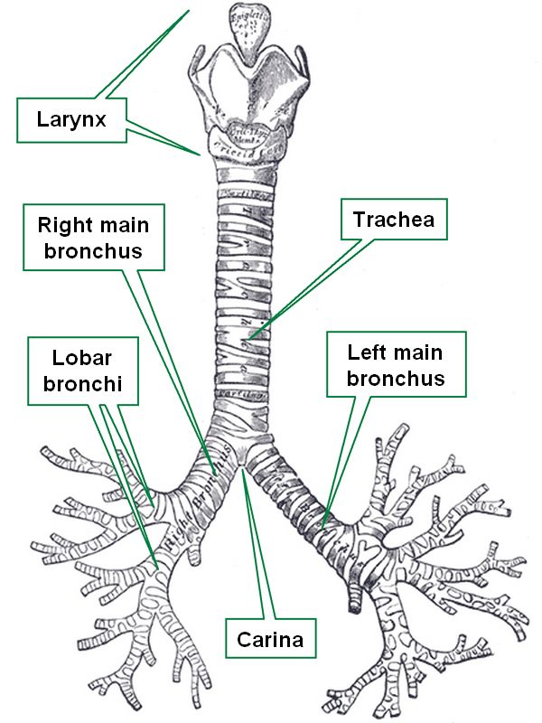
Tracheobronchial tree
The trachea, vernacularly known as the "windpipe", is in the adult a 4 inch (10.1 cm) membranous tube which contains incomplete, almost circular rings of cartilage. Some authors describe between fourteen to sixteen of these incomplete rings of cartilage in the trachea.
These semicircular rings are open-ended posteriorly, where the trachea only has a membrane, thus being flattened posteriorly. This posterior aspect of the trachea is in close anatomical relationship with the thoracic esophagus.
The trachea begins at the inferior end of the larynx, just about the level of the sixth cervical vertebra, and descends in the superior mediastinum to the level of the sternal angle (of Louis) close to the superior border of the fifth thoracic vertebra, where it divides into two main bronchi, each entering the ipsilateral lung at the level of the pulmonary hilum.
The word [trachea] arises from the Greek [trakhus] meaning "rough". This is because in the original description of this structure there was the belief that all arteries were full of air (Lat. air = ar/aria). The trachea, being rough was called the "rough artery" or "tracheartery", which was later amended, eliminating the "artery" part. In Latin the term used for the trachea was [arteria aspera], also meaning "rough artery".
The internal lower border of the bifurcation of the trachea into the left and right main bronchi is knows as the carina, where the carinal lymphatic nodes are found.
Sources:
1 "Tratado de Anatomia Humana" Testut et Latarjet 8 Ed. 1931 Salvat Editores, Spain
2. "Anatomy of the Human Body" Henry Gray 1918. Philadelphia: Lea & Febiger
Image modified by CAA, Inc. Original image courtesy of bartleby.com
- Details

Posterior view of the contents of the thoracic cavity.
The descending aorta, also known as the thoracic aorta is the third portion of the aorta. It is found distal to the aortic arch in the posterior portion of the inferior mediastinum, and proximal to the abdominal aorta.
The descending aorta is situated between the lungs and has a close relation with the thoracic esophagus and the thoracic duct.
The descending aorta gives off 10 pairs of posterior intercostal arteries. The first intercostal artery, known as the supreme intercostal artery arises as a branch of the costocervical trunk of the subclavian artery. The last pair of arteries to arise from the descending aorta are the subcostal arteries, which pass under the 12th rib. The descending aorta also gives a number of smaller branches to the esophagus and the main stem bronchi.
The descending aorta ends at the aortic hiatus, where it continues as the abdominal aorta.
Sources:
1 "Tratado de Anatomia Humana" Testut et Latarjet 8 Ed. 1931 Salvat Editores, Spain
2. "Anatomy of the Human Body" Henry Gray 1918. Philadelphia: Lea & Febiger
Image modified by CAA, Inc. Original image courtesy of bartleby.com
- Details
First used by Galen of Pergamon (129AD - 200AD) the word [etiology] is of Greek origin. [αiτία] or [aitia], meaning "cause" or "reason", and [λογία] or [-ology], meaning "study of". Etiology is the "study of cause". In medicine this word is used to denote the study of the causes for a pathology, disease, or condition.
Because of its Greek origin, Skinner (1970) states that the proper way of spelling this term should be aetiology, but the initial "a" has been discarded through use.
There are many pathologies of unknown origin. In this case the word to use would be [idiopathic] or [idiopathy] meaning "of unknown cause"
- Details
The suffix [-(o)cele] arises from the Greek [κήλη] meaning "dilation" or "pouching". In medical terminology this suffix is used to mean "hernia", "bulging", or a "dilation". In older times a synonym was used for hernia: "rupture".
Dr. Aaron Ruhalter would state the proper description for an "ocele" is that of a prolapse, that is, a herniation without a hernia sac.
Examples the use of [ocele] are:
- Orchiocele: The prefix [orchi-] means "testicle" or "scrotum". Refers to a scrotal or testicular bulging, a scrotal hernia
- Hydrocele: The root term [-hydr-] means "water". A watery dilation. Usually used to refer to the accumulation of fluids in the scrotum
- Hydatidocele: Refers to a dilated cyst containing hydatids, the larval form of the tapeworm Echinococcus granulosus or Echinococcus multilocularis
- Enterocele: The root term [-enter-] means "intestine" or "small intestine". The bulging of the small intestine into the vagina because of weakness of the vaginal wall
- Cystocele: The root term [-cyst-] means "bladder" or "sac". The bulging of the urinary bladder into the vaginal canal because of weakness of the vaginal wall
- Cystourethrocele: A combination of root terms; [-cyst-] means "bladder" or "sac" and [-urethr-] means "urethra". The bulging of the urinary bladder and urethra into the vaginal canal
- Myelomeningocele: A combination of root terms; [-myel-] means "spinal cord" (also "bone marrow") and [-mening-] means "menynx". The herniation of the spinal cords and its meningeal coverings into the back, creating a bulge
- Varicocele: The root term [-varic-] means "varix" or "sac". A bulging of the skin caused by varices