Medical Terminology Daily (MTD) is a blog sponsored by Clinical Anatomy Associates, Inc. as a service to the medical community. We post anatomical, medical or surgical terms, their meaning and usage, as well as biographical notes on anatomists, surgeons, and researchers through the ages. Be warned that some of the images used depict human anatomical specimens.
You are welcome to submit questions and suggestions using our "Contact Us" form. The information on this blog follows the terms on our "Privacy and Security Statement" and cannot be construed as medical guidance or instructions for treatment.
We have 759 guests and no members online

Jean George Bachmann
(1877 – 1959)
French physician–physiologist whose experimental work in the early twentieth century provided the first clear functional description of a preferential interatrial conduction pathway. This structure, eponymically named “Bachmann’s bundle”, plays a central role in normal atrial activation and in the pathophysiology of interatrial block and atrial arrhythmias.
As a young man, Bachmann served as a merchant sailor, crossing the Atlantic multiple times. He emigrated to the United States in 1902 and earned his medical degree at the top of his class from Jefferson Medical College in Philadelphia in 1907. He stayed at this Medical College as a demonstrator and physiologist. In 1910, he joined Emory University in Atlanta. Between 1917 -1918 he served as a medical officer in the US Army. He retired from Emory in 1947 and continued his private medical practice until his death in 1959.
On the personal side, Bachmann was a man of many talents: a polyglot, he was fluent in German, French, Spanish and English. He was a chef in his own right and occasionally worked as a chef in international hotels. In fact, he paid his tuition at Jefferson Medical College, working both as a chef and as a language tutor.
The intrinsic cardiac conduction system was a major focus of cardiovascular research in the late nineteenth and early twentieth centuries. The atrioventricular (AV) node was discovered and described by Sunao Tawara and Karl Albert Aschoff in 1906, and the sinoatrial node by Arthur Keith and Martin Flack in 1907.
While the connections that distribute the electrical impulse from the AV node to the ventricles were known through the works of Wilhelm His Jr, in 1893 and Jan Evangelista Purkinje in 1839, the mechanism by which electrical impulses spread between the atria remained uncertain.
In 1916 Bachmann published a paper titled “The Inter-Auricular Time Interval” in the American Journal of Physiology. Bachmann measured activation times between the right and left atria and demonstrated that interruption of a distinct anterior interatrial muscular band resulted in delayed left atrial activation. He concluded that this band constituted the principal route for rapid interatrial conduction.
Subsequent anatomical and electrophysiological studies confirmed the importance of the structure described by Bachmann, which came to bear his name. Bachmann’s bundle is now recognized as a key determinant of atrial activation patterns, and its dysfunction is associated with interatrial block, atrial fibrillation, and abnormal P-wave morphology. His work remains foundational in both basic cardiac anatomy and clinical electrophysiology.
Sources and references
1. Bachmann G. “The inter-auricular time interval”. Am J Physiol. 1916;41:309–320.
2. Hurst JW. “Profiles in Cardiology: Jean George Bachmann (1877–1959)”. Clin Cardiol. 1987;10:185–187.
3. Lemery R, Guiraudon G, Veinot JP. “Anatomic description of Bachmann’s bundle and its relation to the atrial septum”. Am J Cardiol. 2003;91:148–152.
4. "Remembering the canonical discoverers of the core components of the mammalian cardiac conduction system: Keith and Flack, Aschoff and Tawara, His, and Purkinje" Icilio Cavero and Henry Holzgrefe Advances in Physiology Education 2022 46:4, 549-579.
5. Knol WG, de Vos CB, Crijns HJGM, et al. “The Bachmann bundle and interatrial conduction” Heart Rhythm. 2019;16:127–133.
6. “Iatrogenic biatrial flutter. The role of the Bachmann’s bundle” Constán E.; García F., Linde, A.. Complejo Hospitalario de Jaén, Jaén. Spain
7. Keith A, Flack M. The form and nature of the muscular connections between the primary divisions of the vertebrate heart. J Anat Physiol 41: 172–189, 1907.
"Clinical Anatomy Associates, Inc., and the contributors of "Medical Terminology Daily" wish to thank all individuals who donate their bodies and tissues for the advancement of education and research”.
Click here for more information
- Details
This article is part of the series "A Moment in History" where we honor those who have contributed to the growth of medical knowledge in the areas of anatomy, medicine, surgery, and medical research.

William Cowper
William Cowper (1666 – 1710). English barber-surgeon and anatomist, William Cowper (sometimes known as William Cooper) was born in Petersfield, Hampshire. After apprenticeship with famous barber-surgeons, Cowmper was admitted as a Freeman to the Company of Barber Surgeons in 1691 after which he had a successful career as a surgeon and an anatomist in London.
Cowper opened the first private school of anatomy in London. In 1694, Cowper published “Myotomia Reformata “, a great textbook with a description of all the muscles. A controversy arose because Cowper used the illustrations from another book by Govert Bidloo (1649 – 1713). The controversy became a problem for Cowper who had to answer questions on the subject to the Royal academy, where Cowper became a member in 1699. Cowper alleged that Bidloo’s plates, although greatly illustrated had a poor description which he improved.
In 1699 Cowper published the “Philosophical Transactions” where he describes the bulbourethral glands which are today eponymically tied to his name. An infection of these glands is called a “Cowperitis”.
The bulbourethral glands had already been described by Jean M?ry (1645– 1722) and Cowper did not claim to discover these glands. As in the case with many eponyms, the name attached to a structure is not necessarily the one who discovered it. Today, many do not remember that Cowper's name is also used to describe "Cowper's ligament", that portion of the fascia lata that is attached to the iliac crest.
Sources:
1. “Two eponymous surgeons: William Cowper and Francois Poupart” Ellis, H. Brit J Hosp Med (2009) 70:4, 225
2. "Cowper, William (1666–1710)" Kornell, M, Oxford Dictionary of National Biography, Oxford University Press, 2004
3. "Medical Discoveries, who and when" Schmidt, JE: C Thomas Pub. 1959
Original image in the public domain, courtesy of "Images from the History of Medicine" at www.nih.gov
- Details
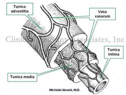
[UPDATED] The term [vasa vasorum] comes from the Latin [vasa] or [vas] meaning "a vessel", as in a container. Vasa vasorum means "the vessels of the vessels". The term "vasa vasorum" is plural. The singular form is "vas vasis".
In arteries and veins, only the inner layer of the vessel, the endothelium can receive nutrition and oxygen from the blood that is carried by the vessel. Arteries and veins have their own blood supply network, their own arteries and veins. These are the vasa vasorum.
The image shows the internal structure of an artery with its three layers, tunica intima (named after Xavier Bichat), tunica media, and tunica adventitia, as well as the vasa vasorum. Click on the image for a larger depiction.
NOTE: My personal thanks to Michiaki Akashi, M.D. for allowing us to use his artwork in this article. Dr. Akashi works as a surgeon and pathologist in the Saga Prefectural Hospital Koseikan in Saga, Japan. Dr. Miranda
- Details
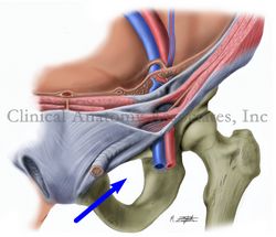
The [obturator foramen] is a bilateral and roughly triangular opening in the lower portion of the pelvis bound by the following bones: ilium, ischium, and pubic bone. The obturator foramen is closed off by the obturator membrane, a though, tendinous membrane that leaves a small superior opening (obturator canal) that allows passageway of the obturator nerve, obturator artery, and obturator vein. These neurovascular structures provide nerve and blood supply to the anterior compartment of the thigh.
The obturator membrane serves as a point of attachment to the external obturator and internal obturator muscles.
The obturator foramen is important for transobturator sling surgery used in cases of urinary incontinency.
Image property of: CAA.Inc. Artist: M. Zuptich
- Details
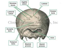
The word [lambda] is Greek and is represented by the symbols [Λ] and [λ]. Because of the inverted "V" shape and inverted "Y" shape of these symbols, it was used by Galen of Pergamon (129AD - 200AD) to denote the junction of the sagital suture (interparietal articulation) and the occipitoparietal suture, also known as the "lamboid" suture. The lambda can also be described as the point of junction of the occipital bone with the parietal bones.
Although Galen named this junction originally, it was Vesalius (1514- 1564) who brought this name into modern anatomy. Later, Peter Paul Broca (1824- 1880) used this landmark as a craniometric point.
Image in the public domain, modified from Toldt's "Atlas of Human Anatomy", 1903.
- Details
This article is part of the series "A Moment in History" where we honor those who have contributed to the growth of medical knowledge in the areas of anatomy, medicine, surgery, and medical research.
Leonardo Botallus (c.1530- ??) Italian anatomist and physician,Leonardo Botallus (also known as Botalli, Botallo, or Botal) was born circa 1530 in the region of Piedmont. Botallus studied medicine at the italian university of Pavia, where he was a student under Gabrielle Fallopius. Botallus graduated circa 1553. He was an avid advocate of bloodletting, causing him to direct his anatomical studies towards the subject of the vascular system. Although he did not discover the foramen ovale and the ductus arteriosus, he mentions these structures by those names in both his posthumous publications " De via sanguinis a dextro in sinistro cordis ventriculum" (1640) and "Opera Omnia Medica et Chirurgica" (1660)
In France Botallus became physician to King Charles IX. Not much more is known about Botallus, and history fails to record his date and place of death, as well as his image. The image depicted here is from his 1660 publication. Click on the image for a larger depiction.
Botallus' name is eponymically remembered in the following structures:
- Foramen of Botallus: The foramen ovale, an opening found in the fetus in the region of the fossa ovalis that closes upon birth
- Duct of Botallus: A communicating vessel between the left pulmonary artery and the proximal region of the descending aorta, part of fetal circulation, also known as the ductus arteriosus
- Ligament of Botallus: The closed ductus arteriosus in the adult
Sources:
1. "History of medicine; a correlative text, arranged according to subjects" Mettler, C Ch. 1947 The Blakiston Co
2. "Stedmans Medical Eponyms" Forbis, P; Bartolucci, SL. Williams & Wilkins 1998
3. "The origin of Medical Terms" Skinner, AH, 1970
4. "The First Closure of the Persistent Ductus Arteriosus" Alexi-Meskishvili,V; B?ttcher, W. Ann Thorac Surg 2010;90:349 –56
NOTE: There is no known image of Botallus. Skinner's "Origin of Medical Terms" shows one, but we could not confirm the origin of the image. If you know or have one, let us know!
- Details
This term arises from the Latin [lumbus] meaning "loin". The "loins" region of the body is the area lateral to the umbilicus, wrapping around posteriorly. In fact, there are two abdominal regions known as the "lumbar regions".
Galen of Pergamon(129AD - 200AD) named and described the lumbar vertebrae.


