Medical Terminology Daily (MTD) is a blog sponsored by Clinical Anatomy Associates, Inc. as a service to the medical community. We post anatomical, medical or surgical terms, their meaning and usage, as well as biographical notes on anatomists, surgeons, and researchers through the ages. Be warned that some of the images used depict human anatomical specimens.
You are welcome to submit questions and suggestions using our "Contact Us" form. The information on this blog follows the terms on our "Privacy and Security Statement" and cannot be construed as medical guidance or instructions for treatment.
We have 166 guests and no members online

Jean George Bachmann
(1877 – 1959)
French physician–physiologist whose experimental work in the early twentieth century provided the first clear functional description of a preferential interatrial conduction pathway. This structure, eponymically named “Bachmann’s bundle”, plays a central role in normal atrial activation and in the pathophysiology of interatrial block and atrial arrhythmias.
As a young man, Bachmann served as a merchant sailor, crossing the Atlantic multiple times. He emigrated to the United States in 1902 and earned his medical degree at the top of his class from Jefferson Medical College in Philadelphia in 1907. He stayed at this Medical College as a demonstrator and physiologist. In 1910, he joined Emory University in Atlanta. Between 1917 -1918 he served as a medical officer in the US Army. He retired from Emory in 1947 and continued his private medical practice until his death in 1959.
On the personal side, Bachmann was a man of many talents: a polyglot, he was fluent in German, French, Spanish and English. He was a chef in his own right and occasionally worked as a chef in international hotels. In fact, he paid his tuition at Jefferson Medical College, working both as a chef and as a language tutor.
The intrinsic cardiac conduction system was a major focus of cardiovascular research in the late nineteenth and early twentieth centuries. The atrioventricular (AV) node was discovered and described by Sunao Tawara and Karl Albert Aschoff in 1906, and the sinoatrial node by Arthur Keith and Martin Flack in 1907.
While the connections that distribute the electrical impulse from the AV node to the ventricles were known through the works of Wilhelm His Jr, in 1893 and Jan Evangelista Purkinje in 1839, the mechanism by which electrical impulses spread between the atria remained uncertain.
In 1916 Bachmann published a paper titled “The Inter-Auricular Time Interval” in the American Journal of Physiology. Bachmann measured activation times between the right and left atria and demonstrated that interruption of a distinct anterior interatrial muscular band resulted in delayed left atrial activation. He concluded that this band constituted the principal route for rapid interatrial conduction.
Subsequent anatomical and electrophysiological studies confirmed the importance of the structure described by Bachmann, which came to bear his name. Bachmann’s bundle is now recognized as a key determinant of atrial activation patterns, and its dysfunction is associated with interatrial block, atrial fibrillation, and abnormal P-wave morphology. His work remains foundational in both basic cardiac anatomy and clinical electrophysiology.
Sources and references
1. Bachmann G. “The inter-auricular time interval”. Am J Physiol. 1916;41:309–320.
2. Hurst JW. “Profiles in Cardiology: Jean George Bachmann (1877–1959)”. Clin Cardiol. 1987;10:185–187.
3. Lemery R, Guiraudon G, Veinot JP. “Anatomic description of Bachmann’s bundle and its relation to the atrial septum”. Am J Cardiol. 2003;91:148–152.
4. "Remembering the canonical discoverers of the core components of the mammalian cardiac conduction system: Keith and Flack, Aschoff and Tawara, His, and Purkinje" Icilio Cavero and Henry Holzgrefe Advances in Physiology Education 2022 46:4, 549-579.
5. Knol WG, de Vos CB, Crijns HJGM, et al. “The Bachmann bundle and interatrial conduction” Heart Rhythm. 2019;16:127–133.
6. “Iatrogenic biatrial flutter. The role of the Bachmann’s bundle” Constán E.; García F., Linde, A.. Complejo Hospitalario de Jaén, Jaén. Spain
7. Keith A, Flack M. The form and nature of the muscular connections between the primary divisions of the vertebrate heart. J Anat Physiol 41: 172–189, 1907.
"Clinical Anatomy Associates, Inc., and the contributors of "Medical Terminology Daily" wish to thank all individuals who donate their bodies and tissues for the advancement of education and research”.
Click here for more information
- Details
|
The prefix [-trans-] originates from the Latin and means "trough", "across", or "beyond". This prefix is quite used in vernacular language too. Applications of this prefix include:
|
|
| Back to MTD Main Page | Subscribe to MTD |
- Details
The medical term [schizophrenia] is formed by the combination of two root terms of Greek origin. The term [-schiz] comes from the Greek word [σχίσις] and means "to tear" or "to separate". In Medicine today its meaning is that of "a cleft", a "split", or "a separation". The second root term [-phren-] has two meanings, "diaphragm", and "mind". The suffix [-ia] means "condition". Thus analyzed, the word [schizophrenia] means "a condition of a split mind".
Schizophrenia is a chronic, severe, and disabling brain disorder where the patient may hear voices other people do not hear. They may believe other people are reading their minds, controlling their thoughts, or plotting to harm them. Schizophrenic patients may not make sense when they talk. They may sit for hours without moving or talking. Sometimes people with schizophrenia seem perfectly fine until they talk about what they are really thinking
Some of the patients may lose total touch with reality, presenting with hallucination, delusions, or catatonia. Others may present with depression-like symptoms such as monotony, and reclusion. Schizophrenic patients also present with memory problems and inability to carry instructions.
For more information on this condition, please click here to go to the National Institute of Mental Health website on the topic.
- Details
This article is part of the series "A Moment in History" where we honor those who have contributed to the growth of medical knowledge in the areas of anatomy, medicine, surgery, and medical research.

Francisco Javier Balmis
The Balmis Expedition (1803 -1806) The “Royal Philanthropic Vaccine Expedition”, otherwise known as the "Balmis expedition" is a little known chapter in the history of the eradication of smallpox from this world.
One of the unintended consequences of the Spanish invasion of the New World and the work of the “Conquistadores” was the introduction of smallpox to a virgin population. Other viruses were also introduced, so that between smallpox, measles, rubella, etc. it is said that in 1520 almost 50% of the population of Mexico died because of a biological infection.
Edward Jenner (1749 – 1823) discovered in 1796 that someone infected with cowpox would be protected against smallpox. The vaccine and the expansion of its use brought some relief to Europe, but the damage caused by smallpox in the Spanish colonies was catastrophic. Smallpox in its more virulent variety carried at the time a mortality rate close to 30%, leaving those who survived the virus with skin pockmarks or blinded for life. It is estimated that towards the end of the 18th century, 400,000 people died in Europe because of smallpox, and one third of the survivors were left blind.
A solution was needed, so in 1803 the Spanish king Carlos IV commissioned Don Francisco Javier de Balmis i Berenguer (1753 – 1819), a Spanish phyisician, to find a solution to the smallpox problem. While planning what was later to be known as the “Royal Philanthropic Vaccine Expedition” Balmis received critical contributions from Don Antonio de Gimbernat y Arbós (of Gimbernat’s ligament).
The main problem to this expedition was that there was no refrigeration, and the vaccine production and storage as we know it today had not been invented. The procedure was simple: Inoculate the live virus of cowpox from someone who was infected with cowpox. The solution was brilliant. A group of 10 physicians and 25 orphaned children were recruited along with nurses, and having at least one infected child on board each ship, the expedition sailed on November 30, 1803. During the long voyage, child after child were sequentially infected with the smallpox virus so that four months later, on March 19, 1804, the expedition landed in Venezuela with a child with the virus.
From here, the same method was used to distribute the vaccine North and South, to cover all of the Spanish territories until 1810. The original expedition is known as the “Balmis expedition”, and Balmis returned to Spain in 1806. Other expeditions were named after other leaders (Salvany, Justiniano, Grajales y Bolaños, etc.) but all carried the original strain brought to the Americas by Balmis. Although the original 25 children were granted the title of “special children of the Spanish nation”, no one knows how many children were used in the end, or what was their eventual fate transplanted away from their homes.
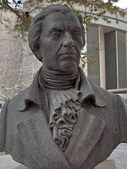
The image in this article, courtesy of Wikipedia, is a bust found at the Medical College of the Miguel Hernandez University in San Juan de Alicante, Spain
Sources:
1. “La viruela, aliado oculto en la conquista española” Sanchez-Silva, DJ. INFORMED 2007; 9 (12) 581-587
2. “La expedición de Balmis” Laval ER Rev Chil Infect 2003; 107-108
3. “Antonio de Gimbernat, 1734-1816” Matheson NM. Proc R Soc Med 1949; 42: 407-10.
4. “La vuelta al mundo de la expedición de la vacuna (1803-1810)” Diaz de Yraola, G. (2003)
5. “La segunda expedicion de Balmis” Tuells, J.; Duro-Torrijos, JL G Med Mex 2013 (149) 377-84
Original image in the Public Domain courtesy of Wikipedia
- Details
The prefix [eu-] is of Greek origin and means "good", or "well". It is found in medical and everyday terms as follow:
- Euphoria: The root term [-phor-] means "to bear", "to carry" or "oneself" Euphoria is "feeling good about oneself". The antonym would be "dysphoria"
- Euphonic:The root term [-phon-] means "sound", or "voice". To "sound good"
- Eugenic: Good genes
- Euthanasia: From the Greek word [θάνατος] (thanatos) meaning "death". A good death
- Eulogy: The root term [-log-] means "word". A "good word", a "good speech"
- Eunym: A good name
Note: The links to Google Translate include an icon that will allow you to hear the pronunciation of the word
- Details
The term [dysphoria] is composed of the prefix [dys-] meaning "abnormal, the root term [-phor-] meaning "to bear" or "to carry", and the suffix [-ia] meaning "condition" or "situation". The root term requires a little more explanation. Initially the term was used to denote "enduring", later "productive" or "fertile" and in modern medical terminology, the meaning of mental health, as in to "carry oneself" or "well-being".
Thus explained, dysphoria means "a condition of abnormal feeling about oneself". The term is used in depressive mood disorders and is characterized by anxiety, depression, sleeping disorder, etc.
The opposite of dysphoria is [euphoria] from the Greek suffix [eu-] meaning "good" or "well". Euphoria is "feeling good"
- Details
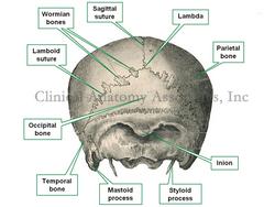
Wormian bones are small flat bones found within the suture joints of the cranium. They are also known as "intrasutural bones" (see image). These bones will vary in number and in size per individual, and are not rare to find.
These bones are eponymic, named after Olao Claus Worm Sr. (1588 - 1654), a Danish professor of Medicine and Physiology. Known by his Latinized name Olaus Wormius, he described the embryology of these bones. The eponym was created by his nephew Thomas Bartholin (1616 - 1680).
An interesting variation of a Wormian bone is the [Inca bone] also known as the Os Incae. This is an interparietal bone which can present many variations, its importance being that it can be misdiagnosed as a skull fracture, specially in children.
Sources:
1. "Interparietal bone: a case report"Mirsa, BD. J Anat Soc India (1960) 9; 39.
2. "Variations of the interparietal bone in man" Pal,GP J Anat (1987)152; 205-208.
3. "Radiological case of the month" Parente, K et al Arch Ped Adolesc Med (2001) 155;731-732
4. "The Origin of Medical Terms" Skinner, HA 1970 Hafner Publishing Co.
Article image in public domain, modified from Toldt's "Atlas of Human Anatomy", 1903


