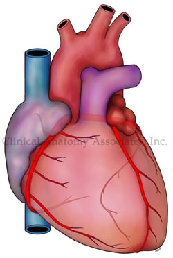
Personal Note: A few weeks ago, I came across a very interesting article in Spanish by Dr. Jose Manuel Revuelta from the University of Cantabria, Spain. The article (in Spanish) talked about the “small brain inside the heart”. One of my interests in the anatomy of the heart is the intrinsic system called the cardiac “ganglionated plexuses”, “ganglionated plexi”, or “GPs”.
It is our proposal that this nervous system inside the heart, which works autonomously (if needed to or forced to) and also dependent, of the autonomic nervous system, is responsible for the intrinsic working of the heart and its dysfunction is probably the root of cardiac dysrhythmias. We have several seminars on this topic here.
In a recent publication with Dr. Randall K. Wolf, we explained both the Wolf Procedure and the anatomical basis that underline atrial fibrillation.
The concept of the "small brain of the heart" is not new. It has been mentioned by Woolard (1926), Armour (1997), Pauza (2000), and others. These authors and others are referenced in this publication. Unfortunately, the diffusion of the concept of an intrinsic, interconnected mesh of clusters of neurons within the heart and other organs that have rhythmic activity, has many names used by the media. That is why you can find articles on the "little brain of the gut", the "gut brain", the "enteric nervous system", the "little brain of the heart", etc.
Dr. Revuelta's article shows that the interest on the GPs continues on, and more research is being done on this topic. He has graciously granted us permission to translate and publish his article in “Medical Terminology Daily”. He has also expressed interest in publishing some of his articles in this blog. Dr. Miranda
The “Little Brain” Inside the Heart
Dr. José Manuel Revuelta Soba
Professor of Surgery. Professor Emeritus, University of Cantabria, Spain
In December 2024, the prestigious journal Nature Communications published that a group of researchers from the Karolinska Institute (Sweden) and Columbia University (United States) have discovered that the heart contains a small autonomous brain.
In general, scientific knowledge has been relating cardiac activity to the brain, as the only organ that regulates its functioning. This intimate bidirectional relationship regulates the adaptation of its rhythm and contractile force to changing energy demands, through impulses and signals transmitted by the autonomic nervous system. However, the heart surprises us again with new properties that go beyond what is known.
Autonomic nervous system
The neurovegetative nervous system of the human being is involuntary, comprising the sympathetic and parasympathetic nervous systems, essential for the functioning of the organism. They act in conjunction with the enteric nervous system, which also has involuntary action and regulates the activity of the gastrointestinal tract. The complex interactions between these autonomous systems, which have opposing actions, maintain cardiovascular homeostasis, that is, they provide the appropriate amount of oxygenated blood to the organs and tissues according to their demands.
The sympathetic nervous system regulates, among other functions, the body's response to any danger perceived as a threat to physical or mental health, known as the "fight or flight" reaction, described in 1915 by the physiologist Walter B. Cannon in the United States. This instinctive reaction leads to the immediate release of certain chemical substances into the blood, such as adrenaline and noradrenaline. These hormones act as neurotransmitters that produce an increase in the contractile force of the heart, tachycardia, contraction of blood vessels, hypertension and dilation of the airways. These neurotransmitters are released in the brain, facilitating the diffusion of their messages through the extensive network that forms this autonomous nervous system, increasing the state of alertness and eliminating any feeling of drowsiness. By producing an immediate tachycardia, it improves the supply of oxygen to the organs and tissues, enlarges the pupils and reduces the digestion of food to save energy and make it available for this reaction to an unexpected danger.
The parasympathetic nervous system controls the relaxation of the body at the end of the stress caused by the sudden “fight or flight” reaction, once the threat has passed, restoring the normal functioning of the organism. Its main function is the conservation and storage of energy through the release of acetylcholine, a powerful neurotransmitter, discovered by the English physiologist Henry H. Dale, for which he was awarded the Nobel Prize in Medicine in 1936. This substance produces vasodilation, reduction of blood pressure, decreased heart rate and increased intestinal motility.
The enteric nervous system has the exclusive mission of regulating the functioning of the gastrointestinal tract, which is completely covered by hundreds of millions of nerve fibers that transmit brain messages for digestive mobility and function, modifying the volume of blood flow via vasoconstriction or vasodilation.
The small brain of the heart
The innervation of the heart is more complex than previously thought, conditioned by messages from the autonomic nervous system and others from the organ itself. In the early 1990s, scientists described that the heart contained some neurons similar to those in the human brain, which led to speculation about the possible existence of independent neuronal activity within the heart that mediated its functioning and rhythm. This fascinating discovery soon became a priority objective of scientific research.
In 2021, James S. Schwaber and R. Vadigepalli, researchers at Thomas Jefferson University in Philadelphia, performed a three-dimensional mapping of the heart's neural center. They found that the heart receives constant information from the brain about the internal and external state of its environment, adjusting heart rate, blood pressure, and cardiac output. However, these messages also came from the heart's own neural system, called the "little brain," behaving as if there were an internal loop, something similar to what systems engineers call a programmable logic controller or PLC. Most of these neurons are located near the aortic and pulmonary valves, with their largest neuronal cluster (74 percent) located in the area of the sinoatrial plexus, on the upper lateral wall of the right atrium, in immediate relation to the mouth of the superior vena cava.
Using mathematical models, they observed that when this peculiar neural programmable logic controller was activated, it perfectly adjusted the heart's response to the various impulses and signals from the brain to improve cardiac performance, making its work more efficient. Without the presence of this "little brain" it would be impossible to eliminate or correct the possible errors and damage contained in some brain messages, so the heart could become erratic, causing irregular heartbeats or arrhythmias, as well as defects in its contractility.
As these scientists analyze their 3D heart models, obtained from various mammals, new questions arise about the actions of this "brain of the heart", its internal organization, its influence on the contractile force and rhythm of the heart, as well as its coordination and responses to the constant messages from the brain. Currently, these three-dimensional maps are being used to better understand how the vagus nerve connects with cardiac neurons, opening new opportunities for the greater integration of systems engineering into the field of cardiology.
Recent findings from the Karolinska Institutet and Columbia University have revealed that the heart does indeed have its own “mini-brain,” containing a nervous system that self-regulates its rhythm and function according to demand. This complex neurological center is made up of various types of neurons with different functions, some of which function as cardiac pacemakers.
This important research project was carried out in the zebrafish, an animal model that has great similarities with the human heart, both in terms of its heart rate and its general functioning. These scientists mapped the composition, organization and functions of neurons within this small intracardiac brain, using a combination of anatomical methods, electrophysiological techniques and neuronal RNA sequencing. They carried out a complete molecular and functional classification of intracardiac neurons, revealing a complex neuronal diversity within the heart itself.
This intracardiac neurological center is not part of the autonomic nervous system governed by the brain, contrary to what was believed. The data obtained in this interesting scientific investigation show that this “small brain” is made up of several types of independent sensory neurons with clear neurochemical and functional diversity. This population of neuronal cells allows the expression of various genes that encode various neurotransmitter receptors (glycine, glutamate, adrenergic, inotropic, GABA, muscarinic, serotonergic receptors, etc.), suggesting a complex network of neurotransmission specific to the heart, which ignores its total dependence on central orders from the brain.
“We were surprised to see the complexity of this small brain inside the heart, which has a key role in maintaining and controlling the heartbeat, similar to how the brain regulates other rhythmic functions such as locomotion and breathing. Better understanding this nervous system could lead to new insights into heart disease and help develop new treatments, such as for arrhythmias. We will continue to investigate how the heart’s brain interacts with the real brain to regulate cardiac functions under different conditions, such as exercise, stress or disease”, explains Konstantinos Ampatzis, a senior researcher at the Department of Neuroscience at Karolinska Institutet, Sweden, who led the study.
Future electrophysiological, pharmacological and molecular research will be critical to better understand the tangled interactions and complex regulatory mechanisms of the internal neurotransmission of this autonomous “small brain” and thus understand its overall regulation of cardiac rhythm, contraction and output in the face of the multiple physical and mental changes to which we are exposed throughout life.
“There is a wisdom of the head and a wisdom of the heart"
Charles Dickens (1812-1870), English writer
“Facts do not cease to exist because they are ignored”
Aldous Huxley (1894-1963), English writer and philosopher.
Sources:
1. “Decoding the molecular, cellular, and functional heterogeneity of zebrafish intracardiac nervous system”. Pedroni, A; Yilmaz, E; Del Vecchio,L; et al. Nature Communications, 2024; 15 (1) DOI: 10.1038/s41467-024-54830-w
2. Bodily changes in pain, hunger, fear, and rage; an account of recent researches into the function of emotional excitement” Cannon, W. 1915. D. Appleton and Co. USA.
3. “Mapping the little brain at the heart by an interdisciplinary systems biology team” Vadigepalli, Rajanikanth et al. iScience, Volume 24, Issue 5, 102433. https://doi.org/10.1016/j.isci.2021.102433
4. A comprehensive integrated anatomical and molecular atlas of rat intrinsic cardiac nervous system. Achanta S. et al. iScience 2020 Jun 26;23(6):101140 doi: 10.1016/j.isci.2020.101140
5. "Minimally Invasive Surgical Treatment of Atrial Fibrillation: A New Look at an Old Problem" Randall K. Wolf, Efrain A. Miranda, Operative Techniques in Thoracic and Cardiovascular Surgery, 2024, doi.org/10.1053/j.optechstcvs.2024.10.003
6 Valenza, G., Matić, Z. & Catrambone, V. The brain–heart axis: integrative cooperation of neural, mechanical and biochemical pathways. Nat Rev Cardiol 22, 537–550 (2025).


