![]()
Medical Terminology Daily (MTD) is a blog sponsored by Clinical Anatomy Associates, Inc. as a service to the medical community. We post anatomical, medical or surgical terms, their meaning and usage, as well as biographical notes on anatomists, surgeons, and researchers through the ages. Be warned that some of the images used depict human anatomical specimens.
You are welcome to submit questions and suggestions using our "Contact Us" form. The information on this blog follows the terms on our "Privacy and Security Statement" and cannot be construed as medical guidance or instructions for treatment.
We have 1795 guests online
")
Marcia Crocker Noyes
(1869 – 1946)
Further to my comment on old books and research that started with an interesting bookplate (Ex-Libris). I continued my research and found that the person in charge of the Osler library bookplate was a fascinating individual that today maybe a ghost in the MedChi library and building in Baltimore... This is certainly an article that can be called "A Moment in History"
Marcia Crocker Noyes was the librarian at The Maryland State Medical Society from 1896 to 1946 and was a founding member of the Medical Library Association.[1][2][3]
Sir William Osler, MD. a famous Johns Hopkins surgeon was a noted bibliophile and had a large personal collection of books on various topics. When he became the President of MedChi in 1896, he was dismayed at the condition of the library and knew that with the right person and some stewardship, it could become a significant collection. Sir William asked his friend, Dr. Bernard Steiner, a physician and President of the Enoch Pratt Free Library in Baltimore for suggestions of a librarian, and Dr. Steiner recommended Marcia Crocker Noyes. A native of New York, and a graduate of Hunter College, Marcia had moved to Baltimore for a lengthy visit with her sister, and took a “temporary” position at the Pratt Library, which turned into three years. Although she had no medical experience or background, she was enthusiastic, and most importantly, she was willing to move into the apartment provided for the librarian, who needed to be available 24 hours a day.
The image in this article is Ms. Noyes on her first year on the job. Marcia developed a book classification system for medical books, based on the Index Medicus, and called it the Classification for Medical Literature. The system uses the alphabet with capital letters for the major divisions of medicine and lower-case ones for the sub-sections. The system was used for many years, but it's now dated and the Faculty's original shelving scheme was never changed. The card catalogs still reflect her classification and many of the cards are written in Marcia's back-slanting handwriting.
Marcia knew enough to ask the Faculty's members about medical questions, terminology and literature. She gradually won over the predominantly male membership and they became her greatest allies; Sir William at the start, and then for nearly 40 years, Dr. John Ruhräh, a wealthy pediatrician with no immediate family of his own. She made a point of attending almost every Faculty function, and in 1904, under guidelines from the American Medical Association, Marcia was made the Faculty Secretary. For much of her first 10 years, she was the Faculty's only full-time employee, only being assisted by Mr. Caution, the Faculty's janitor. Later in life Marcia would say that she hired him because of his name!
Within ten years, the library had outgrown its space, and plans, spearheaded by Marcia and Sir William before his move to Oxford, were made to build a headquarters building, mainly to house the library's growing collection of medical books and journals.
Marcia was instrumental in the design and building of the new headquarters. She travelled to Philadelphia, New York and Boston to look at their medical society buildings, and eventually, the Philadelphia architectural firm, Ellicott & Emmart was selected to design and build the new Faculty building. Every detail of the building held her imprimatur, from the graceful staircase, to the light-filled reading room, and all of the myriad details of the millwork, marble tesserae, and most of all, the four-story cast iron stacks. She was on-site, climbing up unfinished staircases, checking out the progress of the building, which was built in less than one year at a cost of $90,000.
Among the features of the new building was a fourth-floor apartment for her. She referred to it as the "first penthouse in Baltimore" and it had a garden and rooftop terrace. The library collection eventually grew to more than 65,000 volumes from medical and specialty societies around the world. Journals were traded back and forth, and physicians eagerly anticipated the arrival of each new issue. At the same time, Marcia was involved in the Medical Library Association as one of eight founding members. The MLA promotes medical libraries and the exchange of information. One of the earliest mandates of the MLA was the Exchange, a distribution and trade service for those who had duplicates or little-used books in their collections. Initially, the Exchange was run out of the Philadelphia medical society, but in 1900 it was moved to Baltimore and Marcia oversaw it. Several hundred periodicals and journals were received and sent each month, a huge amount of work for a tiny staff. In 1904, the Faculty had run out of room to manage the Exchange, so it was moved to the Medical Society of the Kings County (Brooklyn). But without Marcia's excellent administrative skills, it floundered and in 1908, the MLA asked Marcia to take charge once again.
In 1909, when the new Faculty building opened, there was enough room to run the Exchange and with the help of MLA Treasurer, noted bibliophile and close friend, Dr. John Ruhräh, it once again became successful. Additionally, Marcia and Dr. Ruhräh combined forces to revive the MLA's bulletin, which had all but ceased publication in 1908, taking the Exchange with it. This duo maintained editorial control from 1911 until 1926. In 1934, around the time of Dr. Ruhräh's death, Marcia became the first “unmedicated” professional to head the MLA. During her tenure, the MLA incorporated, the first seal was adopted, and the annual meeting was held in Baltimore. Marcia wanted to write the history of the MLA once she retired from full-time work at the Faculty, but her health was beginning to fail. She had back problems and had suffered a serious burn on her shoulder as a young woman, possibly from her time running a summer camp, Camp Seyon, for young ladies in the Adirondack Mountains. In 1946, a celebration was planned to honor Marcia's 50 years at the Faculty. But she was adamant that the physicians wait until November, the actual date of her 50 years. However, they knew she was gravely ill, and might not make it until then, so a huge party was held in April. More than 250 physicians attended the celebration, but the ones she was closest to in the early years, were long gone. She was presented with a suitcase, a sum of money to use for travelling, and her favorite painting of Dr. John Philip Smith, a founder of the Medical College in Winchester, Virginia. It was painted by Edward Caledon Smith, a Virginia painter who had been a student of the painter Thomas Sully.[4] She adored this painting and vowed, jokingly, to take it with her wherever she went.
The painting was not to stay with her for very long, for she died in November 1946, and left it to the Faculty in her will. Her funeral was held in the Faculty's Osler Hall, named for her dear friend. More than 60 physicians served as her pallbearers, and she was buried at Baltimore's Green Mount Cemetery. In 1948, the MLA decided to establish an award in the name of Marcia Crocker Noyes. It was for outstanding achievement in medical library field and was to be awarded every two years, or when a truly worthy candidate was submitted. In 2014, the Faculty began giving a bouquet of flowers to the winner of the award in Marcia's name, and in honor of her work. Much evidence exists for this tradition, as we know that the physicians, especially Drs. Osler and Ruhräh, frequently gave her bouquets of flowers. Marcia also cultivated flower gardens at the Faculty and decorated the rooms with her work.
Today, the MedChi building is open for tours and if the rumors are to be believed Ms. Marcia Crocker Noyes is still at work in her beloved library as the "resident ghost" [1][5]

NOTE: This article has been modified from the original Wikipedia article on Marcia Crocker Noyes. The article itself is well-written with interesting images of the subject. I would encourage you to visit it. The second insert is from book 00736 in my personal library and shows in pencil, the incredibly small handwriting of Marsha C. Noyes.
Sources:
1. "Marcia, Marcia, Marcia" MedChi Archives blog.
2. "Marcia C. Noyes, Medical Librarian" (PDF). Bulletin of the Medical Library Association. 35 (1): 108–109. 1947. PMC 194645
3. Smith, Bernie Todd (1974). "Marcia Crocker Noyes, Medical Librarian: The Shaping of a Career" (PDF). Bulletin of the Medical Library Association. 62 (3): 314–324. PMC 198800Freely accessible. PMID 4619344.
4. Edward Caledon BRUCE (1825-1901)"
5. Behind the scenes tour MedChiBuilding
"Clinical Anatomy Associates, Inc., and the contributors of "Medical Terminology Daily" wish to thank all individuals who donate their bodies and tissues for the advancement of education and research”.
Click here for more information


- Details

Click for a larger image
The word [pinna] is Latin and means "feather". It also means "wing". The variation [penna] as in the case of [pennate], means "winged". It refer to the external ear, or auricle. It appears that the use of the term [pinna] for ear arises from the ear-like or winged extensions of Viking and medieval helmets.
The ear has three components, the internal, middle, and external ear. The external ear is composed of the external acoustic canal and the pinna. The pinna is composed of fibrocartilage covered with skin, and has several ligaments and small muscles related to it. These muscles are extrinsic (between the pinna and the skull) and intrinsic (within the pinna) All these muscles have limited capabilities in the human.
The pinna receives blood supply from the anterior and posterior auricular arteries, and a small branch of the occipital artery. The nerve supply is by way of the great auricular nerve, the auricular branch of the vagus nerve, the auriculotemporal branch of the mandibular nerve, and the lesser occipital nerve.
The external (lateral) anatomy of the pinna is complicated and very detailed, with potential anatomical variations. Click on the image for a higher detail. The medial aspect of the pinna presents elevations which correspond to the depressions (fossae) on its lateral surface and they are named, eminentia conchae, eminentia triangularis, eminentia scaphoides, etc.
Image property of: CAA, Inc. Artist: Dr. Miranda
- Details
The prefix [circum-] is Latin and means "around" or "about". It is used in medical terms such as:
- Circumcision: the root term [-cis-] meaning to "cut". To cut around
- Circumflex: the root term [flex] for [flexion] meaning to "bend". Bends around, as in "circumflex artery"
- Circumambulation: a patient that walks in circles
Also in everyday terms such as:
- Circumlocution: to talk around a subject
- Circumnavigation: To sail or navigate around
- Circumscribe: to write in circles or around a subject
- Details

The word [flexion] comes from the Latin [flexere] meaning "to bend". In anatomy, flexion is the reduction in the angle between two bodily components that are communicated by a type of joint.
By contrast, [extension] refers to the opposite action, that is, the increase in the angle between two bodily components that are communicated by a type of joint.
The image shows flexion of the head, the upper extremity, and the lower extremity. Hover over the image to see extension of the same structures.
Excessive flexion (hyperflexion) or extension (hyperextension) of a joint can lead to potential pathology as would be the case of hyperextension of the neck as a result of a car crash (whiplash injury)
Note that in a human in the anatomical position, flexion of the upper extremity is an anterior movement, while flexion of the lower extremity is a posterior movement. You could make a case that in these image the upper extremity is performing an anteflexion (anterior flexion) while the lower extremity is performing a retroflexion (posterior flexion).
In the upper and lower extremities there are whole groups of muscles that, because of their action, are called flexors or extensors.
- Details

Terminal ileum, cecum,
and vermiform appendix
The word [vermis] is Latin and means "worm". The term [vermiform appendix] means "the worm-shaped appendage", and refers to a worm-like appendage that is related to the cecum, a segment of the right colon.
This structure was first described by Jacobo Berengario da Carpi in 1524, and it was Andreas Vesalius who first described it as an appendix, and suggested it looked like a worm. It has been called the [vermix] and the [cecal appendix]
The vermiform appendix* has the same four layers found in most of the abdominal digestive tract and is attached to the cecum at the point where the three tenia coli (libera, mesenterica, and omentalis) meet. The length of the vermiform appendix is variable. On average about 2.5 to 3 inches, it can be as long as 10 inches in length, with one recorded case of a 13 inch appendix!**
The location of the vermiform appendix is also subject to anatomical variation, being found in a retrocecal position in 65% of the cases. For more information on this organ's anatomical variations, click here.
The vermiform appendix is an intraperitoneal structure, as it has a peritoneal extension called the mesoappendix. Within the mesoappendix are the appendiceal arteries and veins. The appendiceal artery is usually a branch of the ileocolic artery.
Sources:
1. "The Origin of Medical Terms" Skinner, HA 1970 Hafner Publishing Co.
2. "Medical Meanings - A Glossary of Word Origins" Haubrich, WD. ACP Philadelphia
3 "Tratado de Anatomia Humana" Testut et Latarjet 8 Ed. 1931 Salvat Editores, Spain
4. "Anatomy of the Human Body" Henry Gray 1918. Philadelphia: Lea & Febiger Image modified by CAA, Inc. Original image by Henry Vandyke Carter, MD., courtesy of bartleby.com
*. It is not proper to call this structure the "appendix", as there are many appendices in the human body.
**. Personal note: The longest vermiform appendix I have personally seen was 8 inches (20.3 cm) in length, retrocolic, and the tip of the organ was actually retrohepatic!. Dr. Miranda.
- Details
This article is part of the series "A Moment in History" where we honor those who have contributed to the growth of medical knowledge in the areas of anatomy, medicine, surgery, and medical research.

Dr. Willem Einthoven
Dr. Willem Einthoven (1860 - 1927). Einthoven was Dutch, born on 1860 in the city of Semarang in the island of Java. His father was a physician working for the Dutch military. He started his medical studies at the University of Utrecht, Holland. Having developed an interest in ophthalmology and physiology, he developed his medicine doctorate thesis on stereoscopic color vision.
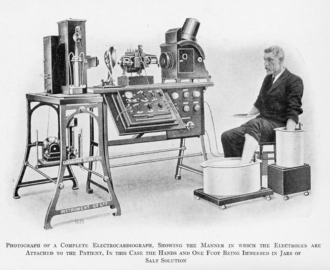
In 1885 Einthoven became a Professor of Physiology at the University of Leiden. Having seen a demonstration of Augustus Waller’s “electrogram” (a device that recorded minute deviations on a mercury column when electrically stimulated) in 1887, he improved it by creating the “string galvanometer”. In 1901 Einthoven published his first recordings of what he called “elektrokardiogramm” (EKG).
The initial device was bulky, heavy, and required the patient to sit with both arms and the left leg in separate buckets of salt water, but it did record the electrical activity of the heart (Click here for an image of one of the first electrocardiographs). Eventually the device was commercialized and history was made. It was Einthoven who used the letter P,Q,R,S, and T in electrocardiography.
In 1924, Willem Einthoven was awarded the Nobel Prize in Physiology.
Sources:
1. "Willem Einthoven (1860-1927): father of electrocardiography". Merritt, C. Tan. SY. Singapore Med J 53:(1) 17
2. "Willem Einthoven (1860-1927)" Davies, M; Hollman, A. Heart. 1997 October; 78(4): 324
3. "Willem Einthoven: The development of the human electrocardiogram" Cajavilcaa, C.,Varonb, J.Resuscitation 76:3 2008; 325–328
Original image courtesy of "Images from the History of Medicine" at www.nih.gov.
- Details

Cerebelum
The word [vermis] is Latin and means "worm".
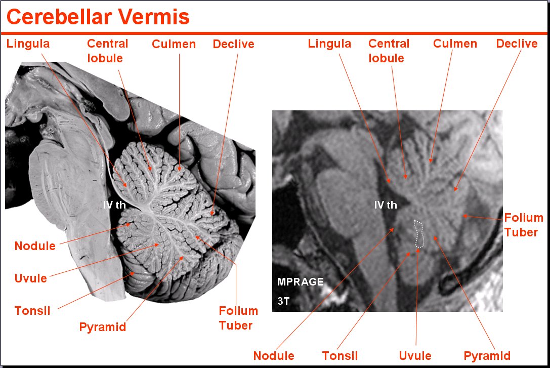
The vermis is the name given by Galen of Pergamon (129AD - 200AD) to the median lobe of the cerebellum, since when seen from the superior aspect, this cerebellar lobe looks like a multisegmented worm. See accompanying image, or click for a larger depiction.
When seen in a median section, the cerebellar vermis looks like a multilobulated leaf with the fourth ventricle of the brain at its base. It is composed of several smaller lobules: Lingula, central, culmen, clivus, tuber vermis, pyramid, uvula, and nodular lobes.
Median section image link courtesy of UCLA Radiology
Sources:
1. "The Origin of Medical Terms" Skinner, HA 1970 Hafner Publishing Co.
2. "Medical Meanings - A Glossary of Word Origins" Haubrich, WD. ACP Philadelphia
3 "Tratado de Anatomia Humana" Testut et Latarjet 8 Ed. 1931 Salvat Editores, Spain
4. "Anatomy of the Human Body" Henry Gray 1918. Philadelphia: Lea & Febiger
Image modified by CAA, Inc. Original image courtesy of bartleby.com