Medical Terminology Daily (MTD) is a blog sponsored by Clinical Anatomy Associates, Inc. as a service to the medical community. We post anatomical, medical or surgical terms, their meaning and usage, as well as biographical notes on anatomists, surgeons, and researchers through the ages. Be warned that some of the images used depict human anatomical specimens.
You are welcome to submit questions and suggestions using our "Contact Us" form. The information on this blog follows the terms on our "Privacy and Security Statement" and cannot be construed as medical guidance or instructions for treatment.
We have 334 guests and no members online

Jean George Bachmann
(1877 – 1959)
French physician–physiologist whose experimental work in the early twentieth century provided the first clear functional description of a preferential interatrial conduction pathway. This structure, eponymically named “Bachmann’s bundle”, plays a central role in normal atrial activation and in the pathophysiology of interatrial block and atrial arrhythmias.
As a young man, Bachmann served as a merchant sailor, crossing the Atlantic multiple times. He emigrated to the United States in 1902 and earned his medical degree at the top of his class from Jefferson Medical College in Philadelphia in 1907. He stayed at this Medical College as a demonstrator and physiologist. In 1910, he joined Emory University in Atlanta. Between 1917 -1918 he served as a medical officer in the US Army. He retired from Emory in 1947 and continued his private medical practice until his death in 1959.
On the personal side, Bachmann was a man of many talents: a polyglot, he was fluent in German, French, Spanish and English. He was a chef in his own right and occasionally worked as a chef in international hotels. In fact, he paid his tuition at Jefferson Medical College, working both as a chef and as a language tutor.
The intrinsic cardiac conduction system was a major focus of cardiovascular research in the late nineteenth and early twentieth centuries. The atrioventricular (AV) node was discovered and described by Sunao Tawara and Karl Albert Aschoff in 1906, and the sinoatrial node by Arthur Keith and Martin Flack in 1907.
While the connections that distribute the electrical impulse from the AV node to the ventricles were known through the works of Wilhelm His Jr, in 1893 and Jan Evangelista Purkinje in 1839, the mechanism by which electrical impulses spread between the atria remained uncertain.
In 1916 Bachmann published a paper titled “The Inter-Auricular Time Interval” in the American Journal of Physiology. Bachmann measured activation times between the right and left atria and demonstrated that interruption of a distinct anterior interatrial muscular band resulted in delayed left atrial activation. He concluded that this band constituted the principal route for rapid interatrial conduction.
Subsequent anatomical and electrophysiological studies confirmed the importance of the structure described by Bachmann, which came to bear his name. Bachmann’s bundle is now recognized as a key determinant of atrial activation patterns, and its dysfunction is associated with interatrial block, atrial fibrillation, and abnormal P-wave morphology. His work remains foundational in both basic cardiac anatomy and clinical electrophysiology.
Sources and references
1. Bachmann G. “The inter-auricular time interval”. Am J Physiol. 1916;41:309–320.
2. Hurst JW. “Profiles in Cardiology: Jean George Bachmann (1877–1959)”. Clin Cardiol. 1987;10:185–187.
3. Lemery R, Guiraudon G, Veinot JP. “Anatomic description of Bachmann’s bundle and its relation to the atrial septum”. Am J Cardiol. 2003;91:148–152.
4. "Remembering the canonical discoverers of the core components of the mammalian cardiac conduction system: Keith and Flack, Aschoff and Tawara, His, and Purkinje" Icilio Cavero and Henry Holzgrefe Advances in Physiology Education 2022 46:4, 549-579.
5. Knol WG, de Vos CB, Crijns HJGM, et al. “The Bachmann bundle and interatrial conduction” Heart Rhythm. 2019;16:127–133.
6. “Iatrogenic biatrial flutter. The role of the Bachmann’s bundle” Constán E.; García F., Linde, A.. Complejo Hospitalario de Jaén, Jaén. Spain
7. Keith A, Flack M. The form and nature of the muscular connections between the primary divisions of the vertebrate heart. J Anat Physiol 41: 172–189, 1907.
"Clinical Anatomy Associates, Inc., and the contributors of "Medical Terminology Daily" wish to thank all individuals who donate their bodies and tissues for the advancement of education and research”.
Click here for more information
- Details
The jejunum is an intraperitoneal organ, it is the second portion of the small intestine and part of the digestive tract. It begins at the duodenojejunal junction where it is related to the ligament of Treitz, and extends 8 to 9 feet, continuing distally with the ileum.
Being intraperitoneal, it is anchored to the posterior abdominal wall by the double-layered mesentery through which the jejunum receives its blood and nerve supply. At the root (base) of the mesentery are the superior mesenteric vessels.
The Latin word [jejunis] means "empty" or "fasting". The Latin term [jejunum] was used by the Romans to denote the first meal of the day, breakfast, when you have an "empty" stomach. The term was associated with this segment of the small intestine, as it is most of the time found empty in cadavers being dissected.
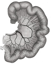
There is no clear anatomical boundary between the jejunum and ileum, as they blend smoothly one into the other. There are several gross changes from jejunum to ileum, one of them being that the complexity of the mesenteric arterial arches increases from proximal to distal. See the accompanying image. Click on it for a larger depiction.
Two interesting side notes: In English, the term for the first meal of the day is self-explanatory: [break - fast], adding to the Roman concept of "fasting" or "jejunum". In Spanish, the term for breakfast is [desayuno], where the word [ayuno] means "fasting", therefore the word [des-ayuno] also means "the end of fasting". Look at the evolution (in Spanish) from [jejunum] to [yeyuno] (the Spanish term for the organ) to [ayuno], meaning "fasting" or "empty".
Sources:
1. "The Origin of Medical Terms" Skinner, HA 1970 Hafner Publishing Co.
2. "Clinically Oriented Anatomy" Moore, KL. 3r Ed. Williams & Wilkins 1992
3 "Tratado de Anatomia Humana" Testut et Latarjet 8 Ed. 1931 Salvat Editores, Spain
4. "Anatomy of the Human Body" Henry Gray 1918. Philadelphia: Lea & Febiger Image modified by CAA, Inc.
Original image by Henry Vandyke Carter, MD., courtesy of bartleby.com
- Details
The word [crus] is Latin (cruris) and refers to the leg, or region of the shin. It is commonly used to mean "leg" or "pillar". The plural form is [crura].
Several authors suggest a relation of [crus] with another Latin term [crux] meaning "cross" as if a cross is formed by two [crura] (legs).
The term crus is widely used in human anatomy:
- crus cerebri: there are two crus cerebri in the anterior aspect of the mesencephalon
- crura of the penis: the posterior aspect of the corpora cavernosa firmly attached to the ischiopubic rami
- crura of the clitoris: the posterior aspect of the corpora cavernosa firmly attached to the ischiopubic rami
- crura fornix cerebrii: the posterior converging bands that form the fornix of the cerebrum
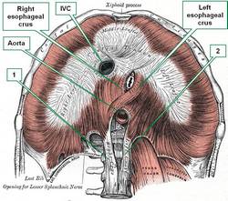
Special mention is deserved by the crura of the diaphragm. There are two pairs of diaphragmatic crura. The esophageal crura (right and left) which bound the passageway of the esophagus from the thorax into the abdomen, the esophageal hiatus. The esophageal crura have a muscular structure. The aortic crura (righ and left) allow for passage of the aorta into the abdomen, and although muscular superiorly, they are mostly tendinous. The accompanying image shows an anteroinferior view of the respiratory diaphragm. Click on the image for a larger picture.
Sources:
1. "The Origin of Medical Terms" Skinner, HA 1970 Hafner Publishing Co.
2. "Medical Meanings - A Glossary of Word Origins" Haubrich, WD. ACP Philadelphia
3 "Tratado de Anatomia Humana" Testut et Latarjet 8 Ed. 1931 Salvat Editores, Spain
4. "Anatomy of the Human Body" Henry Gray 1918. Philadelphia: Lea & Febiger
Image modified by CAA, Inc. Original image by Henry Vandyke Carter, MD., courtesy of bartleby.com
- Details
This article is part of the series "A Moment in History" where we honor those who have contributed to the growth of medical knowledge in the areas of anatomy, medicine, surgery, and medical research.
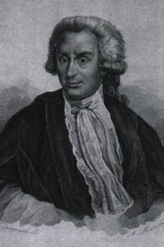
Luigi Galvani
Luigi A. Galvani (1737-1798). Italian anatomist, surgeon, and physiologist, Luigi Aloisio Galvani was born in Bologna in 1737. Although he started his studies to join the church, Galvani followed with medical studies at the University of Bologna, where he became a skilled anatomist and surgeon. On July 15, 1759 Galvani obtained his degree in medicine and philosophy.
He was interested in the effects of electricity on tissues and through observation and experimentation he postulated the existence of "animal electricity", that is, electricity generated within the tissues. He postulated the possibility that nerves carried electricity. His theories led to a passionate controversy with Volta, who denied Galvani's postulates. Galvani's theories would only be confirmed after his death.
Galvani was deeply religious, and when forced by government officials to take an oath of atheism, he refused. He was stripped of his position and was lead to poverty. His position was restored close to his death. In his honor, Andre Ampere (1775-1836) named one of his inventions that measures electricity, the "galvanometer". His name is also present in vernacular English, when we say that a rock star or a movie "galvanizes" an audience, meaning it was "electrifying"!
Sources:
1. "Luigi Galvani" Haas LF J Neurol Neurosurg Psychiatry v.56(10); Oct 1993
2. "Luigi Galvani and the foundations of electrophysiology" Cajavilca C, Varonb,J,Sternbachc GL; Resuscitation 80 (2009) 159–162
- Details
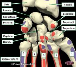
The capitate bone is one of the four bones that comprise the distal row of the carpus or carpal bones that form the wrist. It is the largest of the carpal bones and is placed in the center of the wrist (see image).
Its name originates from the Latin [caput], meaning "head". The capitate bone presents a large, rounded area, called the "head". To complete the homology, the capitate bone also has a narrow segment called the "neck", the rest of the bone called the "body". It is also known as "os capitatum" or "os magnum"
The capitate bone articulates with seven bones, including the scaphoid, lunate, trapezoid, hamate, and the three central metacarpals (2nd, 3rd, and 4th).
The accompanying image shows the anterior (volar) surface of the wrist.
Image modified from the original: 3D Human Anatomy: Regional Edition DVD-ROM" Courtesy of Primal Pictures
- Details
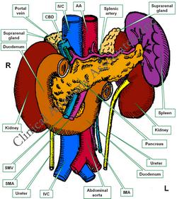
The duodenum is a mostly retroperitoneal organ, part of the digestive tract, and the most proximal portion of the small intestine. This organ is approximately 10 inches in length (24.5 cm). It starts at the pylorus of the stomach, has a "C" shape, curving around the head and the neck of the pancreas, to end at the duodenojejunal junction.
The duodenum is described as having four segments of differing length, usually named numerically:
- First segment: about two inches in length, it is dilated and called the "duodenal ampulla", or "superior duodenum"
- Second segment: about three inches in length, it receives bile and pancreatic juice through the hepatopancreatic ducts and ampullae. It is also called the "descending duodenum"
- Third segment: about four inches in length, it crosses the midline, and is also known as the "horizontal" or "transverse duodenum"
- Fourth segment: one inch in length, this is the shortest segment, it ascends towards the duodenojejunal junction, which is tethered to the diaphragm by a fold of peritoneum around a fibromuscular band called the "ligament of Treitz". At this point the retroperitoneal duodenum becomes the intraperitoneal jejunum. This fourth segment is also called the "ascending duodenum"
The name of the organ is interesting. Most textbooks claim that is originates from the Latin [duodeni], meaning "twelve". The fact is that the duodenum was originally named in Greek [δώδεκα δάχτυλαν] meaning "twelve fingers". If you place both your hands together and add 1/4 of an inch to each side (as if you had an extra finger on each hand) that measures approximately 10 inches. The term was shortened by an incorrect translation to "twelve" by Gerard of Cremona (1114 - 1187) who called it "duodenum", a bad translation, as twelve fingers in Latin is [duodecim digitorum].
While most of the duodenum is retroperitoneal, the first inch of the superior duodenum (first segment) is intraperitoneal as it shares a small portion of the lesser omentum with the stomach and liver.
Sources:
1. "Clinically Oriented Anatomy" Moore, KL. 3r Ed. Williams & Wilkins 1992
2. "The origin of Medical Terms" Skinner, AH, 1970
Image property of: CAA, Inc. Artist: Dr. E. Miranda
- Details
This prefix is derived from the Greek and means "slow". Most everybody knows about [bradycardia] meaning "slow heart", but there is a large number of applications of this prefix as follows:
- Bradytrophia: from the Greek [trophe] meaning "to feed" or "nutrition". Braditrophia is a slow nutritional process
- Bradypnea: from the Greek [pnoia], meaning "breath" or "air". Bradypnea is an abnormally slow breathing rhythm
- Bradylalia: from the Greel [lalein] meaning "to talk". Bradylalia is a slow articulation or formation of words, sometimes also known as [bradyarthria] or [bradyphasia]. See the article on aphasia and dysphasia here
- Bradykinesia: from the Greek [kinesis], meaning "movement". Bradykinesia means "slow movement", also known as [bradypragia]
- Bradycrotic: from the Greek [krotos], meaning "pulse" "or pulsation" A bradycrotic agent slows down the patient's pulse or heart rate.
- Bradytocia: from the Greek [tokos], meaning "birth". Bradytocia is a slow birthing process


