Medical Terminology Daily (MTD) is a blog sponsored by Clinical Anatomy Associates, Inc. as a service to the medical community. We post anatomical, medical or surgical terms, their meaning and usage, as well as biographical notes on anatomists, surgeons, and researchers through the ages. Be warned that some of the images used depict human anatomical specimens.
You are welcome to submit questions and suggestions using our "Contact Us" form. The information on this blog follows the terms on our "Privacy and Security Statement" and cannot be construed as medical guidance or instructions for treatment.
We have 338 guests and no members online

Jean George Bachmann
(1877 – 1959)
French physician–physiologist whose experimental work in the early twentieth century provided the first clear functional description of a preferential interatrial conduction pathway. This structure, eponymically named “Bachmann’s bundle”, plays a central role in normal atrial activation and in the pathophysiology of interatrial block and atrial arrhythmias.
As a young man, Bachmann served as a merchant sailor, crossing the Atlantic multiple times. He emigrated to the United States in 1902 and earned his medical degree at the top of his class from Jefferson Medical College in Philadelphia in 1907. He stayed at this Medical College as a demonstrator and physiologist. In 1910, he joined Emory University in Atlanta. Between 1917 -1918 he served as a medical officer in the US Army. He retired from Emory in 1947 and continued his private medical practice until his death in 1959.
On the personal side, Bachmann was a man of many talents: a polyglot, he was fluent in German, French, Spanish and English. He was a chef in his own right and occasionally worked as a chef in international hotels. In fact, he paid his tuition at Jefferson Medical College, working both as a chef and as a language tutor.
The intrinsic cardiac conduction system was a major focus of cardiovascular research in the late nineteenth and early twentieth centuries. The atrioventricular (AV) node was discovered and described by Sunao Tawara and Karl Albert Aschoff in 1906, and the sinoatrial node by Arthur Keith and Martin Flack in 1907.
While the connections that distribute the electrical impulse from the AV node to the ventricles were known through the works of Wilhelm His Jr, in 1893 and Jan Evangelista Purkinje in 1839, the mechanism by which electrical impulses spread between the atria remained uncertain.
In 1916 Bachmann published a paper titled “The Inter-Auricular Time Interval” in the American Journal of Physiology. Bachmann measured activation times between the right and left atria and demonstrated that interruption of a distinct anterior interatrial muscular band resulted in delayed left atrial activation. He concluded that this band constituted the principal route for rapid interatrial conduction.
Subsequent anatomical and electrophysiological studies confirmed the importance of the structure described by Bachmann, which came to bear his name. Bachmann’s bundle is now recognized as a key determinant of atrial activation patterns, and its dysfunction is associated with interatrial block, atrial fibrillation, and abnormal P-wave morphology. His work remains foundational in both basic cardiac anatomy and clinical electrophysiology.
Sources and references
1. Bachmann G. “The inter-auricular time interval”. Am J Physiol. 1916;41:309–320.
2. Hurst JW. “Profiles in Cardiology: Jean George Bachmann (1877–1959)”. Clin Cardiol. 1987;10:185–187.
3. Lemery R, Guiraudon G, Veinot JP. “Anatomic description of Bachmann’s bundle and its relation to the atrial septum”. Am J Cardiol. 2003;91:148–152.
4. "Remembering the canonical discoverers of the core components of the mammalian cardiac conduction system: Keith and Flack, Aschoff and Tawara, His, and Purkinje" Icilio Cavero and Henry Holzgrefe Advances in Physiology Education 2022 46:4, 549-579.
5. Knol WG, de Vos CB, Crijns HJGM, et al. “The Bachmann bundle and interatrial conduction” Heart Rhythm. 2019;16:127–133.
6. “Iatrogenic biatrial flutter. The role of the Bachmann’s bundle” Constán E.; García F., Linde, A.. Complejo Hospitalario de Jaén, Jaén. Spain
7. Keith A, Flack M. The form and nature of the muscular connections between the primary divisions of the vertebrate heart. J Anat Physiol 41: 172–189, 1907.
"Clinical Anatomy Associates, Inc., and the contributors of "Medical Terminology Daily" wish to thank all individuals who donate their bodies and tissues for the advancement of education and research”.
Click here for more information
- Details
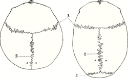
The word [bregma] is Greek and means "the front of the head". It is actually the point of intersection of the the coronal and sagittal sutures. The coronal suture is the articulation or joint between the frontal and parietal bones, and the sagittal suture is the median joint between both parietal bones.
The term was first used in anatomy as a craniometric point by Paul Broca (1824 - 1880). The image shows a superior view of two heads and the location of the coronal and sagittal sutures. The bregma is the point of intersection of these two articulations.
Click on the image for a larger view. 1 = coronal suture 2 = sagittal suture 3 = lambdoid suture. The bregma is the point of intersection of 1 and 2
Original image courtesy of Wikipedia
- Details
This article is part of the series "A Moment in History" where we honor those who have contributed to the growth of medical knowledge in the areas of anatomy, medicine, surgery, and medical research.

Original image courtesy of
Images from the History of Medicine
Marie-Francois Xavier Bichat (1771 - 1802). French physician, surgeon, anatomist and physiologist, Marie-Francois Xavier Bichat was born in the village of Thoirette. His father was a physician, influencing his early instruction and vocation. In Lyon he studied anatomy and surgery. At 28 years of age Bichat was appointed physician to the Hôtel (Hospital) Dieu. His life was influenced by his mentor, Pierre-Joseph Dassault (1738 - 1795). Upon his mentor's death Bichat took upon him to continue and finish his work, while supporting his mentor's family.
Bichat is know for the concept of the body composed of distinct tissues, which he originally called "membranes". Without the aid of the microscope Bichat described 21 different tissues and is considered the founder of the science of histology. His name is preserved in many eponymic structures such as Bichat’s fossa (pterygopalatine fossa), Bichat’s buccal fat pad, Bichat’s foramen (cistern of the vena magna of Galen), Bichat’s ligament (lower fasciculus of the posterior sacroiliac ligament), and Bichat’s tunica intima (tunica intima vasorum).
Xavier Bichat also contributed to a newer description of the humoral physiological theory, later becoming the basis of hematology. He was also interested in the description of life and death, proposing the existence of an "organic life" and an "animal life". An interesting note is that Bichat died because of an infection he acquired while dissecting a cadaver. Remember that at the time, no embalming was used!
Today Bichat's name is almost forgotten, although in some countries the buccal fat pad is still called "Bichat's fat pad" In many Spanish-speaking countries this structure is referred to as "la bola grasa de Bichat", and many still refer to the removal of this fat pad as "Bichectomy". For an image of the before and after of the procedure, click here.
Sources:
1. "Marie-Fran?ois Xavier Bichat (1771-1802) and his contributions to the foundations of pathological anatomy and modern medicine" Shoja M.M., Tubbs R.S., Loukas M., Shokouhi G., Ardalan M.R.(2008) Annals of Anatomy, 190(5),413-420
2. "Physiological Researches on Life and Death" Bichat, Marie-Francois Xavier, 1827. Translated from French by F. Gold. Richardson and Lord, Boston.
3. "A Historical Perspective: Infection from Cadaveric Dissection from the 18th to the 20th Centuries" Shoja, MM et al. Clin Anat (2013) 26:154-160
- Details
This complex medical word is formed by the combination of two root terms: [dacry-] meaning "tear" and [-cyst-], meaning "sac". The combined root [dacryocyst-] means "tear sac" or better, "lacrimal sac" (the Latin word [lacrima] means "tear"). This medical word also has a combined suffix: [-(o)lith], meaning "stone", and [-iasis], meaning "disease or condition".
The word [dacryocystolithiasis] means then, "a condition or pathology of stones (calculi) in the lacrimal sac". The procedure to remove the stones would then be called a [dacryocystolithectomy].
- Details
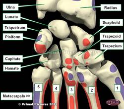
The Hamate bone is one of the four bones that comprise the distal row of the carpus or carpal bones that form the wrist. The name arises from the Latin [hamatus], meaning "hooked". The hamate bone has a distinct hook-like bony process in its volar (anterior) surface, known as the hamulus. This bone is also known as the "unciform bone" (from the Latin [uncus], also meaning "hook") or the os hamatum.
The lunate bone has a wedge-like shape and six surfaces (as a die). It articulates with five bones, including the lunate bone, capitate, triquetrum, and the fourth and fifth metacarpal bones.
The hook of the hamate bone is one of the distal boundaries of the carpal tunnel and serves as a pulley for the tendons of the fourth and fifth flexor tendons. It also serves as one of the points of muscular attachment for the following muscles: flexor carpi ulnaris, flexor digit minimi, and opponens digiti minimi. Because of its projection into the palm of the hand, the hamulus is involved in injuries in sports that require the athlete to use an accessory, as in racquetball, tennis, baseball, golf, etc.
The accompanying image shows the anterior (volar) surface of the wrist.
Image modified from the original: 3D Human Anatomy: Regional Edition DVD-ROM Courtesy of Primal Pictures
- Details

The word itself arises from the Greek. The root term [-phleb-] derives from [φλέβα] (phleba) meaning "vein", and the suffix [-otomy], meaning "to cut" or "to open". Let's not forget that the suffix component [-y] means "process of". So [phlebotomy] is the "process (or action) of cutting open a vein"
For centuries a standard practice in medicine was to "bleed" a patient, by opening a vein under controlled conditions and letting some blood flow. The practice was known as "bloodletting" or phlebotomy. Not in use today, it is said that excessive bloodletting contributed to the death of George Washington, having removed 5 pints of blood in one day!. Today the professionals who draw blood are called "phlebotomists"
The image (circa 1860) depicts one of the only known three photographs of a bloodletting procedure. Observe the lack of aseptic technique.
Image in the public domain, by The Burns Archive, courtesy of Wikipedia.org.
- Details
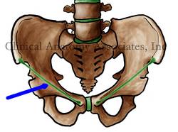
The iliopubic tract is a thickening of the transversalis fascia found in direct relation, immediately posterior to the inguinal (Poupart's) ligament. As the inguinal ligament, the iliopubic tract extends between the anterior superior iliac spine (ASIS) superolaterally, and the pubic tubercle inferomedially.
This obscure structure has been brought up to light because it is one of the anatomical landmarks used in laparoscopic herniorrhaphy. When securing a mesh to reinforce the posterior abdominal wall, and also prevent mesh migration, the surgeon will place sutures, tacks, or staples in this structure. Since the iliopubic tract (posteriorly) and the inguinal ligament (anteriorly) are so close together, they are both secured when doing this procedure.
The image shows the location of the inguinal ligament. The iliopubic tract is immediately posterior to it.
Image property of: CAA, Inc. Artist: David M. Klein


