Medical Terminology Daily (MTD) is a blog sponsored by Clinical Anatomy Associates, Inc. as a service to the medical community. We post anatomical, medical or surgical terms, their meaning and usage, as well as biographical notes on anatomists, surgeons, and researchers through the ages. Be warned that some of the images used depict human anatomical specimens.
You are welcome to submit questions and suggestions using our "Contact Us" form. The information on this blog follows the terms on our "Privacy and Security Statement" and cannot be construed as medical guidance or instructions for treatment.
We have 335 guests and no members online

Jean George Bachmann
(1877 – 1959)
French physician–physiologist whose experimental work in the early twentieth century provided the first clear functional description of a preferential interatrial conduction pathway. This structure, eponymically named “Bachmann’s bundle”, plays a central role in normal atrial activation and in the pathophysiology of interatrial block and atrial arrhythmias.
As a young man, Bachmann served as a merchant sailor, crossing the Atlantic multiple times. He emigrated to the United States in 1902 and earned his medical degree at the top of his class from Jefferson Medical College in Philadelphia in 1907. He stayed at this Medical College as a demonstrator and physiologist. In 1910, he joined Emory University in Atlanta. Between 1917 -1918 he served as a medical officer in the US Army. He retired from Emory in 1947 and continued his private medical practice until his death in 1959.
On the personal side, Bachmann was a man of many talents: a polyglot, he was fluent in German, French, Spanish and English. He was a chef in his own right and occasionally worked as a chef in international hotels. In fact, he paid his tuition at Jefferson Medical College, working both as a chef and as a language tutor.
The intrinsic cardiac conduction system was a major focus of cardiovascular research in the late nineteenth and early twentieth centuries. The atrioventricular (AV) node was discovered and described by Sunao Tawara and Karl Albert Aschoff in 1906, and the sinoatrial node by Arthur Keith and Martin Flack in 1907.
While the connections that distribute the electrical impulse from the AV node to the ventricles were known through the works of Wilhelm His Jr, in 1893 and Jan Evangelista Purkinje in 1839, the mechanism by which electrical impulses spread between the atria remained uncertain.
In 1916 Bachmann published a paper titled “The Inter-Auricular Time Interval” in the American Journal of Physiology. Bachmann measured activation times between the right and left atria and demonstrated that interruption of a distinct anterior interatrial muscular band resulted in delayed left atrial activation. He concluded that this band constituted the principal route for rapid interatrial conduction.
Subsequent anatomical and electrophysiological studies confirmed the importance of the structure described by Bachmann, which came to bear his name. Bachmann’s bundle is now recognized as a key determinant of atrial activation patterns, and its dysfunction is associated with interatrial block, atrial fibrillation, and abnormal P-wave morphology. His work remains foundational in both basic cardiac anatomy and clinical electrophysiology.
Sources and references
1. Bachmann G. “The inter-auricular time interval”. Am J Physiol. 1916;41:309–320.
2. Hurst JW. “Profiles in Cardiology: Jean George Bachmann (1877–1959)”. Clin Cardiol. 1987;10:185–187.
3. Lemery R, Guiraudon G, Veinot JP. “Anatomic description of Bachmann’s bundle and its relation to the atrial septum”. Am J Cardiol. 2003;91:148–152.
4. "Remembering the canonical discoverers of the core components of the mammalian cardiac conduction system: Keith and Flack, Aschoff and Tawara, His, and Purkinje" Icilio Cavero and Henry Holzgrefe Advances in Physiology Education 2022 46:4, 549-579.
5. Knol WG, de Vos CB, Crijns HJGM, et al. “The Bachmann bundle and interatrial conduction” Heart Rhythm. 2019;16:127–133.
6. “Iatrogenic biatrial flutter. The role of the Bachmann’s bundle” Constán E.; García F., Linde, A.. Complejo Hospitalario de Jaén, Jaén. Spain
7. Keith A, Flack M. The form and nature of the muscular connections between the primary divisions of the vertebrate heart. J Anat Physiol 41: 172–189, 1907.
"Clinical Anatomy Associates, Inc., and the contributors of "Medical Terminology Daily" wish to thank all individuals who donate their bodies and tissues for the advancement of education and research”.
Click here for more information
- Details
This article is part of the series "A Moment in History" where we honor those who have contributed to the growth of medical knowledge in the areas of anatomy, medicine, surgery, and medical research.
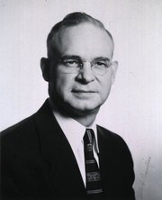
Dr. Otto C. Brantigan
Otto C. Brantigan, MD. (1904-1981) An American surgeon and anatomist, Otto Charles Brantigan was born in Chattanooga, TN in 1904. Having dropped out of high school to help his family and working as a first class machinist, he decided to continue with graduate school. He studied at the Northwestern University in Chicago, where he graduated from the Medical School in 1933. In 1948 he became Chief of Surgery, and eventually became Professor of Surgery, Professor of Thoracic Surgery, and Professor of Anatomy at the Maryland School of Medicine. He retired in 1976 having earned many accolades for his profuse surgical work and publications.
As a surgeon of the times, Dr. Brantigan had a wide area of interest. His over 110 publications and surgical work range from thoracoscopy to vascular, plastic, cardiac, and orthopedic surgery. He is most remembered for the pioneer work he did on chronic obstructive pulmonary disease (COPD), emphysema and lung volume reduction surgery (LVRS), which he presented in 1958. The procedure had (at the time) a very high mortality rate (16 -20%) and Brantigan's work was not readily accepted.
It was not until J. Cooper and his team, revisited the operation proposed by Brantigan that the operation was accepted, now with new surgical stapling and staple line buttressing technology. Dr. Brantigan's name was recognized as a pioneer in lung emphysema surgery, unfortunately 14 years after his death. In 1994 his son, Dr Charles O. Brantigan delivered a beautiful biography of Dr. Otto Brantigan in the same meeting where Cooper presented his results with LVRS.
Personal note: I am proud to own one of the copies of Dr. O.C. Brantigan;s "Clinical Anatomy", a book that I use quite frequently. It is listed in my library catalog. Dr. Miranda.
Sources:
1. "Biography of Otto C Brantigan" C.O. Brantigan 1994 Meeting of the American Association for Thoracic Surgery
2. "LVRS in chronic obstructive pulmonary disease" Davies, L; Calverley, P. Thorax 1996;51(Suppl 2):S29-S34
3. ""Bilateral pneumectomy (volume reduction) for chronic obstructive pulmonary disease" Cooper, J.,The Journal of Thoracic and Cardiovascular Surgery Volume 109, Number 1:106-119
4. "The Surgical Approach to Pulmonary Emphysema" Brantigan, OC; Kress, MB; Mueller, EA. Chest. 1961; 39(5):485-499
5. "History of Emphysema Surgery" Naef, AP. Ann Thorac Surg 1997;64:1506-1508
Original image courtesy of National Institutes of Health.Biography of Dr. Otto Brantigan courtesy of Dr. Charles O. Brantigan.
- Details
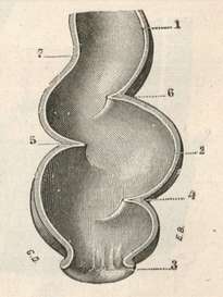
The rectum is the most distal segment of the large intestine, along with the anal canal.
The word [rectum] arises from the Latin [rectus] and means "straight", such as its use in the name "rectus abdominis" for the "straight muscle of the abdomen".
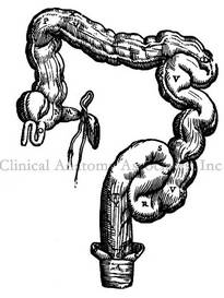
It seems a misnomer, as the rectum of the human species is actually "S" shaped, as seen in the accompanying image. The reason for this discrepancy is that the rectum was named by Galen of Pergamon (129AD - 200 AD) who himself studied this structure in animals such as sheep and goats. In these animals the rectum is indeed straight, and since contradicting Galen was not acceptable (see Michael Servetus), the name has survived until this day. Even Andreas Vesalius has in his 1953 "Fabrica" a depiction of a straight rectum in the human! Click on second image to see a larger depiction of Vesalius' idea of the rectum. Although Vesalius stated that he wanted to show human anatomy as it is, and not as Galen said it should be, here is a demonstration that in 1543 he was still a lukewarm Galenist.
There is an area between the sigmoid and the rectum called the sigmoidorectal junction, although most anatomists call it (wrongly) the rectosigmoid junction (RSJ). This is an anatomically diffuse area with no clear anatomical transition between the sigmoid and the rectum or the RSJ from the rectum.
As the proximal end of the "S" shaped rectum is not clearly discernible from the sigmoidorectal region, there is no clear agreement on the length of the rectum. Authors state that it measures approximately six to seven inches in length (15 - 17 cm), while others measure it as between 8-10 inches. The rectum ends distally at the junction of the rectum with the pelvic diaphragm. It is at this point that the anal canal begins.
The rectum is characterized by three transverse rectal folds, one on the right side, and two on the left side. These folds are know as the "rectal valves" or the "valves of Houston". The middle rectal fold is known to European anatomists as the "valve of Kohlrausch" Their function in maintaining fecal material in place as well as their function in defecation is still under study. The rectal valves also have a high level of anatomical variation and may not be present at all.
Images:
1. "Tratado de Anatomia Humana" Testut et Latarjet 8 Ed. 1931 Salvat Editores, Spain
2. "De Humani Corporis Fabrica, Libri Septem" A. Vesalius 1543 Brussels
Recommended reading: "Transverse Folds of Rectum: Anatomic Study and Clinical Implications" Shafik, A, et al. Clin Anat 14: 196-203 (2001).
- Details
The term "collateral circulation" is generally used to denote a situation where small blood channels dilate and provide blood supply when a pathology creates a stricture and diminishes blood flow (ischemia).
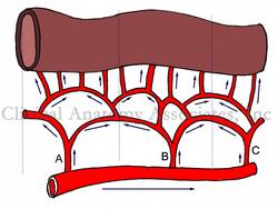
Although the above is correct, the term is also applicable to a normal, non-pathological situation most common in the human body. Please refer to the accompanying image for the following explanation. If needed, click on the image for a larger depiction. In the image, the arrows represent direction of flow.
Most organs or organ segments receive blood supply from more than one source of blood supply. In some cases, like the stomach, there are up to four arteries that provide blood supply to the organ: the right and left gastric arteries, and the right and left gastroepiploic arteries.
In other cases, like the small intestine shown in the image, blood arrives to the organ arising from several arteries (A, B, and C) that themselves arise from a parent structure. Because of hydrodynamics, the vascular territories of each artery (represented by dashed lines) tend not to overlap. If for any reason there is stenosis or blockage in any of these arteries (A,B, or C) blood will flow immediately through an alternate route and the organ will not suffer ischemia or necrosis.
This is extremely important, as these collateral channels maintain blood supply to areas that may be affected by bending, such as the elbow and knee, which have a rich collateral network. Most of the organs in the body, with some exceptions (brain, heart), have collateral circulation.
Collateral circulation is extremely important for surgery, as surgeons can safely remove parts of organs without affecting the blood supply to the organ. This is also true for all gastrointestinal anastomoses.
Image property of: CAA, Inc. Artist: Dr. Miranda
- Details
Histology is the scientific branch that studies tissues.
The root term [-hist-] is used to mean "tissues", but how the term came to be used is somewhat convoluted. It arises from the Greek [histos], which indicates the mast of a ship, it then was used to denote a Greek weaver's loom central mast (where the fabric is woven horizontally), and then it was used to indicate that which was woven [histios], the fabric, or the "tissues". The suffix [-ology] also has Greek origin from [logos] meaning a "book", a "treatise" or "to study".
The concept of the body being formed by different tissues was pioneered by Marie-Francois Xavier Bichat (1771-1802) who called them "membranes" Bichat is considered to be the "father of Histology". The image shows a histological slide of cardiac muscle. Click on the image for a larger depiction.
Original image by S. Girod and A. Becker, courtesy of Wikipedia.
- Details
The root term [-spondyl-] arises from the Greek [spondylos] meaning "vertebra", and suffix [-osis] means "condition", but with the connotation of "many". The word [spondylosis] means " condition of many vertebrae". This does not add much to the use of this word as an indicator of a pathology, but it does indicate that there is excess bone in a vertebral pathology.
Spondylosis is an osteoarthritic degeneration of the vertebrae and the spine characterized by abnormal bony growths on the vertebrae that can impinge on nerves and other structures causing pain and mobility problems. The definition of spondylosis also includes degenerative changes in the intervertebral discs.
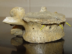
The abnormal growth of portions of the vertebral body, usually forms "bone spurs", also referred to as "spondylophytes". The accompanying image shows a lumbar vertebra with spondylophytes.
Image property of: CAA, Inc. Photographer: David M. Klein
- Details
This article is part of the series "A Moment in History" where we honor those who have contributed to the growth of medical knowledge in the areas of anatomy, medicine, surgery, and medical research.
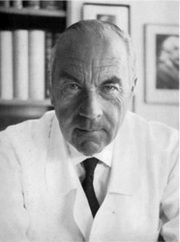
Dr. Rudolph Nissen
Dr. Rudolf Nissen (1896 - 1981). Dr Nissen’s life is extraordinary. Born in the city of Neisse, Germany in 1896, he was the son of a local surgeon. He studied medicine in the Universities of Munich, Marburg, and Breslau. He was the pupil of the Karl Albert Ludwig Aschoff (1866 - 1942) a German physician and cardiovascular researcher. Another of Aschoff's pupil, Dr. Sunao Tawara (1873 - 1952), discovered the atrioventricular node.
Nissen became a professor of surgery in Berlin, and in 1933 moved to Turkey where he was placed in charge of the Department of Surgery of the University of Istanbul. In 1939 he moved to the US, first to the Massachusetts General Hospital and later to the Jewish Hospital in Brooklyn, New York. After becoming a US citizen, he moved again in 1952 to Basel, Switzerland as Chief of the Department of Surgery, where he retired in 1967. He died in 1981.
His contributions to surgery are innumerable. He wrote over 30 books and 450 journal articles. Known for the development in 1956 of what is today known as the “Nissen fundoplication” for esophageal hiatus hernia surgery, Nissen also worked with his assistant, Dr. Mario Rossetti to develop the “floppy Nissen fundoplication”, also known as the “Nissen-Rossetti procedure”. This would be enough to honor this man, still, he (with Sauerbruch) performed the first lung lobectomy and the first pneumonectomy (called then a total pneumonectomy). In 1949 he performed the first esophagectomy with a gastroesophagostomy for lower esophageal cancer.
His personal life is even more interesting. Drafted at 20, he fought in WWI and was wounded several times. In 1933, under the Nazi regime, he was ordered to fire all the Jewish-German assistants under his care. Being Jewish himself, he was told that he would keep his job, Nissen could not take this. He resigned his position and moved out of Germany.
Another little known fact is that he operated on Albert Einstein in 1948. He operated on Einstein because of intestinal cysts. Having found a developing abdominal aortic aneurysm, he reinforced it with cellophane, undoubtedly giving his patient a few extra years to live. Einstein died in 1955.
As a personal side note, our good friend Dr. Aaron Ruhalter scrubbed in with Dr. Nissen while serving as a surgical resident at the Brooklyn Jewish Hospital!
Sources:
1. “Rudolf Nissen: The man behind the fundoplication” Schein et al. Surgery 1999;125:347-53
2. “Rudolf Nissen (1896–1981)-Perspective” Liebermann-Meffert, D. J Gastrointest Surg (2010) 14 (Suppl 1):S58–S61
3. “The Life of Rudolf Nissen: Advancing Surgery Through Science and Principle” Fults, DW; Taussky, P. World J Surg (2011) 35:1402–1408
4. “Total Pneumonectomy” Nissen, R. Ann Thorac Surg 1980; 29:390-394
5. “Historical Development of Pulmonary Surgery” Nissen, R. Am J Surg 80: Jan 1955 9- 15
Image in the public domain, courtesy of the Universitat Basel


