
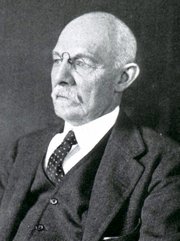




Dr. José Manuel Revuelta
This article was originally published in Spanish by our contributor Dr. Jose Manuel Revuelta Soba, Professor of Surgery and Professor Emeritus at the University of Cantabria.
The article's title (in Spanish) is "El Centro del Placer" (The Center of Pleasure), a discussion of the brain mechanisms regarding pleasure, rewards, and the aging brain.
He has graciously granted us permission to translate and publish his article in “Medical Terminology Daily”. Dr. Miranda.
PLEASURE , BRAIN ACTIVITY, AND THE AGING HEART
Why does drinking cool water on a hot day feel such a pleasurable experience? The answer isn't in our throat, but in the hidden corners of our brain. For millions of years, evolution has perfected an extraordinary biological mechanism: the brain's reward system. This ancient neural circuit was designed with the sole purpose of ensuring the survival of the individual and the continuation of the species, transforming basic biological needs into pure pleasure.
As we age, the signals from this amazing circuit begin to weaken; thirst subsides before cells can rehydrate, food loses its appeal, and sexual desire diminishes. Understanding how this amazing system works will allow us to better understand the intimate secrets of our daily behavior.
The Brain's Reward System
It's not an isolated organ, but a complex neural circuit that connects phylogenetically ancient structures with evolved cortical areas, with the crucial function of triggering thirst, hunger, and sexual desire.
Physiologists have coined a strange term to describe this part of the brain architecture: the mesocorticolimbic dopaminergic pathway. Simply put, it's the brain's most expressive and stimulating reward system. This biological structure consists of the ventral tegmental area, located in the midbrain, the true factory that synthesizes dopamine, a chemical that sends messages from neurons to the nervous system (neurotransmitter). This molecule doesn't generate pleasure itself, but rather desire, that is, the motivation to seek it.
The nucleus accumbens, located in the basal portion of the forebrain, acts as the processing center for pleasure and anticipation, integrating affective and motor information. Meanwhile, other neurons in the prefrontal cortex assign reward value, evaluate risks, and make decisions about when and how to act. These two groups of neurons provide hedonistic and pleasurable sensation through endogenous opioids (endorphins and enkephalins).
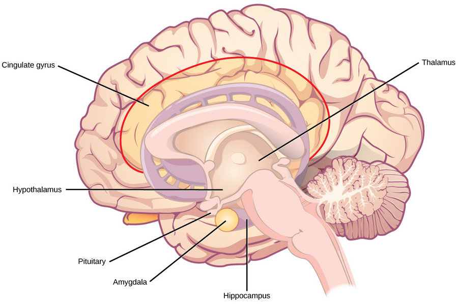 The main components of the limbic system
Subsequently, a group of neurons located in the amygdala and hippocampus contributes to the corresponding emotional charge and memory, allowing us to recall where and how we obtained the pleasurable stimulus. This function is carried out through the release of the hormone oxytocin, a highly efficient neurotransmitter commonly known as the "love hormone." Its blood levels increase exponentially during the ingestion of appetizing liquids or foods, physical contact, or orgasm. The duration and intensity of pleasure varies according to the evolutionary value of the stimulus. This hormone is stored in the pituitary gland (a lentil-sized gland located at the base of the skull), which releases it into the bloodstream. Oxytocin is well known for triggering uterine contractions before childbirth and facilitating breast milk production.
Finally, another group of neurons located in the midline of the brainstem is the main source of serotonin, the "happiness hormone," a neurotransmitter that regulates mood and satiety.
The System's Splendid Rewards
From birth, the body's fluid deficit is readily detected through thirst. When we drink, the pleasurable sensation in the mouth and throat usually lasts only a few minutes; the brain doesn't wait for the water to be absorbed in the intestine, a process that typically takes a little over 10 minutes. Therefore, the desire to drink is interrupted by a rapid inhibitory neural circuit to prevent excessive fluid intake (hyperhydration). This "flow meter," located in the forebrain, deactivates the "thirst neurons" of the subfornical organ, preventing us from overindulging.
Similarly, the sensation of hunger, driven by the physiological need for energy, is regulated by the brain's reward system through the release of dopamine by the hypothalamus, which acts as the main center. However, the stomach also plays a crucial role in triggering this vital drive. Upon detecting a shortage of nutrients, the stomach walls release ghrelin, or the "hunger hormone," either because the stomach is empty or after a prolonged period of fasting. This hormone signals the hypothalamus to stimulate the sensation of hunger and release dopamine into the bloodstream. There is an antagonistic hormone, leptin, or the "satiety hormone," which is produced in adipose tissue (body fat). Leptin acts as a messenger, signaling the hypothalamus to reduce the sensation of hunger and stimulate energy expenditure, thus helping to maintain body weight. While the initial taste pleasure lasts only a few seconds or minutes, once food is in the mouth, the secondary release of dopamine, triggered by the arrival of nutrients in the stomach, generates a feeling of well-being and homeostatic satiety that lasts for 1 to 3 hours, depending on the type of food and other individual factors.
The “sexual urge” constitutes another powerful and intense drive, like thirst or hunger, although it is influenced by a more complex variety of external factors. In general, sexual activity has a triphasic dynamic: a seeking phase, a plateau and orgasm phase, and a relaxation and satiety phase.
Contrary to what was previously believed, dopamine, or the “pleasure molecule,” drives the seeking and anticipation of pleasure, rather than pleasure itself, according to neuroscientist Kent Berridge (University of Michigan). When the brain becomes hyperreactive to dopamine stimuli, it triggers a very strong, compulsive desire to seek pleasure.
During the plateau phase, the brain releases large amounts of dopamine and oxytocin, far exceeding the levels used to satisfy thirst or hunger. This generates an intense feeling of reward, accompanied by euphoria and increased sexual potency (orgasm). Dopamine enhances sexual activity by binding to dopaminergic receptors.
Finally, the relaxation and satiety phase is mediated by other opioids such as oxytocin and prolactin, whose neurobiological effects last from 30 minutes to several hours, generating the pleasant sensation of emotional attachment and trust in the sexual partner.
Pleasure is not a luxury; it has direct benefits for the heart and blood vessels. The moderate, physiological activation of the brain's reward system increases parasympathetic tone, reducing blood pressure and resting heart rate. Oxytocin, released during sexual intercourse or affectionate social interaction, acts on endothelial receptors, stimulating the synthesis of nitric oxide, a potent vasodilator that improves coronary artery perfusion and reduces arterial stiffness. Balanced levels of dopamine and endorphins in the blood buffer the HPA (hypothalamic-pituitary-adrenal) axis, decreasing circulating cortisol (the stress hormone) and reducing chronic vascular inflammation.
When the System Turns Off the Lights
When cool water after a long walk ceases to be appealing, a favorite dish loses its irresistible aroma and displays of affection or intimate contact become just a vague memory, it's not apathy or simply "weariness with life," but rather the brain's chemistry turning off its lights. The brain's reward system, which for decades has driven us to drink, eat, love, and connect with others, gradually weakens over time. Therefore, understanding how this amazing biological machinery ages is essential to protecting overall health and preserving normal heart function in older adults.
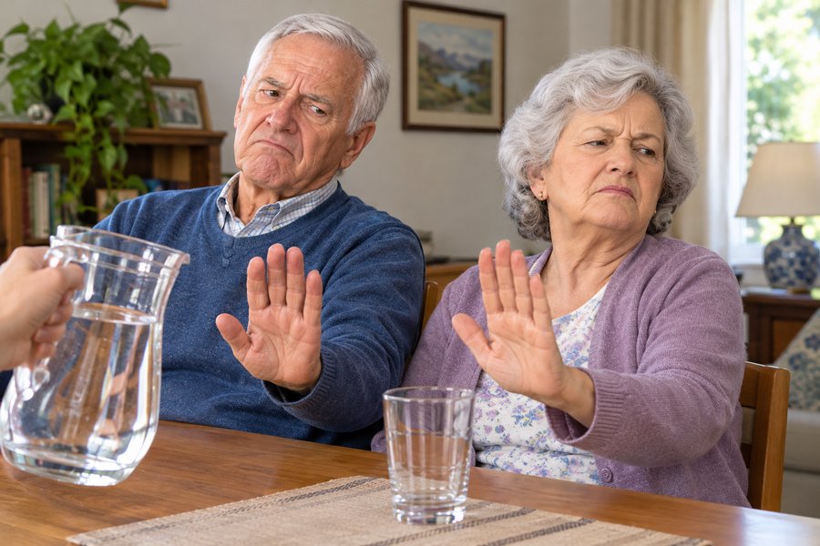 Elderly Couple Refusing Water (AI generated)
When the brain stops releasing bursts of dopamine, making drinking water a highly rewarding experience, progressive involuntary dehydration occurs. If little water is consumed (hypodipsia), plasma volume decreases, making the blood thicker (hemoconcentration), which forces the heart to beat faster to maintain perfusion of the body's organs and tissues. This blood viscosity, induced by chronic dehydration, drastically increases the risk of strokes, heart attacks, and episodes of orthostatic hypotension, a common cause of traumatic falls in the elderly.
Anorexia in old age is not simply a loss of appetite; it is a deprivation of the heart muscle of its necessary amino acids and essential micronutrients. The heart is a predominantly muscular organ, highly sensitive to protein-energy malnutrition, which accelerates cardiac sarcopenia (progressive loss of myocardial mass), decreases its contractile strength, and ultimately leads to heart failure.
The deterioration of the pathways that generate dopamine, oxytocin, and other opioid substances impairs social relationships and extinguishes affective-sexual desire, isolating the individual not only emotionally but also physically. The loss of social rewards activates the hypothalamic-pituitary-adrenal axis, chronically elevating cortisol and circulating catecholamine levels. This inflammatory environment impairs endothelial function (the layer lining the inside of the arteries), accelerating atherosclerosis, and decreasing heart rate variability, a key marker of cardiovascular vulnerability.
Therefore, protecting the sensitivity of the brain's reward system in the elderly, through sensory stimulation, appealing foods, guided hydration routines, and supportive social environments, is not only an act of emotional comfort but also a crucial medical intervention to protect the hearts of the growing senior population.
“Pleasure is not a biological excess, but rather the language evolution uses to ensure we are still alive tomorrow.”
Kent Berridge, Professor of Neuroscience and Psychology, University of Michigan, USA
Sources and references
1. Berridge, K. C., et al. Pleasure systems in the brain. Neuron, 2015; 86(3), 646-664.
2. Zimmerman, C. A., et al. Thirst neurons anticipate the homeostatic consequences of eating and drinking. Nature, 2016; 537(7622), 680-684.
3. Bäckman, L., et al. The correlative triad among aging, dopamine, and cognition: current status and future prospects. Neuroscience & Biobehavioral Reviews, 2006; 30(6), 791-807.
4. Popkin, B. M., et al. Water, hydration, and health. Nutrition Reviews, 2010; 68(8), 439-458.
5. Landi, F., et al. Anorexia of aging: risk factors and clinical consequences. Current Opinion in Clinical Nutrition & Metabolic Care, 2016; 19(1), 14-19.
6. Hyde Z., et al. Prevalence of Sexual Activity and Associated Factors in Men Aged 75 to 95 Years: A Cohort Study. Annals of Internal Medicine, 2010; 153(11), 693-702.
Notes:
1. Attribution Image of the Limbic system: CNX OpenStax, CC BY 4.0 , via Wikimedia Commons. Public Domain
2. Elderly Couple Refusing Water Image (AI generated)