
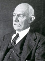




Dr. José Manuel Revuelta
The transplantation of organs from other species—xenotransplantation—has been the subject of intense scientific research for over a century. The progressive global shortage of human organs to meet the growing demand from patients with end-stage heart failure has intensified interest in this potential therapeutic option. According to data from the Organ Procurement and Transplantation Network (OPTN), in the United States more than 100,000 patients are on transplant waiting lists, thousands of whom are waiting for a heart; unfortunately, a significant percentage die before receiving a compatible donor. The Global Observatory on Donation and Transplantation records heart transplants performed annually; more than 8,000 worldwide and around 2,000 in the European Union.
From Primates to Porcine Bioengineering
In the early days of xenotransplantation, the use of organs from non-human primates (chimpanzees and baboons) yielded very limited results, provoking strong ethical objections. Over time, pigs became established as the ideal source of organs due to their anatomical and physiological similarity to humans, their suitability for controlled, pathogen-free rearing, and the possibility of scheduling surgical procedures with ample time to perform the essential and exhaustive immunological and microbiological analyses.
However, the main obstacle to xenotransplantation lies in the immune system rejection of the transplanted organ. As the history of allotransplantation (transplantation of organs between genetically different individuals of the same species) reminds us, initial survival rates were very modest. When Christiaan Barnard performed the first heart transplant on Louis Washkansky in Cape Town (December 3, 1967), the patient survived for 18 days. Three days later, Adrian Kantrowitz performed a heart transplant in New York (Maimonides Medical Center) on a newborn with a complex congenital heart defect, who died hours later; and Norman Shumway, the true father of modern surgical technique, performed a transplant a month later in Stanford (Palo Alto, California) on a 54-year-old patient, who survived for two weeks. After more than a hundred failures in different countries, enthusiasm fell drastically, so that in 1971 only 10 heart transplants were performed worldwide.
Translational research and the discovery of Cyclosporine A in the Sandoz® laboratories (Switzerland) revolutionized this field of medicine, allowing partial control of the immune response, which enabled its significant current development. Surgical experience and early diagnosis of postoperative complications, along with the introduction of new immunosuppressive drugs, have made the current success of allocardiac transplantation possible, achieving survival rates exceeding 80% in the first year and 70% at five years.
In xenotransplantation, the immunological barrier is vastly superior. DNA constitutes a precise biological clock for measuring the phylogenetic distance between species. As physicist Michio Kaku points out, DNA mutates approximately 1% every 1.5 million years, which is why we are separated by 0.5% from Neanderthals (750,000 years), 4% from chimpanzees (6 million years), and an overwhelming distance of tens of millions of years from pigs. This significant evolutionary divergence causes the human immune system (B and T lymphocytes, NK cells, macrophages, granulocytes, and dendritic cells) to recognize porcine tissue as completely foreign and trigger an immediate and violent reaction (hyperacute rejection). This process is characterized by massive thrombosis of the coronary arteries, devastating inflammation, myocardial stiffening, and cardiac arrest within minutes or a few hours.
The “humanized” porcine heart
To overcome hyperacute rejection, it was necessary to genetically “humanize” the pig through cellular bioengineering. The discovery of genome editing technologies, especially the CRISPR/Cas9 tool, as well as the ZFN and TALEN nucleases, made it possible to perform precise modifications to the porcine genome.
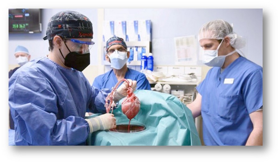
First pig heart ready for Xenotransplant
Surgeon David Cooper (Massachusetts General Hospital/Harvard University) discovered that the primary trigger for hyperacute rejection is the presence of the carbohydrate galactose-α1,3-galactose (α-Gal) on the surface of porcine cells. By inactivating the gene that codes for the alpha-1,3-galactosyltransferase enzyme (Gal Knockout or GalSafe™ pigs), human antibodies immediately cease to recognize porcine tissue as a destructive target.
Biotechnology companies, such as Revivicor Inc. (a subsidiary of United Therapeutics), have developed engineered pigs that incorporate up to 10 combined genetic modifications:
-
Deletion of 3 porcine sugar genes (including α-Gal) to prevent hyperacute rejection and antibody-mediated acute rejection.
-
Insertion of six human genes: two anti-inflammatory genes, two complement protein regulatory genes (CD46 and CD55) to attenuate the antibody response, and two anticoagulant genes (thrombomodulin-TM and EPCR) to prevent thrombus formation in coronary vessels.
-
Inactivation of the porcine growth hormone receptor gene (GGTA1/GH), which prevents the heart from continuing to grow within the human recipient's thorax until it reaches an incompatible size.
In parallel, other international groups are advancing along complementary lines, such as Makana Therapeutics, which is investigating the elimination of three key genes to reduce antibody reactivity. Eckhard Wolf's team at Ludwig Maximilian University of Munich uses New Zealand mini-pig breeds that do not require growth manipulation, while eGenesis (founded by Luhan Yang and George Church at Harvard University) has achieved a milestone by using CRISPR to eliminate dozens of copies of porcine endogenous retroviruses (PERVs) to ensure biosafety.
The first human heart xenotransplants with a porcine donor
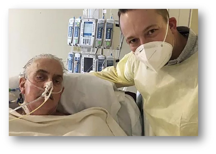
Mr David Bennet First cardiac xenotransplant
On January 7, 2022, surgeon Bartley P. Griffith and his team at the University of Maryland Medical Center (UMMC) performed the first porcine heart xenotransplant in history on 57-year-old David Bennett, who suffered from end-stage heart failure and was ineligible for a conventional transplant or mechanical ventilation. After receiving a GalSafe™ porcine heart and experimental immunosuppressive therapy (which included Kiniksa Pharmaceuticals' monoclonal antibody KPL-404 to block the CD40/CD40L pathway), the heart beat and functioned immediately without hyperacute rejection; the patient survived for two months. Comprehensive post-mortem analysis, published in The New England Journal of Medicine, revealed that the organ showed signs of a massive inflammatory response and vascular damage associated with the reactivation of latent porcine cytomegalovirus (pcMV), a herpesvirus that had not been detected in conventional pre-surgical screening.
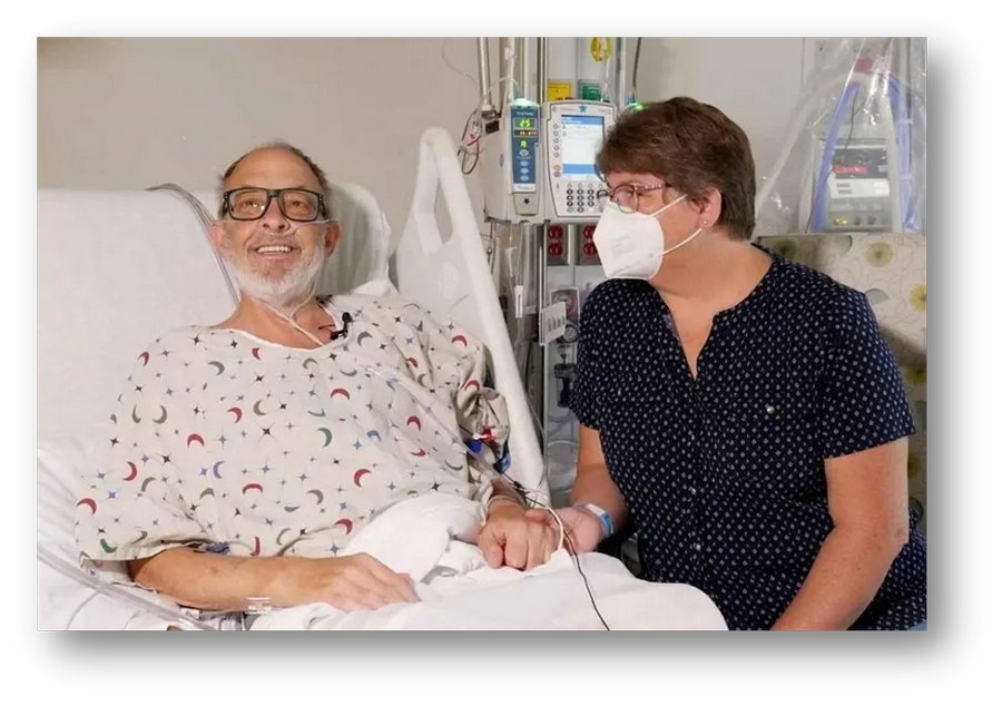
Mr. and Mrs. Lawrence Faucette
In September 2023, the same team at the University of Maryland performed the world's second porcine heart xenotransplant on Lawrence Faucette, a 58-year-old Navy veteran and NIH laboratory technician with end-stage heart disease unsuitable for human transplantation. The porcine heart was preserved prior to implantation in a warmed continuous perfusion system (XVIVO Heart Box®), supervised by Dr. Muhammad Mohiuddin. Ultrasensitive PCR tests were implemented to ensure the absence of pcMV in the donor, and an innovative immunosuppressive therapy based on Tegoprubart® (Eledon Pharmaceuticals), an antibody designed to block the CD154 (CD40L) protein, was employed. The recipient died six weeks after transplantation due to acute rejection. During this period, invaluable data on the hemodynamic function and immune response of the porcine organ in humans were collected.
Complementarily, teams such as those led by Robert Montgomery at New York University (NYU Langone) and Jayme Locke at the University of Alabama at Birmingham have performed kidney and heart xenotransplants in brain-dead individuals with family consent. These studies have provided irrefutable evidence of the short-term diuretic and myocardial contractile function of porcine organs, helping to refine protocols before expanding into widespread clinical trials.
The Zoonosis Barrier and Biosafety
One of the most delicate aspects of xenotransplantation is the risk of zoonosis, that is, the transmission of animal pathogens to humans. Public health history demonstrates that viruses of animal origin (such as HIV, Ebola, avian or swine influenza viruses, and coronaviruses like SARS-CoV-2) can cause serious health emergencies.
In the context of porcine xenotransplantation, the following are of particular concern:
-
Porcine endogenous retroviruses (PERVs): Viral sequences integrated into the pig genome. Although they do not cause disease in the animal, their potential to infect human cells in vitro has been evaluated. Mass inactivation of PERV using CRISPR has demonstrated that it is possible to raise animals free from this risk of transmission.
-
Porcine cytomegalovirus (pcMV): A herpesvirus that, while remaining dormant in the donor, can reactivate under the recipient's immunosuppressive regimen and trigger endothelial dysfunction and acute rejection, mediated by inflammation. The intensification of molecular diagnostics using ultrasensitive PCR and rearing in Designated Pathogen Free (DPF) environments are now indispensable requirements.
Due to these risks, regulatory agencies such as the US FDA and the European EMA require close long-term monitoring and prohibit xenotransplant recipients from donating blood or organs.
The Future of Xenotransplantation
Xenotransplantation research represents one of the greatest multidisciplinary efforts in the history of medicine, bringing together cardiology, cardiovascular surgery, genetic engineering, immunology, virology, and bioethics. Its strategic value lies in its potential to definitively eliminate the global organ shortage crisis. The possibility of having "made-to-order" porcine organs—genetically adapted, pathogen-free, and available on a scheduled basis—would transform end-stage heart failure from a short-term death sentence into a medically treatable condition.
Each of these pioneering steps, with its successes and challenges, but valuable lessons learned, brings us closer to the moment when transplant waiting lists can be managed. The gradual overcoming of species barriers will reaffirm science's capacity to transform the most complex biological dilemmas into real solutions for the preservation of human life.
“Change is rarely comfortable, but in science it is the only path to what once seemed impossible.”
Arno Penzias (1933–2024), astrophysicist and Nobel Laureate in Physics, 1978
References:
1. Yang, L., et al. Genome-wide inactivation of porcine endogenous retroviruses (PERVs). Science, 2015; 350(6264), 1101–1104.
2. Cooper, D. K., et al. Immunological barriers to xenotransplantation. Int J Surg., 2015; 23:211–216.
3. Mohiuddin, M. M., et al. Chimeric 2C10R4 anti-CD40 antibody therapy is critical for long-term survival of GTKO.hCD46.hTBM pig-to-primate cardiac xenograft. Nature Communications, 2016; 7(1), 11138.
4. Griffith, B. P., et al. Genetically modified porcine-to-human cardiac xenotransplantation. New Eng J Med, 2022; 387(1), 35–44.
5. Riella, L.V. Xenotransplantation: The future is here. J Am Soc Nephrol, 2024; 36(2):305–307.
Note: All images in the public domain courtesy of Dr. JM Revuelta.