Medical Terminology Daily (MTD) is a blog sponsored by Clinical Anatomy Associates, Inc. as a service to the medical community. We post anatomical, medical or surgical terms, their meaning and usage, as well as biographical notes on anatomists, surgeons, and researchers through the ages. Be warned that some of the images used depict human anatomical specimens.
You are welcome to submit questions and suggestions using our "Contact Us" form. The information on this blog follows the terms on our "Privacy and Security Statement" and cannot be construed as medical guidance or instructions for treatment.
We have 334 guests and no members online

Jean George Bachmann
(1877 – 1959)
French physician–physiologist whose experimental work in the early twentieth century provided the first clear functional description of a preferential interatrial conduction pathway. This structure, eponymically named “Bachmann’s bundle”, plays a central role in normal atrial activation and in the pathophysiology of interatrial block and atrial arrhythmias.
As a young man, Bachmann served as a merchant sailor, crossing the Atlantic multiple times. He emigrated to the United States in 1902 and earned his medical degree at the top of his class from Jefferson Medical College in Philadelphia in 1907. He stayed at this Medical College as a demonstrator and physiologist. In 1910, he joined Emory University in Atlanta. Between 1917 -1918 he served as a medical officer in the US Army. He retired from Emory in 1947 and continued his private medical practice until his death in 1959.
On the personal side, Bachmann was a man of many talents: a polyglot, he was fluent in German, French, Spanish and English. He was a chef in his own right and occasionally worked as a chef in international hotels. In fact, he paid his tuition at Jefferson Medical College, working both as a chef and as a language tutor.
The intrinsic cardiac conduction system was a major focus of cardiovascular research in the late nineteenth and early twentieth centuries. The atrioventricular (AV) node was discovered and described by Sunao Tawara and Karl Albert Aschoff in 1906, and the sinoatrial node by Arthur Keith and Martin Flack in 1907.
While the connections that distribute the electrical impulse from the AV node to the ventricles were known through the works of Wilhelm His Jr, in 1893 and Jan Evangelista Purkinje in 1839, the mechanism by which electrical impulses spread between the atria remained uncertain.
In 1916 Bachmann published a paper titled “The Inter-Auricular Time Interval” in the American Journal of Physiology. Bachmann measured activation times between the right and left atria and demonstrated that interruption of a distinct anterior interatrial muscular band resulted in delayed left atrial activation. He concluded that this band constituted the principal route for rapid interatrial conduction.
Subsequent anatomical and electrophysiological studies confirmed the importance of the structure described by Bachmann, which came to bear his name. Bachmann’s bundle is now recognized as a key determinant of atrial activation patterns, and its dysfunction is associated with interatrial block, atrial fibrillation, and abnormal P-wave morphology. His work remains foundational in both basic cardiac anatomy and clinical electrophysiology.
Sources and references
1. Bachmann G. “The inter-auricular time interval”. Am J Physiol. 1916;41:309–320.
2. Hurst JW. “Profiles in Cardiology: Jean George Bachmann (1877–1959)”. Clin Cardiol. 1987;10:185–187.
3. Lemery R, Guiraudon G, Veinot JP. “Anatomic description of Bachmann’s bundle and its relation to the atrial septum”. Am J Cardiol. 2003;91:148–152.
4. "Remembering the canonical discoverers of the core components of the mammalian cardiac conduction system: Keith and Flack, Aschoff and Tawara, His, and Purkinje" Icilio Cavero and Henry Holzgrefe Advances in Physiology Education 2022 46:4, 549-579.
5. Knol WG, de Vos CB, Crijns HJGM, et al. “The Bachmann bundle and interatrial conduction” Heart Rhythm. 2019;16:127–133.
6. “Iatrogenic biatrial flutter. The role of the Bachmann’s bundle” Constán E.; García F., Linde, A.. Complejo Hospitalario de Jaén, Jaén. Spain
7. Keith A, Flack M. The form and nature of the muscular connections between the primary divisions of the vertebrate heart. J Anat Physiol 41: 172–189, 1907.
"Clinical Anatomy Associates, Inc., and the contributors of "Medical Terminology Daily" wish to thank all individuals who donate their bodies and tissues for the advancement of education and research”.
Click here for more information
- Details

Left clavicle, superior surface
The clavicle is part of the anterior portion of the shoulder girdle. It is an elongated bone with an "italic S" curvature. The Latin term for clavicle is [clavicula], and it has two root terms: [-clavic-] and [-clav-]. This is why we have the terms [subclavicular], and [subclavian] both meaning the same: "inferior to the clavicle".
The clavicle articulates medially with the manubrium of the sternum (see image on this article) by way of the sternoclavicular joint. This joint contains a meniscus. Laterally, the clavicle articulates with the acromial process or acromium of the scapula.
The clavicle has the muscular insertions of several muscles: sternocleidomastoid, trapezius, pectoralis major, deltoid, subclavius, and sternohyoid.
Sources:
1. "The Origin of Medical Terms" Skinner, HA 1970 Hafner Publishing Co.
2 "Tratado de Anatomia Humana" Testut et Latarjet 8 Ed. 1931 Salvat Editores, Spain
3. "Anatomy of the Human Body" Henry Gray 1918. Philadelphia: Lea & Febiger
Image modified by CAA, Inc. Original image by Henry Vandyke Carter, MD., courtesy of bartleby.com
- Details

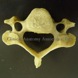
Cervical vertebra, superior view
The term [foramen transversarium] is Latin for "transverse foramen". It refers to bilateral foramina (openings) found lateral to the vertebral body in the cervical vertebrae. These foramina are found only in cervical vertebrae and serve as a good way to identify them.
Through the foramina transversaria (plural form) pass the vertebral artery and vertebral vein. The vertebral artery is one of the first branches to arise off the subclavian arteries. While the vertebral vein passes through all seven foramina transversaria, the vertebral artery does not pass through the foramen transversarium of the seventh cervical vertebra (vertebra prominens).
Image property of:CAA.Inc.Photographer:David M. Klein
- Details
This article is part of the series "A Moment in History" where we honor those who have contributed to the growth of medical knowledge in the areas of anatomy, medicine, surgery, and medical research.

Carl Wernicke
Carl Wernicke (1848-1905). German psychiatrist, neurologist, and neurosurgeon, Carl Wernicke was born in 1848 in the town of Tarnowitz, in what was then Prussia. He studied medicine in Breslau, Poland. In 1817 he became an assistant psychiatrist at a Breslau hospital. Fascinated with the discoveries and publications of Paul Broca on localized brain damage and aphasia, Wernicke left his post for a time to work with Theodor Meynert in Vienna. At that time Meynert was considered an authority in neuropsychiatry. In 1874, soon after his return to Breslau, Wernicke published his ideas and findings in aphasia in a revolutionary publication "The Aphasia Symptom Complex". Wernicke was only 26.
At the time, the general outlook on brain activity was that the brain worked in localized areas. Carl Wernicke's ideas were that although this was true, the functionality of the brain resided in the connections between the different areas of the brain. His ideas were right. Wernicke described what later would be known as "sensory aphasia" or the eponymic "Wernicke's aphasia".
Wernicke was a pioneer in the surgical treatment of hydrocephalus, as well as the surgical treatment of brain abscesses. He published several books, including a brain atlas. Carl Wernicke died as the consequence of a bicycle accident in 1905.
Sources:
1. "Pioneers in Neurology: Carl Wernicke (1848–1905)" Pillmann, F. J Neurol (2003) 250 : 1390–1391
2. "The scientific history of hydrocephalus and its treatment" Aschoff, A.; Ashemi, P.; Kunze, S.Neurosurg Rev (1999) 22:67–93
3. "Aphasia" Marshall,RS; Lazar, RM;PhD, Mohr,JP. Medical Update for Psychiatrists. Elsevier (1998)3;5:132–138
- Details
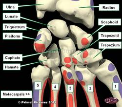
The lunate bone is one of the proximal carpal bones that form the wrist. The name arises from the Latin [luna], meaning "moon". The lunate bone has a deep concavity and crescent-like shape, resembling a crescent moon. This bone is also known as the "semilunar bone" or the os lunatum.
The lunate bone has six surfaces (as a die). It articulates with the scaphoid bone by way of a strong ligament, the scapholunate interosseous ligament. This ligament has several components. Besides the scaphoid bone, the lunate bone articulates with the radius, capitate, hamate, and the triquetrum.
The accompanying image shows the anterior (volar) surface of the wrist. Click on the image for a larger picture.
Image modified from the original: "3D Human Anatomy: Regional Edition DVD-ROM." Courtesy of Primal Pictures
- Details
The term [endarterectomy] is composed by the prefix [end-] (sometimes used as [endo]), meaning "inner" or "internal"; the root term [-arter-], meaning "artery", and the suffix [-ectomy] meaning "removal" or "excision".
An endarterectomy is performed to remove atheromatous plaque that is causing arterial stenosis. Although every artery is a candidate for this procedure, carotid endarterectomy is one of the most frequently peripheral vascular procedures performed.
The plaque causing arterial stricture can be the origin of thrombi (in situ blood clots), or emboli (loose blood clots that travel with the flood of blood). These emboli can be the cause of cerebral transient ischemic attacks (TIA's).
In an endarterectomy the objective is to remove the inner layer of the vessel containing the atheroma. This layer is the tunica intima of the vessel. The problem when performing the procedure is to maintain perfusion of the brain which receives much of its blood supply trough the internal carotid artery. The brain also receives blood through the vertebral arteries and the contralateral internal carotid artery. All these vessels are interconnected at the base of the brain by the arterial circle of Willis, named after Thomas Willis (1621 - 1675)
For a YouTube video of the procedure: CLICK HERE (9 minutes)
For an image of the carotid plaque in situ: CLICK HERE
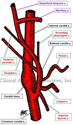
The image shows an anterior view of the right-side carotid system.
Images property of: CAA.Inc. Artist:Dr. E. Miranda
- Details
A [hordeolum] is an inflamed Meibomian gland (also known as a tarsal gland). These glands are found at the edge of the eyelids and their lipid product (meibum) helps seal the eyes and prevent tear evaporation. The vernacular term for an hordeolum is "stye" or "sty".
The term arises from the Latin [hordeum] meaning "barley". It refers to the appearance of the inflamed gland to a barleycorn. The eponymic term "Meibomiam gland" honors Heinrich Meibom (1638-1700), a German anatomist who first described the tarsal glands of the eye.
The removal of a hordeolum is called a "hordeolectomy"
The accompanying image shows a five-day old hordeolum.
Image courtesy of Wikipedia


