


Medical Terminology Daily - Est. 2012

Medical Terminology Daily (MTD) is a blog sponsored by Clinical Anatomy Associates, Inc. as a service to the medical community. We post anatomical, medical or surgical terms, their meaning and usage, as well as biographical notes on anatomists, surgeons, and researchers through the ages. Be warned that some of the images used depict human anatomical specimens.

You are welcome to submit questions and suggestions using our "Contact Us" form. The information on this blog follows the terms on our "Privacy and Security Statement" and cannot be construed as medical guidance or instructions for treatment.


We have 2004 guests online


A Moment in History

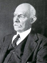
William S. Halsted, MD

William S. Halsted, MD
(1852 – 1922)

American anatomist, teacher, and surgeon, William Stewart Halsted was born in New York City, USA to a wealthy family of English origin. His father was involved in charitable work and Governor and trustee to a city hospital. Not a brilliant student initially, Halsted took an undergraduate in Liberal Arts in Yale, CT., after which he entered the Medical College of Physicians at the Columbia College, where he excelled.

As a second-year medical student Halsted applied and obtained a position in surgery at a local hospital. In here he learned about Lister’s antiseptic technique and became an adamant proponent of it to reduce infection. In 1877 Halsted obtained his MD. After a short time as House Physician at the New York Hospital, Halsted traveled to Europe to further his education studying for two years at the Universities of Vienna, Leipzig, and W?rzburg.

Besides being at the forefront of surgical and antiseptic techniques (introducing the use of rubber gloves in surgery), Halsted was extremely concerned with the way medical students were taught in the US. He pioneered bedside clinical round discussions with the medical students after two years of basic sciences studies. Halsted developed the idea of a patient chart; he also developed the residency program for medical students in use today.

Halsted is probably the most influential researcher and surgeon at the turn of the century. He dedicated time to the study of intestinal anastomoses and the use of silk as a suture material. His experimental work in 1887 proved that the inclusion of the submucosa layer in an anastomosis was mandatory, as well that a single layered anastomosis was enough to attain closure. Perhaps Halsted’s most important contribution was the application and use of the scientific method to surgical questions. Halsted’s principles, also known as  "Halsted's Rules of Surgery", set the standards used today in surgical suturing and surgical stapling.

He also pioneered the development and surgical techniques for radical mastectomy as a treatment for breast cancer.

As a side effect of this studied in anesthesia and the use of cocaine for anesthesia, Halsted became addicted to this substance, a problem that followed him through the years. Without impairing his capacity as a researcher and a surgeon, Halsted eventually recovered. He died in Baltimore in 1922 as a complication to surgery.

Sources:
1. Dubay, A. D., & Franz, G. M. (2003). Acute Wound Healing: The Biology of Acute Wound Failure. Surg Clin NA, 83, 463-481.
2. Halsted, W. S. (1887). Circular Suture of the Intestine - An Experimental Study. Am J Med Sci, 436-461.
3. “William Stewart Halsted: his life and contributions to surgery” Osborne, P. Lancet Oncol 2007; 8: 256–65
4. “William Stewart Halsted: Surgical pioneer” Burress, P Endoc Today (2010), 8: (2) 22
5. “William Stewart Halsted (1852–1922) Neurological stamp” Haas, LF J Neurol Neurosurg Psych 2000;69:641
Original image courtesy of "Images from the History of Medicine" at  www.nih.gov


 "Clinical Anatomy Associates, Inc., and the contributors of "Medical Terminology Daily" wish to thank all individuals who donate their bodies and tissues for the advancement of education and research”.

Click here for more information


bookplateink.com


This article is part of the series "A Moment in History" where we honor those who have contributed to the growth of medical knowledge in the areas of anatomy, medicine, surgery, and medical research.

John Marshall
John Marshall (1818 – 1891)

John Marshall (1818 – 1891) British anatomist and surgeon. Initially, he apprenticed with Dr. Wales in Wisbech. In 1838 he entered the University College, London, where he studied with William Sharpey (1802 – 1880), who was then teaching physiology. In 1849 Marshall became a Fellow of the Royal College of Surgeons of England.

In 1845 he became a demonstrator of anatomy at University College, London and in 1847 with the help of Jones Quain (1796 – 1865) and William Sharpey he was appointed as assistant surgeon.

Marshall was appointed professor of surgery at University College in 1866, and in 1884 he was appointed consulting surgeon to University College Hospital.

He was known as a great artist and used his abilities to teach anatomy with the help of blackboards. He lectured on anatomy to art students, and one of his books is titled “Anatomy for Artists” (London, 1878), which was received with great enthusiasm by his art students.

John Marshall wrote several books and works, mentioned in the “Sources” section of this article, but he is probably better known for his association with the eponymic “Ligament of Marshall”. In 1850, he published a detailed description (36 pages) of the embryology of the cardinal veins, the superior vena cava, and the presence of a persistent “left superior vena cava” with a description of the oblique vein of the left atrium, the obliterated Duct of Cuvier, and the fold of pericardium over this duct that today carries his name.

The paper was published in the journal “Philosophical Transactions of the Royal Society”, and the title was “On the Development of the Great Anterior Veins Man and Mammalia; including an Account of certain remnants of Fœtal Structure found in the Adult, a Comparative View of these Great Veins the different and an Analysis of their occasional peculiarities in the Human Subject”. The sketches that accompany this seminal paper were drawn by him.

Plate 1 Marshall

Marshall Plate 5

 If you are interested in reading this paper, it is available online at the Royal Society website. For the actual publication in PDF format, click here. 

Marshall also is recognized for being one of the first to use electrocautery in surgery, as well as being a pioneer in the removal of lower extremity varicose veins. He was the first to demonstrate that cholera can be spread through contaminated drinking water.

Ligament of Marshall (yellow arrow)

The image shows the ligament of Marshall (yellow arrow), the left pulmonary artery (LPA), and the left superior pulmonary vein (LSPV).

Personal note: My thanks to an attendee to one of my cardiovascular lectures. He asked who was Marshall of the "ligament of Marshall:... Here is the answer. Dr. Miranda.

Sources and publications:
1. “A Description of the Human Body, its Structure and Functions” London, 1860, 4to, with folio plates; 4th ed. 1883.
2. “Anatomy for Artists” London, 1878, royal 8vo; 2nd ed. 1883; 3rd edit. 1890.
3. “A Rule of Proportion for the Human Figure” 1878, fol.
4 “A Series of Life-size Anatomical Diagrams” seven sheets.
5. “Physiological Diagrams” life size, eleven sheets.
6. “On the Relations between the Weight of the Brain and its Parts, and the Stature and Mass of the Body” 1892, J Anat Physiol
7. “The Brain of the late George Grote” 1892, J Anat Phys
8. “On the Development of the Great Anterior Veins Man and Mammalia; including an Account of certain remnants of Fœtal Structure found in the Adult, a Comparative View of these Great Veins the different and an Analysis of their occasional peculiarities in the Human Subject” 1850 Phil Trans R Soc 140:133 - 170
9. Portrait of J. Marshall by Alphonse Legros, Courtesy of Wikipedia. Public Domain.