Medical Terminology Daily (MTD) is a blog sponsored by Clinical Anatomy Associates, Inc. as a service to the medical community. We post anatomical, medical or surgical terms, their meaning and usage, as well as biographical notes on anatomists, surgeons, and researchers through the ages. Be warned that some of the images used depict human anatomical specimens.
You are welcome to submit questions and suggestions using our "Contact Us" form. The information on this blog follows the terms on our "Privacy and Security Statement" and cannot be construed as medical guidance or instructions for treatment.
We have 924 guests online

Georg Eduard Von Rindfleisch
(1836 – 1908)
German pathologist and histologist of Bavarian nobility ancestry. Rindfleisch studied medicine in Würzburg, Berlin, and Heidelberg, earning his MD in 1859 with the thesis “De Vasorum Genesi” (on the generation of vessels) under the tutelage of Rudolf Virchow (1821 - 1902). He then continued as a assistant to Virchow in a newly founded institute in Berlin. He then moved to Breslau in 1861 as an assistant to Rudolf Heidenhain (1834–1897), becoming a professor of pathological anatomy. In 1865 he became full professor in Bonn and in 1874 in Würzburg, where a new pathological institute was built according to his design (completed in 1878), where he worked until his retirement in 1906.
He was the first to describe the inflammatory background of multiple sclerosis in 1863, when he noted that demyelinated lesions have in their center small vessels that are surrounded by a leukocyte inflammatory infiltrate.
After extensive investigations, he suspected an infectious origin of tuberculosis - even before Robert Koch's detection of the tuberculosis bacillus in 1892. Rindfleisch 's special achievement is the description of the morphologically conspicuous macrophages in typhoid inflammation. His distinction between myocardial infarction and myocarditis in 1890 is also of lasting importance.
Associated eponyms
"Rindfleisch's folds": Usually a single semilunar fold of the serous surface of the pericardium around the origin of the aorta. Also known as the plica semilunaris aortæ.
"Rindfleisch's cells": Historical (and obsolete) name for eosinophilic leukocytes.
Personal note: G. Rindfleisch’s book “Traité D' Histologie Pathologique” 2nd edition (1873) is now part of my library. This book was translated from German to French by Dr. Frédéric Gross (1844-1927) , Associate Professor of the Medicine Faculty in Nancy, France. The book is dedicated to Dr. Theodore Billroth (1829-1894), an important surgeon whose pioneering work on subtotal gastrectomies paved the way for today’s robotic bariatric surgery. Dr. Miranda.
Sources:
1. "Stedmans Medical Eponyms" Forbis, P.; Bartolucci, SL; 1998 Williams and Wilkins
2. "Rindfleisch, Georg Eduard von (bayerischer Adel?)" Deutsche Biographie
3. "The pathology of multiple sclerosis and its evolution" Lassmann H. (1999) Philos Trans R Soc Lond B Biol Sci. 354 (1390): 1635–40.
4. “Traité D' Histologie Pathologique” G.E.
Rindfleisch 2nd Ed (1873) Ballieres et Fils. Paris, Translated by F Gross
"Clinical Anatomy Associates, Inc., and the contributors of "Medical Terminology Daily" wish to thank all individuals who donate their bodies and tissues for the advancement of education and research”.
Click here for more information
- Details

This is a Greek compound word. [ana-] meaning "through or complete", the root term [-stom-] from [stoma], meaning "mouth or opening" and the suffix [-osis] meaning "condition". Usually [-osis] refers to a disease, but in this case it refers to an action or process. The plural form for [anastomosis] is [anastomoses].
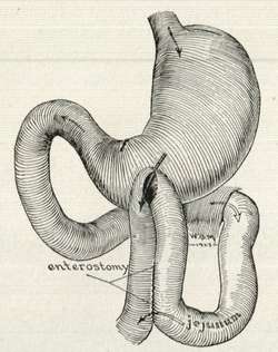
The word [anastomosis] then refers to the "process or action of creating a complete opening". In reality an anastomosis is the process of creating a permanent opening between two structures which allows for drainage or flow from one structure into the other. The suffix [-ostomy], being the term for "drainage" is the best suffix to describe an anastomosis. An anastomosis can be the result of a surgical procedure or found as a natural occurrence, such as the anastomoses found in the arterial circle of Willis or an abnormal fistula.
The accompanying image shows an early 1900's depiction of an anterior gastrojejunostomy. The term [gastrojejunostomy] is then the "creation of a drainage opening (a common mouth or opening) between the stomach and the jejunum, the second portion of the small intestine.
It is our belief and core competency, that surgical medical device sales representatives and managers should be completely familiar with this medical term, as well as the many types of anastomoses that can be performed surgically. To this end, we have developed a video on the History of Surgical Stapling. At the end of a video you can see a demonstration of the use of these devices to create an anastomosis
- Details
The [falx cerebri] is a falciform or sickle-shaped median extension of the dura mater found within the cranial cavity. It is narrow anteriorly, where it attaches to the crista galli of the ethmoid bone and widens posteriorly, where it ends when it meets another dura mater extension, the tentorium cerebelli.
The falx cerebri is found in the interhemispheric fissure of the brain, separating both cerebral hemispheres. Its inferior free edge is closely related to the corpus callosum.
Reduplications of the dura mater forming the falx cerebri creates venous channels known as "sinuses". The superior border of the falx cerebri, where it meets the dura mater covering the brain forms the [superior longitudinal sinus]. The inferior free border of the falx cerebri forms the [inferior longitudinal sinus], and at the junction of the falx cerebri with the tentorium cerebelli we find the [longitudinal sinus].
The Latin word [falx] (plural [falcis]) means "scythe" or "sickle".
Original image, public domain, courtesy of Wikipedia
- Details
UPDATED: The root for this word comes from the Latin [falx] (plural [falcis]) meaning "scythe" or "sickle". [Falciform] means "shaped like a sickle or a scythe". The term [falx cerebri] is used to name the sickle-shaped median extension of the dura mater that separates both cerebral hemispheres.
There are several falciform structures in the human body, such as the falciform ligament of the lung, more commonly known as the pulmonary ligament, and the falciform margin of the saphenous opening, the edge of a foramen in the femoral fascia to allow passage of the greater saphenous vein.
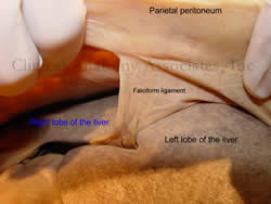
The most commonly known use of the term is the "falciform ligament of the liver", a sickle-shaped extension of peritoneum that connects the liver capsule (Glisson's) to the peritoneal covering of the abdominal wall (parietal peritoneum). At the free edge of the falciform ligament and covered by peritoneum run the round ligament of the liver (an embryological remnant of fetal circulation) and the paraumbilical veins.
- Details
The medical term [vaccine] originates from the Latin term [vacca] meaning "cow". The Latin adjectival form is [vaccinus].
The word originated from the work of Edward Jenner (1749 - 1823) who, based in observation, developed an injection of cowpox virus and injected them into patients, rendering them inmune to smallpox. This extraordinary discovery was the basis for the Balmis Expedition to the New World.
Since the cowpox virus was extracted initally from cows (Lat:vacca), and later from human cowpox sores, and Latin was at the time the preferred language of medical communication, the word "vaccina" was created. Evolution of the word led to the English [vaccine] and Spanish [vacuna]
- Details
The prefix [-ect(o)-] originates from the Greek [εκτός] meaning "outside". In medical terminology it is used to mean "outside", "external", or "superficial". Applications of this prefix include:
- Ectoderm: "derm" means "skin". This term is used in embryology to denote the external embryonic layer that will give origin to the skin and the nervous system
- Ectopic: "topic" means "place" or "location". Outside its place
Note: The links to Google Translate in these articles include an icon that will allow you to hear the Greek or Latin pronunciation of the word.
- Details
The term [volar] is used in human anatomy and orthopedics to denote either the palm of the hand or the sole of the feet. The volar surface of the hand is the anterior aspect or palm, whereas the volar surface of the feet is the inferior aspect or sole of the foot. How we came to use this word as such is a strange trip through the evolution and use of words.
The word [volar] originates from the Latin[vola] meaning "to fly". If you look online the definition of many for [volar] is "The hollow of the palms of the hands and soles of the feet". Very far from "to fly". The word was first used for the palms of the hands when mimicking a a flying bird. The hollow of the hands forces the air, hence [vola]. In fact, in Spanish the word for "to fly" is "volar" and in Italian, "volare". The term [volar] was then used by extension for the soles of the feet.
The term to refer to the palms of the hands and soles of the feet is in disuse except in orthopedics and can be found in some anatomy books. Overtime the word has been changed to [palmar] in the hand, and to [plantar] in the feet. This is why we have a muscle called the "palmaris longus" and we talk about the "plantar fascia".
Note: The links to Google Translate in these articles include an icon that will allow you to hear the Greek or Latin pronunciation of the word.


