")


This article is part of the series "A Moment in History" where we honor those who have contributed to the growth of medical knowledge in the areas of anatomy, medicine, surgery, and medical research.

Philippo Verheyen
Philippo Verheyen (1648 – 1710). Flemish physician and anatomist, Philipo (Philip) Verheyen was born in the city of Verrebroek in Belgium, worked as a farmer in his early years and was probably expected to be a farmer for the rest of his life. The local Catholic parish pastor Johannes Jaspars, recognizing his intelligence, started teaching him Latin and letters with the intention of having Philip become a priest.
In 1672, he was sent to the Trinity College in Leuven where he started his studies in arts and later in theology. He continued his studies in Theology at the Collegium Sancti Spiritus (College of the Holy Spirit). During the latter part of his studies Verhayen was already wearing a priest’s collar. In 1675 Verheyen suffered an injury that lead to gangrene and eventual amputation of his left leg. This injury prevented him from taking his final vows as a priest.
Verheyen enrolled at the University of Leuven College Of Medicine where after 3 years he became a physician. Unhappy with his mostly theoretical medical knowledge Verhayen moved to Leyden to continue his studies with Frederick Ryus and others.
In 1683 Verheyen returns to Leuven where he presents his doctoral thesis which is accepted. In August 1669 elected Rector Magnificus at the University of Leyden, one of the highest honors ever bestowed on him. This is why Verheyen's statue is among the personalities honored at the town hall in Leuven, Belgium.
Verheyen was a prolific writer including several books and manuscripts. His main work “Corporis Humani Anatomiae”, a two book work, had twelve editions, some in other languages besides Latin. This book became one of the most used anatomy books until the middle of the 18th century. Interestingly, Verheyen relates his life and studies at the beginning of his book in a chapter call Compendium Vitae (CV). Only the first edition was published while Verheyen was alive.
The following excerpt of his book "Corporis Humani Anatomiae Liber Primus" reads : " ... QuintusAuricularis, quia cum minimum sit, auribus expurgandis est aptissimus" translates as"..the fifth (finger, called) Auricularis, because how small it is, is most suitable to clean the ears". Incredibly, anatomists at that time called the fifth digit "digitus auricularis".
Verheyen is credited with the creation of the eponym the “Achilles tendon” which denominates the common tendon for the gastrocnemius and soleus muscle, although at the time he called it the “Chorda Achillis”. He also described the kidneys in detail, especially the arterial “stars” found on the surface of the kidney, which are today known as the “Stars of Verheyen”.
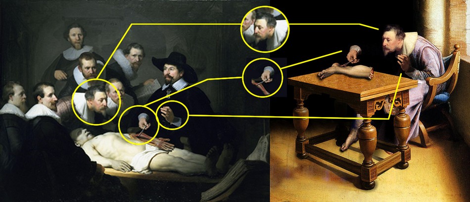
In 2005 an article appeared on the World Wide Web with a painting showing Verhayen dissecting his own amputated leg. This initially “anonymous” oil pintingis actually a Photoshop-edited image which incorporates parts of the painting “The Anatomy Lesson of Dr. Nicolaes Tulp” by Rembrandt. This work was made by ShrestaRit Premnath. This is a conceptual artwork depicting the concept of amputation, but not an actual oil painting, as shown by the image analysis depicted at the bottom of this article.
The story of Verheyen keeping and dissecting his own leg is most probably a myth for several reasons. Verhayen was poor and did not have enough money to go to Leyden to have his leg amputated there as the myth suggests. Preservation techniques were not too good at the time and when Verheyen had his leg amputated he had yet to enter medical school and be introduced to the art of human anatomy. Granted, he had dissected animals before in the farm, but it is doubtful that this myth is true.

"Verheyen dissecting his own leg"

Verheyen's stars on the surface of a human kidney.

Verheyen's statue at Louven's Town Hall.

Title page of Verheyen's Corporis Humani Anatomiae.
Click on the image for a larger version.
The image above shows a comparison between Rembrandt's painting “The Anatomy Lesson of Dr. Nicolaes Tulp” and an "anonymous" oil painting that appeared on the Internet circa 2005.
This oil painting include elements from Rembrandt's work. First, the head of "Verheyen" has been copied and flipped horizontally, This causes "Verheyen's" neck position to be awkward for the job of dissection. Second, the right hand holding the dissector and Achilles' tendon is Dr. Tulp's hand that has been slightly rotated, including the instrument anf the cuff. Third, the left hand is also that of Dr. Tulp, but not the cuff. If you look closely you will notice that both cuffs are different!
I have not been able to find the original painting on which these elements were added. My guess is that the left leg depicted on the table is also added. Interestingly, the table shows perspective problems.
In 2023 I had the honor of being invited by the University of Antwerp in Belgium to speak at the 2023 Vesalius Triennial Meeting in the city of Antwerp, Belgium. While there I visited Verrebroek, the city where Verheyen was born.
Personal note: I am proud to have in my library catalog a copy of the second edition of Philippo Verheyen's “Corporis Humani Anatomiae, Liber Primus”, published in 1710 in Brussels by the printer house of Fraters Serstevens. The book is signed by an Ex-Libris by "Hanolet". I have not been able to find more information on this prior owner of this book. An image of the title page is depicted in this article. Dr. Miranda
Sources
1. “Philip Verheyen (1648-1710) and his Corporis Humani Anatomiae” Suy R. Acta chir belg, 2007, 107, 343-354
2. “Achilles tendon: the 305th anniversary of the French priority on the introduction of the famous anatomical eponym” Musil, V. et al Surg Radiol Anat (2011) 33:421–427
3. "Philip Verheyen" Research and movie by digitalstories.be by Hedwige Daenens
Original image courtesy of the National Library of Medicine