
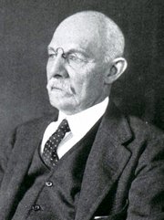



This article is part of the series "A Moment in History" where we honor those who have contributed to the growth of medical knowledge in the areas of anatomy, medicine, surgery, and medical research.
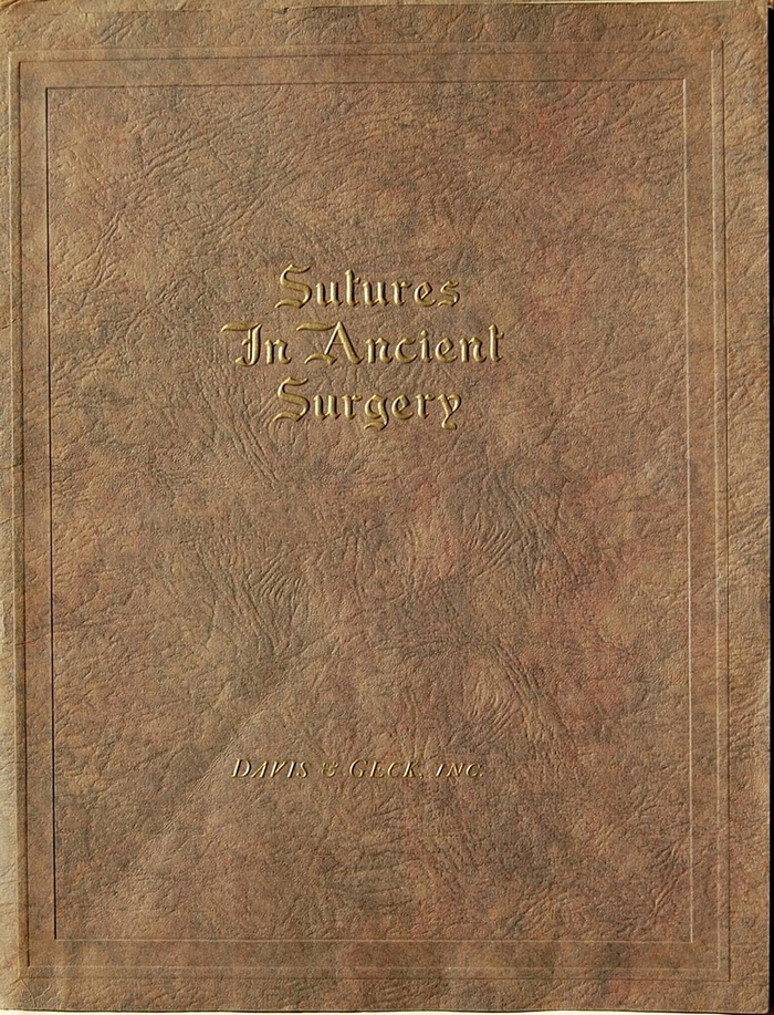
Folder of "Sutures in Ancient Surgery"
This article is the continuation of The interesting story of “Sutures in Ancient Surgery” (1).
3. The content of the publication, images, and controversy
Each tableaux vivant depicted a scene of history of surgery, with a short description of the individual and their accomplishments. In general, these descriptions are accurate, but very short. The objective was to allow the doctor to read the text, admire the image and leave time for the medical representative to deliver their presentation of a particular product.
Each page was printed in black and white, on a high quality paper measuring 12 by 9 ¼ inches. The paper itself was originally lightly yellowed to denote that this was an historical image. In fact, many doctors believed that these were reproductions of antique paintings, which they were not! The image itself was 6 ½ by 5 ¼ inches.
In some images it is clear that the background was painted. In fact, the image of Hyeronimus Fabricius Ab Aquapendente (157-1619) published in 1927, shamelessly copies part of the background of the 1555 title page of the Fabrica of Andreas Vesalius (1514-1564), or one of the many plagiarisms of the same book.
The controversy today lies with a number of women depicted with their nude breasts. In one case, (“Aspasia”, published in 1933) the photograph depicts five women including Aspasia, all of them showing a naked breast. Although “Sutures in Ancient Surgery” is criticized for this easy depiction of nudity, only 30% fall in this category.
Adding Aspasia of Miletus to this collection of physicians also brought criticism, not only for the nudity, but for the fact that Aspasia is a legendary figure whose life is in question as well as her place in Greek society. Some say that she was a prostitute and the regent of a house of ill repute, other say that she was a teacher of philosophy along with Socrates and that she was married to Pericles. Others say that she never existed, yet her name and life has been used in a novelized two-tome book. Why was she selected for this collection? No one knows.
Following are some images from "Sutures in Ancient Surgery".
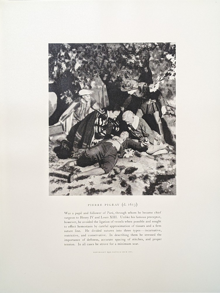 Pierre Pigray (d.1613)
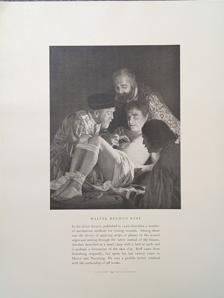
Walter Herman Ryff
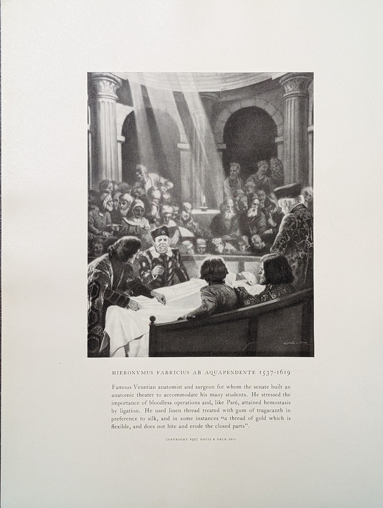 Fabricius Ab Aquapendente (1537-1619)
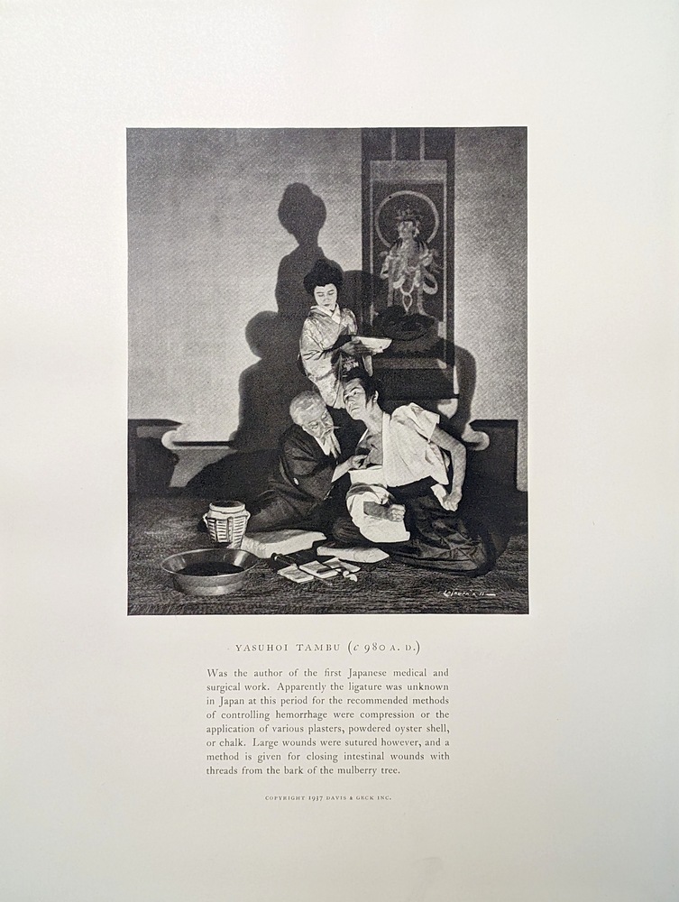
Yasuhoi Tambu (c. 980 A.D.)
4. Missing prints
Because these prints were presented as loose-leaf items to the doctors, many of them have been lost. Some historians comment that doctors would frame these images and use them in their offices, so a complete collection of the 83 photographs is extremely difficult to find. In once case, it is known that a hospital had the complete collection framed in one of its hallways (Sobieszek, 1988) which hospital, or what happened to that collection is unknown.
Today, the “Sutures in Ancient Surgery” folders found for sale in antique libraries have a varying number of prints, all of them incomplete. Some universities list them in their libraries and in one case, they have only three prints in their collection!
My research has been able to find the names of 76 of the tableaux vivants in this collection, but I have not been able to find the names of seven of these prints.
The folder that I received from Jack Kennedy contains 75 prints, and I am looking for the last eight including that of Teodorico Borgognoni, year of printing unknown.
If you have any or know of someone that owns a few in this collection, please contact me. I prepared an PDF file with the prints I have as well as those I do not. You can download it here.
Once again, thanks to Jack Kennedy for his addition to my collection. It has been an interesting research and I look forward to the next one!!
Sources:
1. UConn library Archives and Special Collections: Davis and Geck company records.
2. SNAC-Social Network and Archival Context: Davis and Geck Company.
3. “Soak it up: Examining bioabsorbable polymers in healthcare” Medical Plastic News
4. “The striking social tableaux vivants of Lejaren à Hillier” J.T.H. Connor. Hetkoen International
5. “Photography view; when tableaux vivants flowered in the magazines” Gene Thorton. The New York Times. March 2, 1986.
6.” Tableaux Vivants: A Long History of Recreating Art” Karolina Dzimira-Zarzycka. Daily Art May 1, 2020
7. “Medical History’s Moment in Art Photography (1920 to 1950): How Lejaren à Hillier and Valentino Sarra Created a Fashion for Scenes of Early Surgery” Journal of the History of Medicine and Allied Sciences, Volume 72, Issue 4, October 2017, Pages 381–421
8. “Aspasia” Wiki,org
9. “The Art of Persuasion: A History of Advertising Photography” Sobieszek, Robert A. 1988
10. “Aspasia” A Romance of Art and Love in Ancient Hellas” Mary J Safford German) Robert Hamerling (Translator) 1882. NY. W.S. Gottsberger, Publisher.
11. “Lejaren A. Hillier Photography Collection” University of Texas, Harry Ramson Center.