Sponsor   

Medical Terminology Daily (MTD) is a blog sponsored by Clinical Anatomy Associates, Inc. as a service to the medical community, medical students, and the medical industry. We post anatomical, medical or surgical terms, their meaning and usage, as well as biographical notes on anatomists, surgeons, and researchers through the ages. Be warned that some of the images used depict human anatomical specimens.

Click on the link below to subscribe to the MTD newsletter. If you think an article could be interesting to somebody else, feel free to forward the link of the article. Should you want to use the information on the article, please follow the CAA, Inc Privacy and Security Statement found at the bottom of this page. 

You are welcome to submit questions and suggestions using our "Contact Us" form. The information on this blog follows the terms on our "Privacy and Security Statement"  and cannot be construed as medical guidance or instructions for treatment. 


We have 155 guests and no members online


A Moment in History

Self-portrait, Henry Vandyke Carter, MD (Public Domain)

Henry Vandyke Carter, MD
(1831 – 1897)

English physician, surgeon, medical artist, and a pioneer in leprosy and mycetoma studies.  HV Carter was born in Yorkshire in 1831. He was the son of Henry Barlow Carter, a well-known artist and it is possible that he honed his natural talents with his father. His mother picked his middle name after a famous painter, Anthony Van Dyck. This is probably why his name is sometimes shown as Henry Van Dyke Carter, although the most common presentation of his middle name is Vandyke.

Having problems to finance his medical studies, HV Carter trained as an apothecary and later as an anatomical demonstrator at St. George’s Hospital in London, where he met Henry Gray (1872-1861), who was at the time the anatomical lecturer. Having seen the quality of HV Carter’s drawings, Henry Gray teamed with him to produce one of the most popular and longer-lived anatomy books in history: “Gray’s Anatomy”, which was first published in late 1857.  The book itself, about which many papers have been written, was immediately accepted and praised because of the clarity of the text as well as the incredible drawings of Henry Vandyke Carter.

While working on the book’s drawings, HV Carter continued his studies and received his MD in 1856.

In spite of initially being offered a co-authorship of the book, Dr. Carter was relegated to the position of illustrator by Henry Gray and never saw the royalties that the book could have generated for him. For all his work and dedication, Dr. Carter only received a one-time payment of 150 pounds. Dr.  Carter never worked again with Gray, who died of smallpox only a few years later.

Frustrated, Dr. Carter took the exams for the India Medical Service.  In 1858 he joined as an Assistant Surgeon and later became a professor of anatomy and physiology. Even later he served as a Civil Surgeon. During his tenure with the India Medical Service he attained the ranks of Surgeon, Surgeon-Major, Surgeon-Lieutenant-Colonel, and Brigade-Surgeon.

Dr. Carter dedicated the rest of his life to the study of leprosy, and other ailments typical of India at that time. He held several important offices, including that of Dean of the Medical School of the University of Bombay. In 1890, after his retirement, he was appointed Honorary Physician to the Queen.

Dr. Henry Vandyke Carter died of tuberculosis in 1897.

Personal note: Had history been different, this famous book would have been called “Gray and Carter’s Anatomy” and Dr. Carter never gone to India. His legacy is still seen in the images of the thousands of copies of “Gray’s Anatomy” throughout the world and the many reproductions of his work available on the Internet. We are proud to use some of his images in this blog. The image accompanying this article is a self-portrait of Dr. Carter. Click on the image for a larger depiction. Dr. Miranda

Sources:
1. “Obituary: Henry Vandyke Carter” Br Med J (1897);1:1256-7
2. “The Anatomist: A True Story of ‘Gray’s Anatomy” Hayes W. (2007) USA: Ballantine
3. “A Glimpse of Our Past: Henry Gray’s Anatomy” Pearce, JMS. J Clin Anat (2009) 22:291–295
4. “Henry Gray and Henry Vandyke Carter: Creators of a famous textbook” Roberts S. J Med Biogr (2000) 8:206–212.
5. “Henry Vandyke Carter and his meritorious works in India” Tappa, DM et al. Indian J Dermatol Venereol Leprol (2011) 77:101-3


"Clinical Anatomy Associates, Inc., and the contributors of "Medical Terminology Daily" wish to thank all individuals who donate their bodies and tissues for the advancement of education and research”.

Click here for more information


Rare & Collectible Books at AbeBooks.com 

bookplateink.com


Anatomical variations (2)

"No anatomical structure has the moral obligation to be where they are supposed to be"

Not only may an anatomical structure be absent, such as in the case of renal aplasia or agenesis, or in the case of a non-existent circumflex coronary artery, but sometimes extra structures can be found. Such is the case where a kidney can present two or even three ureters, all functional. Double inferior vena cavae, cervical ribs, lumbar ribs, the list goes on and on!

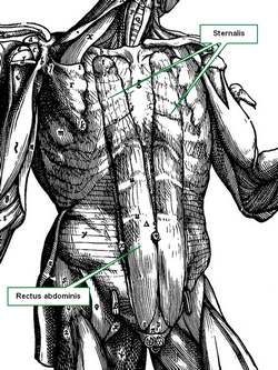
Muscles can be added to this list, again, with absence of a muscle, or with new and completely unexpected attachments. An example of this is the presence of a continuation of the rectus abdominis muscle into the chest region, a variation called a sternalis muscle.

The accompanying image shows the sternalis muscle in one of the "muscle plates" of De Humani Corporis Fabrica Libri Septem, published in 1543 by Andreas Vesalius. This image was criticized by showing a muscle that does not exist, although Vesalius clearly stated in the text of his book that this was an anatomical variation that he had seen.

For many decades surgeons had to operate and "see what they could find". There were the days of the exploratory laparotomy. After the discovery of the application of X-rays by Wilhem Konrad Roentgen (1845 - 1923) and the incredible advances in imaging techniques including CT-scan, MRI, PET, etc, the surgeon is now not usually surprised by anatomical variations.

Sternalis muscle (Andreas Vesalius 1543)
There are areas in the body that have an high rate of anatomical variation, such as the hepatobiliary region, which includes the "Triangle of Calot". In this area, the standard anatomy is found only in 64% of the cases! In the rest, expect the unexpected. Lahey (1948) states "...the fact that cholecystectomy is a dangerous operation. It is dangerous unless one realizes.... that anomalous anatomy is very common". Today the dangers are less, because of better visualization and technology, but anatomical variations are still there.

Another area where anatomical variations are extremely important is the heart's coronary circulation. Anatomical variations can cause different cardiac dominance. Normal anatomy states that there are two coronary arteries, yet, up to five separate coronary arteries arising directly from the ascending aorta have been described! There is one variation where the left coronary arises from the right coronary artery, effectively having only one artery arise from the aorta and being in charge of all the arterial supply to the heart. What happens if this single artery stenoses? Bear in mind that this is not an "anomalous" vessel, it is just an anatomical variation.

Sources:
1. Lahey DH, discussing the paper "Partial Hepatectomy with Intrahepatic Cholangiojejunostomy" by Wilson H, and Gillespie CE, Ann Surg. 1949 June; 129(6): 756–765
2. "Renal aplasia is the predominant cause of congenital solitary kidneys" Hiraoka, M et al Kidney Int. 2002 May;61(5):1840-4.

This article is the second in a series of three; Click here for the first article
TO CONTINUE READING THE NEXT ARTICLE: CLICK HERE

Back to MTD Main Page Subscribe to MTD