Medical Terminology Daily (MTD) is a blog sponsored by Clinical Anatomy Associates, Inc. as a service to the medical community. We post anatomical, medical or surgical terms, their meaning and usage, as well as biographical notes on anatomists, surgeons, and researchers through the ages. Be warned that some of the images used depict human anatomical specimens.

You are welcome to submit questions and suggestions using our "Contact Us" form. The information on this blog follows the terms on our "Privacy and Security Statement" and cannot be construed as medical guidance or instructions for treatment.

We have 91 guests online


A Moment in History

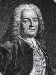
Jean-Louis Petit

Jean Louis Petit
(1674 – 1750)

French surgeon and anatomist, Jean Louis Petit was born in Paris in on March 13, 1674.  His family rented an apartment at his house to Alexis Littre (1658 – 1726), a French anatomist. Petit became an apprentice of Littre at seven years of age, helping him in the dissections for his lectures and at an early age became the assistant in charge of the anatomic amphitheater.

Because of Petit’s dedication to anatomy and medicine, in 1690 at the age of sixteen, became a disciple of a famous Paris surgeon, Castel.

In 1692, Petit entered the French army and performed surgery in two military campaigns. By 1693 he started delivering lectures and was accepted as a great surgeon, being invited to the most difficult operations.  In 1700 he was appointed Chief Surgeon of the Military School in Paris and in the same year he received the degree of Master of Surgery from the Faculty of Paris.

In 1715 he was made a member of the Royal Academy of Sciences and an honorary member of the Royal Society of London. He was appointed by the King as the first Director General of the Royal Academy of Surgery when it was founded in 1731.

Petit’s written works are of historical importance.  “Traite des Maladies des Os” ( A Treatise on Bone Diseases);  “Traite des Maladies Chirurgicales et des Operation” (A Treatise on Surgical Diseases and their Operations” This last book was published posthumously in 1774. He also published a monograph on hemorrhage, another on lachrymal fistula, and others.

He was one of the first to perform choIecystotomy and mastoidotomy. His original tourniquet design for amputations saved many in the battlefield and the design of the same surgical instrument today has not changed much since its invention by him.

His name is remembered in the lumbar triangle, also called the "triangle of Petit", and the abdominal hernia that can ensue through that area of weakness, the lumbar hernia or "Petit's hernia".

Sources:
1. “Jean Louis Petit – A Sketch of his Life, Character, and Writings” Hayne, AP San Fran Western Lancet 1875 4: 446-454
2. “Oeuvres compl?tes de Jean-Louis Petit” 1837 Imprimerie de F. Chapoulaud
3. Extraits de l'eloge de Jean-Louis Petit Ius dans Ia seance publique de I' Academie royale de chirurgie du 26 mai 1750” Louis A. Chirurgie 2001: 126 : 475- 81


 "Clinical Anatomy Associates, Inc., and the contributors of "Medical Terminology Daily" wish to thank all individuals who donate their bodies and tissues for the advancement of education and research”.

Click here for more information


abebooks banner

bookplateink.com


Hamate

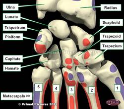
The Hamate bone is one of the four bones that comprise the distal row of the carpus or carpal bones that form the wrist. The name arises from the Latin [hamatus], meaning "hooked". The hamate bone has a distinct hook-like bony process in its volar (anterior) surface, known as the hamulus. This bone is also known as the "unciform bone" (from the Latin [uncus], also meaning "hook") or the os hamatum.

The lunate bone has a wedge-like shape and six surfaces (as a die). It articulates with five bones, including the lunate bone, capitate, triquetrum, and the fourth and fifth metacarpal bones.

The hook of the hamate bone is one of the distal boundaries of the carpal tunnel and serves as a pulley for the tendons of the fourth and fifth flexor tendons. It also serves as one of the points of muscular attachment for the following muscles: flexor carpi ulnaris, flexor digit minimi, and opponens digiti minimi. Because of its projection into the palm of the hand, the hamulus is involved in injuries in sports that require the athlete to use an accessory, as in racquetball, tennis, baseball, golf, etc.

The accompanying image shows the anterior (volar) surface of the wrist. Click on the image for a larger picture.

Scaphoid bone - anterior (volar) view of the wrist

Image modified from the original: "3D Human Anatomy: Regional Edition DVD-ROM." Courtesy of Primal Pictures


Back to MTD Main Page Subscribe to MTD

Phlebotomy

The word itself arises from the Greek. The root term [-phleb-] derives from [φλέβα] (phleba) meaning "vein", and the suffix [-otomy], meaning "to cut" or "to open". Let's not forget that the suffix component  [-y] means "process of". So [phlebotomy] is the "process (or action) of cutting open a vein"

For centuries a standard practice in medicine was to "bleed" a patient, by opening a vein under controlled conditions and letting some blood flow. The practice was known as "bloodletting" or phlebotomy. Not in use today, it is said that excessive bloodletting contributed to the death of George Washington, having removed 5 pints of blood in one day!. Today the professionals who draw blood are called "phlebotomists"

The image (circa 1860) depicts one of the only known three photographs of a bloodletting procedure. Observe the lack of aseptic technique.


Image by The Burns Archive, courtesy of Wikipedia.org.

Back to MTD Main Page Subscribe to MTD

Iliopubic tract

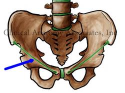
The iliopubic tract is a thickening of the transversalis fascia found in direct relation, immediately posterior to the inguinal (Poupart's) ligament. As the inguinal ligament, the iliopubic tract extends between the anterior superior iliac spine (ASIS) superolaterally, and the pubic tubercle inferomedially. 

This obscure structure has been brought up to light because it is one of the anatomical landmarks used in laparoscopic herniorrhaphy. When securing a mesh to reinforce the posterior abdominal wall, and also prevent mesh migration, the surgeon will place sutures, tacks, or staples in this structure. Since the iliopubic tract (posteriorly) and the inguinal ligament (anteriorly) are so close together, they are both secured when doing this procedure.

Inguinal ligamentImage property of: CAA.Inc.Artist: D.M. Klein
The image shows the location of the inguinal ligament. The iliopubic tract is immediately posterior to it. 
Back to MTD Main Page Subscribe to MTD

Clavicle

Left clavicle, superior surface. Public domain
Left clavicle, superior surface

The clavicle is part of the anterior portion of the shoulder girdle. It is an elongated bone with an "italic S" curvature. The Latin term for clavicle is [clavicula], and it has two root terms: [-clavic-] and [-clav-]. This is why we have the terms [subclavicular], and [subclavian] both meaning the same: "inferior to the clavicle".

The clavicle articulates medially with the manubrium of the sternum (see image on this article) by way of the sternoclavicular joint. This joint contains a meniscus. Laterally, the clavicle articulates with the acromial process or acromium of the scapula.

The clavicle has the muscular insertions of several muscles: sternocleidomastoid, trapezius, pectoralis major, deltoid, subclavius, and sternohyoid.

Sources:
1. "The Origin of Medical Terms" Skinner, HA 1970 Hafner Publishing Co.
2 "Tratado de Anatomia Humana" Testut et Latarjet 8 Ed. 1931 Salvat Editores, Spain
3. "Anatomy of the Human Body" Henry Gray 1918. Philadelphia: Lea & Febiger
Image modified by CAA, Inc. Original image by Henry Vandyke Carter, MD., courtesy of bartleby.com


Foramen transversarium

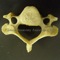
The term [foramen transversarium] is Latin for "transverse foramen". It refers to bilateral foramina (openings) found lateral to the vertebral body in the cervical vertebrae. These foramina are found only in cervical vertebrae and serve as a good way to identify them.

Through the foramina transversaria (plural form) pass the vertebral artery and vertebral vein. The vertebral artery is one of the first branches to arise off the subclavian arteries. While the vertebral vein passes through all seven foramina transversaria, the vertebral artery does not pass through the foramen transversarium of the seventh cervical vertebra (vertebra prominens).

Image property of: CAA.Inc. Photographer: David M. Klein

Vertebra
Back to MTD Main Page Subscribe to MTD

Carl Wernicke


This article is part of the series "A Moment in History" where we honor those who have contributed to the growth of medical knowledge in the areas of anatomy, medicine, surgery, and medical research.To search all the articles in this series, click here.

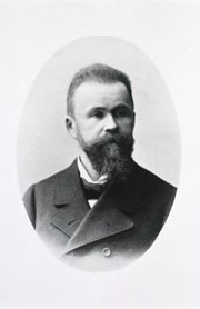
Carl Wernicke (1848-1905). German psychiatrist, neurologist, and neurosurgeon, Carl Wernicke was born in 1848 in the town of Tarnowitz, in what was then Prussia. He studied medicine in Breslau, Poland. In 1817 he became an assistant psychiatrist at a Breslau hospital. Fascinated with the discoveries and publications of Paul Broca on localized brain damage and aphasia, Wernicke left his post for a time to work with Theodor Meynert in Vienna. At that time Meynert was considered an authority in neuropsychiatry. In 1874, soon after his return to Breslau, Wernicke published his ideas and findings in aphasia in a revolutionary  publication "The Aphasia Symptom Complex". Wernicke was only 26.

At the time, the general outlook on brain activity was that the brain worked in localized areas. Carl Wernicke's ideas were that although this was true, the functionality of the brain resided in the connections between the different areas of the brain. His ideas were right. Wernicke described what later would be known as "sensory aphasia" or the eponymic "Wernicke's aphasia".

Wernicke was a pioneer in the surgical treatment of hydrocephalus, as well as the surgical treatment of brain abcesses. He published several books, including a brain atlas. Carl Wernicke died as the consequence of a bycicle accident in 1905.

Sources:
1. "Pioneers in Neurology: Carl Wernicke (1848–1905)" Pillmann, F. J Neurol (2003) 250 : 1390–1391
2. "The scientific history of hydrocephalus and its treatment" Aschoff, A.; Ashemi, P.; Kunze, S.Neurosurg Rev (1999) 22:67–93
3. "Aphasia" Marshall,RS; Lazar, RM;PhD, Mohr,JP. Medical Update for Psychiatrists. Elsevier (1998)3;5:132–138


Original image courtesy of
Images from the History of Medicine.

Back to MTD Main Page Subscribe to MTD