Medical Terminology Daily (MTD) is a blog sponsored by Clinical Anatomy Associates, Inc. as a service to the medical community. We post anatomical, medical or surgical terms, their meaning and usage, as well as biographical notes on anatomists, surgeons, and researchers through the ages. Be warned that some of the images used depict human anatomical specimens.

You are welcome to submit questions and suggestions using our "Contact Us" form. The information on this blog follows the terms on our "Privacy and Security Statement" and cannot be construed as medical guidance or instructions for treatment.


We have 404 guests online


A Moment in History

Jean-Louis Petit

Jean Louis Petit
(1674 – 1750)

French surgeon and anatomist, Jean Louis Petit was born in Paris in on March 13, 1674.  His family rented an apartment at his house to Alexis Littre (1658 – 1726), a French anatomist. Petit became an apprentice of Littre at seven years of age, helping him in the dissections for his lectures and at an early age became the assistant in charge of the anatomic amphitheater.

Because of Petit’s dedication to anatomy and medicine, in 1690 at the age of sixteen, became a disciple of a famous Paris surgeon, Castel.

In 1692, Petit entered the French army and performed surgery in two military campaigns. By 1693 he started delivering lectures and was accepted as a great surgeon, being invited to the most difficult operations.  In 1700 he was appointed Chief Surgeon of the Military School in Paris and in the same year he received the degree of Master of Surgery from the Faculty of Paris.

In 1715 he was made a member of the Royal Academy of Sciences and an honorary member of the Royal Society of London. He was appointed by the King as the first Director General of the Royal Academy of Surgery when it was founded in 1731.

Petit’s written works are of historical importance.  “Traite des Maladies des Os” ( A Treatise on Bone Diseases);  “Traite des Maladies Chirurgicales et des Operation” (A Treatise on Surgical Diseases and their Operations” This last book was published posthumously in 1774. He also published a monograph on hemorrhage, another on lachrymal fistula, and others.

He was one of the first to perform choIecystotomy and mastoidotomy. His original tourniquet design for amputations saved many in the battlefield and the design of the same surgical instrument today has not changed much since its invention by him.

His name is remembered in the lumbar triangle, also called the "triangle of Petit", and the abdominal hernia that can ensue through that area of weakness, the lumbar hernia or "Petit's hernia".

Sources:
1. “Jean Louis Petit – A Sketch of his Life, Character, and Writings” Hayne, AP San Fran Western Lancet 1875 4: 446-454
2. “Oeuvres compl?tes de Jean-Louis Petit” 1837 Imprimerie de F. Chapoulaud
3. Extraits de l'eloge de Jean-Louis Petit Ius dans Ia seance publique de I' Academie royale de chirurgie du 26 mai 1750” Louis A. Chirurgie 2001: 126 : 475- 81


 "Clinical Anatomy Associates, Inc., and the contributors of "Medical Terminology Daily" wish to thank all individuals who donate their bodies and tissues for the advancement of education and research”.

Click here for more information


abebooks banner

bookplateink.com


Sinus Rhythm

Normal electrocardiogram - sinus rhythm
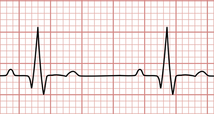
Sinus rhythm electrocardiogram

Sinus Rhythm (SR), also known as Normal Sinus Rhythm (NSR) is the term used to denote normal cardiac activity. A normal beating heart is in sinus rhythm.

In the normal beating of the heart the left and right atria contract followed by the contraction of the left and right ventricles. These atrial and ventricular contractions are separated by a lag time of 1/10th of a second which allows the atria to relax (atrial diastole) while the ventricles contract (ventricular systole). The addition of the normal action of the four heart valves (tricuspid, mitral, aortic, and pulmonary) allows the heart to work as a blood pump and circulate blood within the heart and through the body and lungs.

SR is coordinated by a complex binary conduction system. The first part of the conduction system is composed of heart muscle cells (cardiomyocytes) which are characterized by automaticity. It is important to state here something that is known, but many times ignored: This portion of the conduction system is not composed of nerves... it is formed by specialized cardiac muscle cells! The description of this cardiomyocyte-based system can be read in a separate article here.

The second component of the conduction system is the cardiac autonomic nervous system (CANS), which acts as a moderator (accelerating or slowing) the cardiomyocyte-based conduction system. A separate article will be written soon and linked here. Suffice it to say (for now) that the CANS has two components, an extrinsic component formed by nerves and ganglia outside the heart, and an intrinsic component of nerves and ganglia that forms a complex network within the superficial aspect of the heart muscle and fat surrounding the heart. These are the ganglionated plexuses, and their dysfunction can cause abnormal heart rhythm.

When these two components and their thousands of cardiac muscle cells, ganglia, and nerves are working property, the heart is in sinus rhythm, and beats between 60 to a 100 times per minute.

Personal note: The term Normal Sinus Rhythm (NSR) is redundant. By definition, if the heart is in SR, it is normal. On the other hand, the term Abnormal Sinus Rhythm is also not correct… if the heartbeat is abnormal, the heart is not in SR! (Dr. Miranda)