Medical Terminology Daily (MTD) is a blog sponsored by Clinical Anatomy Associates, Inc. as a service to the medical community. We post anatomical, medical or surgical terms, their meaning and usage, as well as biographical notes on anatomists, surgeons, and researchers through the ages. Be warned that some of the images used depict human anatomical specimens.

You are welcome to submit questions and suggestions using our "Contact Us" form. The information on this blog follows the terms on our "Privacy and Security Statement" and cannot be construed as medical guidance or instructions for treatment.


We have 79 guests online


A Moment in History

Jean-Louis Petit

Jean Louis Petit
(1674 – 1750)

French surgeon and anatomist, Jean Louis Petit was born in Paris in on March 13, 1674.  His family rented an apartment at his house to Alexis Littre (1658 – 1726), a French anatomist. Petit became an apprentice of Littre at seven years of age, helping him in the dissections for his lectures and at an early age became the assistant in charge of the anatomic amphitheater.

Because of Petit’s dedication to anatomy and medicine, in 1690 at the age of sixteen, became a disciple of a famous Paris surgeon, Castel.

In 1692, Petit entered the French army and performed surgery in two military campaigns. By 1693 he started delivering lectures and was accepted as a great surgeon, being invited to the most difficult operations.  In 1700 he was appointed Chief Surgeon of the Military School in Paris and in the same year he received the degree of Master of Surgery from the Faculty of Paris.

In 1715 he was made a member of the Royal Academy of Sciences and an honorary member of the Royal Society of London. He was appointed by the King as the first Director General of the Royal Academy of Surgery when it was founded in 1731.

Petit’s written works are of historical importance.  “Traite des Maladies des Os” ( A Treatise on Bone Diseases);  “Traite des Maladies Chirurgicales et des Operation” (A Treatise on Surgical Diseases and their Operations” This last book was published posthumously in 1774. He also published a monograph on hemorrhage, another on lachrymal fistula, and others.

He was one of the first to perform choIecystotomy and mastoidotomy. His original tourniquet design for amputations saved many in the battlefield and the design of the same surgical instrument today has not changed much since its invention by him.

His name is remembered in the lumbar triangle, also called the "triangle of Petit", and the abdominal hernia that can ensue through that area of weakness, the lumbar hernia or "Petit's hernia".

Sources:
1. “Jean Louis Petit – A Sketch of his Life, Character, and Writings” Hayne, AP San Fran Western Lancet 1875 4: 446-454
2. “Oeuvres compl?tes de Jean-Louis Petit” 1837 Imprimerie de F. Chapoulaud
3. Extraits de l'eloge de Jean-Louis Petit Ius dans Ia seance publique de I' Academie royale de chirurgie du 26 mai 1750” Louis A. Chirurgie 2001: 126 : 475- 81


 "Clinical Anatomy Associates, Inc., and the contributors of "Medical Terminology Daily" wish to thank all individuals who donate their bodies and tissues for the advancement of education and research”.

Click here for more information


abebooks banner

bookplateink.com


Esophageal hiatus

Esophagogastric junction

UPDATED:  The esophageal hiatus is one of the seven hiatuses found in the respiratory diaphragm allowing passage of structures between the thorax and abdomen. As it name implies, the esophageal hiatus is the passageway for the esophagus. It also allows passage of the anterior and posterior vagus nerves, (CN X).

The hiatus is bound by two muscular crura, both of which arise from the right tendinous aortic crus. Since the intraabdominal pressure is higher than the intrathoracic pressure, there is a series of structures at the phrenoesophagogastric junction to close the esophageal hiatus.

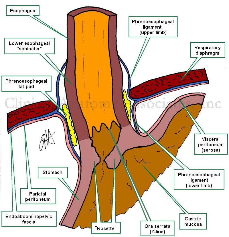
The infradiaphragmatic parietal peritoneum reflects off the diaphragm towards the stomach to form its serosa layer (visceral peritoneum). At the same time the infradiaphragmatic fascia, also known as the  endoabdominopelvic fascia, splits into two components or limbs. These are the superior and inferior phrenoesophageal ligaments or phrenoesophageal membranes. (the root [-phren-] means "diaphragm"). These phrenoesophageal ligaments create a disc-like plug between the abdomen and the thorax. This "plug" is reinforced by a circular infradiaphragmatic fat pad. The phrenoesophageal ligaments are reinforced on their thoracic aspect by the endothoracic fascia.

The lower esophagus has a dilation (evident in the image) called the "esophageal ampulla", in relation to this dilation the circular muscle layer of the esophagus slightly thickens creating the so-called "lower esophageal sphincter". This area is not a true anatomical sphincter, but rather is a functional sphincter. 

The esophagogastric mucosal junction shows a marked transition in the shape of a wavy line. This is called the Z-line or the ora serrata. Extensions of the gastric mucosa and submucosa inferior to the ora serrata create a valve-like flap called the "gastroesophageal flap valve". When viewing this mucosal flap through and endoscope, it looks corrugated and flower-like, hence it is also called the "rosette". 

The congenital or pathological dilation of the esophageal hiatus can predispose to esophageal hiatus hernia.

Sources:
1 "Tratado de Anatomia Humana" Testut et Latarjet 8 Ed. 1931 Salvat Editores, Spain
2. "Anatomy of the Human Body" Henry Gray 1918. Philadelphia: Lea & Febiger
Original image by Dr. E. Miranda